. 2004 Sep 23;3(1):8.
doi: 10.1186/1476-5926-3-8.

Overview of the diagnostic value of biochemical markers of liver fibrosis (FibroTest, HCV FibroSure) and necrosis (ActiTest) in patients with chronic hepatitis C

Affiliations

Overview of the diagnostic value of biochemical markers of liver fibrosis (FibroTest, HCV FibroSure) and necrosis (ActiTest) in patients with chronic hepatitis C

Thierry Poynard et al. Comp Hepatol. .

Abstract

SUMMARY: BACKGROUND: Recent studies strongly suggest that due to the limitations and risks of biopsy, as well as the improvement of the diagnostic accuracy of biochemical markers, liver biopsy should no longer be considered mandatory in patients with chronic hepatitis C. In 2001, FibroTest ActiTest (FT-AT), a panel of biochemical markers, was found to have high diagnostic value for fibrosis (FT range 0.00-1.00) and necroinflammatory histological activity (AT range 0.00-1.00). The aim was to summarize the diagnostic value of these tests from the scientific literature; to respond to frequently asked questions by performing original new analyses (including the range of diagnostic values, a comparison with other markers, the impact of genotype and viral load, and the diagnostic value in intermediate levels of injury); and to develop a system of conversion between the biochemical and biopsy estimates of liver injury. RESULTS: A total of 16 publications were identified. An integrated database was constructed using 1,570 individual data, to which applied analytical recommendations. The control group consisted of 300 prospectively studied blood donors. For the diagnosis of significant fibrosis by the METAVIR scoring system, the areas under the receiver operating characteristics curves (AUROC) ranged from 0.73 to 0.87. For the diagnosis of significant histological activity, the AUROCs ranged from 0.75 to 0.86. At a cut off of 0.31, the FT negative predictive value for excluding significant fibrosis (prevalence 0.31) was 91%. At a cut off of 0.36, the ActiTest negative predictive value for excluding significant necrosis (prevalence 0.41) was 85%. In three studies there was a direct comparison in the same patients of FT versus other biochemical markers, including hyaluronic acid, the Forns index, and the APRI index. All the comparisons favored FT (P < 0.05). There were no differences between the AUROCs of FT-AT according to genotype or viral load. The AUROCs of FT-AT for consecutive stages of fibrosis and grades of necrosis were the same for both moderate and extreme stages and grades. A conversion table was constructed between the continuous FT-AT values (0.00 to 1.00) and the expected semi-quantitative fibrosis stages (F0 to F4) and necrosis grades (A0 to A3). CONCLUSIONS: Based on these results, the use of the biochemical markers of liver fibrosis (FibroTest) and necrosis (ActiTest) can be recommended as an alternative to liver biopsy for the assessment of liver injury in patients with chronic hepatitis C. In clinical practice, liver biopsy should be recommended only as a second line test, i.e., in case of high risk of error of biochemical tests.


Figures

Figure 1
Figure 1
Meta-analysis of the AUROC observed in published studies of FibroTest diagnostic value. AUROCs were all significantly higher for FibroTest than the random 0.50 value (upper panel) (P < 0.001). AUROCs of FibroTest were significantly higher then AUROCs of other fibrosis markers (lower panel) (P < 0.05).
Figure 2
Figure 2
Diagnostic values of FibroTest according to genotype and viral load. Graph A: AUROCs of FibroTest for the diagnosis of significant fibrosis, according to HCV genotypes. There were no significant differences: Genotype 1, n = 684, AUROC = 0.76, 95% Confidence Interval (95CI) = 0.72–0.79; genotype 2, n = 140, AUROC = 0.79, 95CI = 0.70–0.85; genotype 3, n = 143 AUROC = 0.76, 95CI = 0.67–0.83; other genotype, n = 46, AUROC = 0.72, 95CI = 0.52–0.85. Graph B: AUROCs of ActiTest for the diagnosis of significant necrosis, according to HCV genotypes. There were no significant differences: Genotype 1, n = 684, AUROC = 0.81, 95% Confidence Interval (95CI) = 0.77–0.84; genotype 2, n = 140, AUROC = 0.90, 95CI = 0.83–0.94; genotype 3, n = 143, AUROC = 0.79, 95CI = 0.71–0.85; other genotype, n = 46, AUROC = 0.76, 95CI = 0.57–0.87. Graph C: AUROCs of FibroTest for the diagnosis of significant fibrosis, according to serum HCV viral load. There were no significant differences: High viral load, n = 215, AUROC = 0.71, 95% Confidence Interval (95CI) = 0.64–0.78; Low viral load, n = 183, AUROC = 0.73, 95CI = 0.65–0.80. Graph D: AUROCs of ActiTest for the diagnosis of significant necrosis, according to serum HCV viral load. There were no significant differences: High viral load, n = 215, AUROC = 0.74, 95% Confidence Interval (95CI) = 0.64–0.82; Low viral load, n = 183, AUROC = 0.75, 95CI = 0.65–0.82.
Figure 3
Figure 3
Conversion between FibroTest and fibrosis stages, and between ActiTest and necroinflammatory activity grades – Graphs. Graph A: FibroTest values according to status, from blood donors to patients with cirrhosis (n = 1570). Graph B: ActiTest values according to status, from blood donors to patients with severe necrosis (n = 1570). F0 = no fibrosis, F1 = portal fibrosis, F2 = some septa, F3 = many septa, F4 = cirrhosis, A0 = no necroinflammatory activity, A1 = minimal activity, A2 = moderate activity, A3 = severe activity. (Consensus conferences recommend treatment in patients with either F2 stage or A2 grade.) Notched box plots showing the relationship between FibroTest and the stage of fibrosis (A) and between ActiTest and the grade of activity (B). The horizontal line inside each box represents the median, and the width of each box the median ± 1.57 interquartile range/√n (to assess the 95% level of significance between group medians). Failure of the shaded boxes to overlap signifies statistical significance (P < 0.05). The horizontal lines above and below each box encompass the interquartile range (from 25th to 75th percentile), and the vertical lines from the ends of the box encompass the adjacent values (upper: 75th percentile plus 1.5 times interquartile range, lower 25th percentile minus 1.5 times interquartile range).
Figure 4
Figure 4
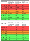
Conversion between FibroTest and fibrosis stages, and between ActiTest and necroinflammatory activity grades – Panels. Conversion between FibroTest and fibrosis stages using METAVIR, Knodell and Ishak fibrosis scoring systems (upper panel). Conversion between ActiTest and activity grades using METAVIR, Knodell and Ishak necroinflammatory activity scoring systems (lower panel).

References

    1. Afdhal NH. Diagnosing fibrosis in hepatitis C: is the pendulum swinging from biopsy to blood tests? Hepatology. 2003;37:972–974. doi: 10.1053/jhep.2003.50223. - DOI - PubMed
    1. Dienstag J. The role of liver biopsy in chronic hepatitis C. Hepatology. 2002;36:S152–S160. doi: 10.1053/jhep.2002.36381. - DOI - PubMed
    1. Bravo AA, Sheth SG, Chopra S. Liver biopsy. N Engl J Med. 2001;344:495–500. doi: 10.1056/NEJM200102153440706. - DOI - PubMed
    1. Regev A, Berho M, Jeffers LJ, Milikowski C, Molina EG, Pyrsopoulos NT, Feng ZZ, Reddy KR, Schiff ER. Sampling error and intraobserver variation in liver biopsy in patients with chronic HCV infection. Am J Gastroenterol. 2002;97:2614–2618. - PubMed
    1. Colloredo G, Guido M, Sonzogni A, Leandro G. Impact of liver biopsy size on histological evaluation of chronic viral hepatitis: the smaller the sample, the milder the disease. J Hepatol. 2003;39:239–244. doi: 10.1016/S0168-8278(03)00191-0. - DOI - PubMed

LinkOut - more resources