. 2004 Oct 5;101(40):14326-32.
doi: 10.1073/pnas.0405941101. Epub 2004 Sep 23.

Efficient reversal of Alzheimer's disease fibril formation and elimination of neurotoxicity by a small molecule

Affiliations

Efficient reversal of Alzheimer's disease fibril formation and elimination of neurotoxicity by a small molecule

Barbara J Blanchard et al. Proc Natl Acad Sci U S A. .

Abstract

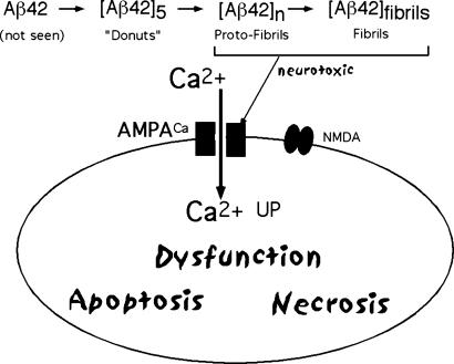
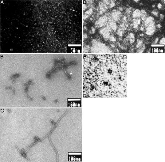
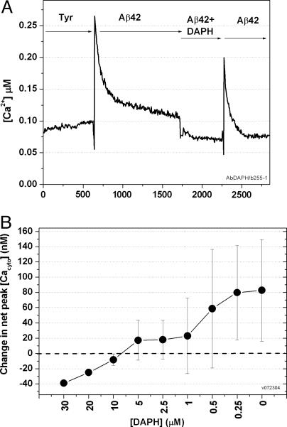
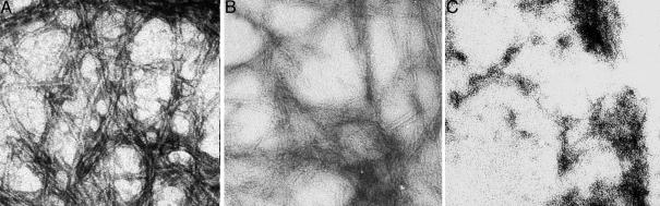
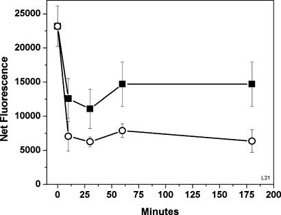
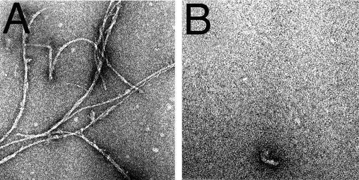
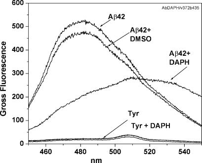
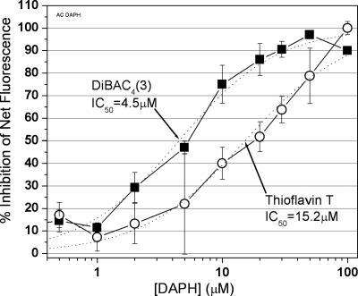
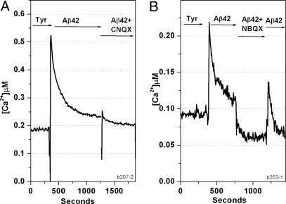
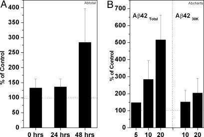
The Abeta1-42 peptide that is overproduced in Alzheimer's disease (AD) from a large precursor protein has a normal amino acid sequence but, when liberated, misfolds at neutral pH to form "protofibrils" and fibrils that are rich in beta-sheets. We find that these protofibrils or fibrils are toxic to certain neuronal cells that carry Ca-permeant alpha-amino-3-hydroxy-5-methyl-4-isoxazolepropionic acid (AMPA) receptors. Disrupting the structure of the Abeta1-42 fibrils and protofibrils might lead to the discovery of molecules that would be very useful in the treatment of AD. A high-throughput screen of a library of >3,000 small molecules with known "biological activity" was set up to find compounds that efficiently decrease the beta-sheet content of aggregating Abeta1-42. Lead compounds were characterized by using thioflavin T (ThT) as a beta-sheet assay. The most effective of six compounds found was 4,5-dianilinophthalimide (DAPH) under the following conditions: DAPH at low micromolar concentrations abolishes or greatly reduces previously existing fully formed Abeta1-42 fibrils, producing instead amorphous materials without fibrils but apparently containing some protofibrils and smaller forms. Coincubation of the Abeta1-42 peptide with DAPH produces either amorphous materials or empty fields. Coincubation of DAPH and Abeta1-42 greatly reduces the beta-sheet content, as measured with ThT fluorescence, and produces a novel fluorescent complex with ThT. When the Abeta1-42 peptide was coincubated with DAPH at very low micromolar concentrations, the neuronal toxicity mentioned above (Ca(2+) influx) was eliminated. Clearly, DAPH is a promising candidate for AD therapy.


Figures

Fig. 1.
Fig. 1.
Schematic representation of neurotoxicity. Misfolding of peptide Aβ42 produces a neurotoxic molecule.
Fig. 2.
Fig. 2.
Time course of aggregation of Aβ4230k at 10 μM in Tyrode's solution/2 mM Ca (pH 7.4) incubated at 37°C for the indicated times; negative-staining EM is shown. (A) t = 0 h. Arrow indicates one of the many donuts shown, which is enlarged in E. A larger and rarer aggregate is also shown. (B) t = 6 h. The “beads” are roughly the same size as the beads that make up the donuts in A and E. (C) t = 16 h. (D) t = 48 h. (E) t = 0 h. A portion of A (arrow) is enlarged to show a donut. The image is black/white-inverted for clarity.
Fig. 10.
Fig. 10.
DAPH eliminates Ca influx of Aβ42Total in CATH.a cells. (A) As described in the legend to Fig. 8, solutions were sequentially exchanged with 10 μM aggregated Aβ42Total, then Aβ42Total plus DAPH (coincubated at 10 μM plus 10 μM), and then Aβ42Total. Because DAPH is soluble only in DMSO, DMSO was present in all solutions. This experiment was repeated three times. (B) Effect of DAPH (30-0.25 μM) on Ca influx. The protocol used was as in A, except with DAPH concentrations ranging from 30-0.25 μM. (Variance is high; see Materials and Methods.) The corresponding concentration of DMSO was used for the controls.
Fig. 3.
Fig. 3.
DAPH reverses Aβ4230k fibrils. (A) Aβ4230k at 10 μM in Tyrode's solution/2 mM Ca (pH 7.4), incubated at 37°C for 24 h. (B) Incubation continued for another 24 h in the presence of vehicle (1% DMSO). (C) Incubation continued for another 24 h in the presence of 10 μM DAPH in DMSO.
Fig. 4.
Fig. 4.
Time course of reversal of fibril, formed by incubating 10 μM Aβ42Total in Tyrode's solution/2 mM Ca (pH 7.4) at 37° for 24 h. The β-sheet content was measured with ThT (see Materials and Methods). To obtain net values, the fluorescence of ThT in Tyrode's buffer was subtracted. ▪, Aggregated Aβ42Total plus 10 μM DAPH; ○, aggregated Aβ42Total plus 20 μM DAPH. The 0-h time points shown were Aβ42Total with only DMSO (the vehicle).
Fig. 5.
Fig. 5.
DAPH prevents Aβ4230k fibril formation. Aβ4230k was incubated without (A) and with (B) DAPH at a 10:10 μM dilution in Tyrode's solution/2 mM Ca (pH 7.4) at 37°C for 24 h. Samples were prepared by using negative-staining EM.
Fig. 6.
Fig. 6.
ThT spectrum of Aβ42Total plus DAPH suggests a new β-sheet conformation. To measure β-sheet content, ThT was added to Aβ42Total plus DAPH and coincubated at a dilution of 10:10 μM for 48 h at 37°C. An emission spectrum at Ex = 435 nm was measured with an f4500 spectrofluorimeter.
Fig. 7.
Fig. 7.
The IC50 value for the inhibition by DAPH of Aβ42Total, measured with ThT fluorescence, is 15.2 μM DAPH. Each data point is the mean of four experiments of 10 μM aggregated Aβ42Total plus DMSO, with varying DAPH concentrations. A logistics curve was fitted and then used to obtain the IC50 value. Parallel experiments using DiBAC4 (3) fluorescence are also shown; this IC50 value is ≈4.5 μM DAPH.
Fig. 8.
Fig. 8.
AMPA antagonists NBQX and CNQX eliminate Ca influx. CATH.a cells on a coverslip were loaded with the Ca-sensitive fluorescent dye fura-2 and measured at Ex = 340 and 380 nm and Em = 510 nm by using a microscope-photometry system obtained from Photon Technology International. Solutions were exchanged sequentially with 10 μM aggregated Aβ42Total, then Aβ42Total plus 50 μM CNQX (A) or 50 μM NBQX (B), and then Aβ42Total again. The NBQX effect is partially reversible.
Fig. 9.
Fig. 9.
Ca influx by aggregated Aβ42Total into CATH.a cells is concentration-dependent. (A) We incubated 10 μM aggregated Aβ42 Total at 37°C for 0, 24, and 48 h. (B) Aβ42Total at 5,10, and 20 μM (n = 1, 15, and 3, respectively) and Aβ4230k at 10 and 20 μM (n = 5 and 4, respectively) were incubated for 48 h at 37°C, then applied to neuronal CATH.a cells, and cytosolic Ca was measured by fura-2 fluorescence (n = 1, 15, and 3, respectively).

References

    1. Glenner, G. G. & Wong, C. W. (1984) Biochem. Biophys. Res. Commun. 122, 1131-1135. - PubMed
    1. Sisodia, S. S., Koo, E. H., Beyreuther, K., Unterbeck, A. & Price, D. L. (1990) Science 248, 492-495. - PubMed
    1. Yankner, B. A., Duffy, L. K. & Kirschner, D. A. (1990) Science 250, 279-282. - PubMed
    1. Lorenzo, A. & Yankner, B. A. (1994) Proc. Natl. Acad. Sci. USA 91, 12243-12247. - PMC - PubMed
    1. Ferreira, A., Lu, Q., Orecchio, L. & Kosik, K. S. (1997) Mol. Cell. Neurosci. 9, 220-234. - PubMed

Publication types

MeSH terms