

Improved venous suppression and spatial resolution with SENSE in elliptical centric 3D contrast-enhanced MR angiography
- PMID: 15389954
- PMCID: PMC2702220
- DOI: 10.1002/mrm.20216
Improved venous suppression and spatial resolution with SENSE in elliptical centric 3D contrast-enhanced MR angiography
Abstract
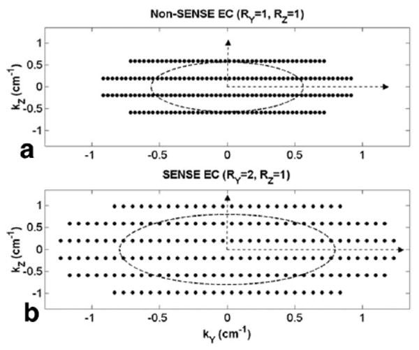
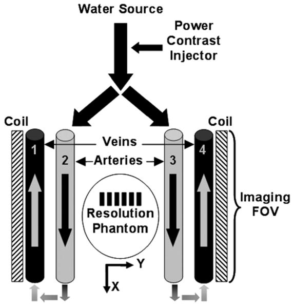
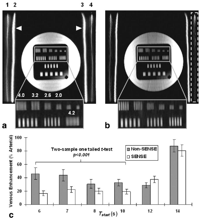
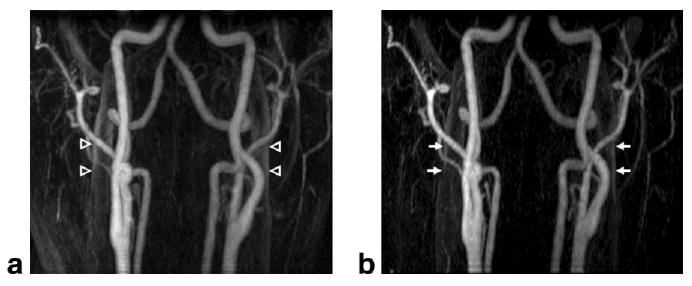
The elliptical centric (EC) view order samples a 3DFT acquisition from the center of k-space outward, and when applied to contrast-enhanced MR angiography (CE-MRA) provides intrinsic venous suppression. This is because the veins enhance several seconds after the scan is initiated, and are thus encoded solely by noncentral k-space frequencies. A separate method, sensitivity encoding (SENSE), accelerates the k-space sampling rate by reducing the phase FOV or, equivalently, by increasing the k-space sampling interval, and has been used to increase spatiotemporal resolution. We hypothesized that by combining SENSE with EC, sampling of central k-space would be accelerated and the k-space radius at which the veins first showed contrast enhancement would be increased over a reference scan, thus providing improved venous suppression and spatial resolution without additional scan time. This hypothesis was studied with the use of phantom and carotid CE-MRA experiments, and the results demonstrated an approximate 25% reduction in venous signal when SENSE was used.
Figures
References
-
- Prince MR, Yucel EK, Kaufman JA, Harrison DC, Geller SC. Dynamic gadolinium-enhanced three-dimensional abdominal MR arteriography. J Magn Reson Imaging. 1993;3:877–881. - PubMed
-
- Prince MR. Gadolinium-enhanced MR aortography. Radiology. 1994;191:155–164. - PubMed
-
- Prince MR, Grist TM, Debatin JF. 3D contrast MR angiography. 3rd. Berlin: Springer; 2003.
-
- Wilman AH, Riederer SJ. Improved centric phase encoding orders for three-dimensional magnetization-prepared MR angiography. Magn Reson Med. 1996;36:384–392. - PubMed
-
- Wilman AH, Riederer SJ, King BF, Debbins JP, Rossman PJ, Ehman RL. Fluoroscopically triggered contrast-enhanced three-dimensional MR angiography with elliptical centric view order: application to the renal arteries. Radiology. 1997;205:137–146. - PubMed
Publication types
MeSH terms
Substances
Grants and funding
LinkOut - more resources
Full Text Sources
