. 2005 Jan 15;385(Pt 2):605-12.
doi: 10.1042/BJ20041330.

Expression, purification, characterization and structure of Pseudomonas aeruginosa arylamine N-acetyltransferase

Affiliations

Expression, purification, characterization and structure of Pseudomonas aeruginosa arylamine N-acetyltransferase

Isaac M Westwood et al. Biochem J. .

Abstract

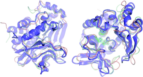
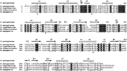
The gene for NAT (arylamine N-acetyltransferase) from Pseudomonas aeruginosa (panat) has been cloned from genomic DNA, and the gene product (PANAT) expressed as an N-terminal histidine-tagged protein in Escherichia coli and purified via nickel ion affinity chromatography. The specific activities of PANAT against a broad range of substrates have been investigated and compared with those of other prokaryotic NAT enzymes. For most arylamine substrates identified, PANAT exhibits in vitro specific activities typically one order of magnitude greater than those of recombinant NAT enzymes from Mycobacterium smegmatis or Salmonella typhimurium. Among the substrates of PANAT so far identified are the anti-tubercular drug isoniazid, 5-aminosalicylate (a drug used in the treatment of inflammatory bowel disease), as well as important environmental pollutants such as 3,4-dichloroaniline and 2-aminofluorene. As well as acetylating common NAT substrates, PANAT is unique among the prokaryotic NATs so far studied in acetylating the folate precursor 4-aminobenzoic acid and the folate catabolite 4-aminobenzoylglutamate. The recombinant protein has been expressed in sufficient quantity to allow protein crystallization, and we have subsequently determined the 1.95 A structure of PANAT by X-ray crystallography.


Figures

Figure 1
Figure 1. Endogenous expression of PANAT and purification of the recombinant enzyme
(A) Ethidium bromide-stained 1.8% (w/v) agarose gel showing the 855 bp fragment obtained after RT-PCR using total RNA isolated from Pseudomonas aeruginosa PA01 (lane 2). The absence of genomic DNA contamination was confirmed using a negative control with no reverse transcriptase in the RT-PCR experiment (lane 1). (B) Western blot showing PANAT detected in P. aeruginosa lysate using an antibody raised against STNAT at 1:10000 dilution (lane 1). Recombinant pure PANAT with a hexa-histidine tag (1.0 μg) was detected using the anti-STNAT antibody at a 1:50000 dilution (lane 2). (C) Purification of PANAT. Coomassie Blue-stained SDS/12%-PAGE gel showing the single-step purification of PANAT as described in the Experimental section. Lanes: M, markers; 1, insoluble lysate; 2, soluble lysate; 3, unbound lysate; 4, 10 mM imidazole wash; 5, 25 mM imidazole wash; 6, 125 mM imidazole wash; 7, 250 mM imidazole wash; 8, PANAT after 2 months at 4 °C.
Figure 2
Figure 2. Comparison of the specific activities of PANAT with those of other bacterial NATs
Shown is the rate of hydrolysis of AcCoA (nmol of CoA·min−1·mg of protein−1) in the presence of AcCoA (400 μM), substrate (500 μM) and NAT enzyme. PANAT from P. aeruginosa (black bars), MSNAT (grey bars) and STNAT (unshaded bars) were compared. MSNAT and STNAT data are literature values [17]. The same method was used for all three enzymes. Each bar represents the mean±S.D. from triplicate determinations. Abbreviations are as described in Table 1.
Figure 3
Figure 3. X-ray crystal structure of PANAT
Orthogonal views of the X-ray crystal structures of PANAT (blue/purple), MSNAT (orange) and STNAT (green) superimposed using the program O [32]. All images were generated using Aesop [M. E. M. Noble, unpublished work; details available from M. E. M. N. on request (martin@biop.ox.ac.uk)]. Representative final 2FobsFcal·αcalc electron density for the active-site triad of PANAT is contoured at 0.2e-Å-3 in green.
Figure 4
Figure 4. Multiple sequence alignment of selected bacterial NATs
The sequence alignment was constructed using ClustalX [51] and ESPript [52]. The secondary structural elements were identified from the PANAT structure using ESPript. α-Helices, η-helices, β-sheets and strict β-turns are denoted α, η, β and TT respectively. Grey stars indicate side chains for which multiple conformations were modelled. SwissProt accession numbers: P. aeruginosa, Q9HUY3; M. smegmatis, O86309; S. typhimurium, Q00267; M. tuberculosis, P96848. Similar amino acids are highlighted in boxes, and completely conserved residues are indicated by white lettering on a dark background.
Figure 5
Figure 5. Molecular surface and active-site architecture of PANAT and MSNAT
Molecular surfaces were calculated for both PANAT (A) and MSNAT (B) using the program Aesop [M. E. M. Noble, unpublished work; details available from M. E. M. N. on request (martin@biop.ox.ac.uk)]. Hydrophobicity was then calculated for each surface and underlying protein using the program GRID [50]. Surfaces are coloured on identical scales of increasing hydrophobicity from grey through green to yellow. Underlying secondary structural elements of PANAT (blue) and MSNAT (orange) and the active-site cysteine (ball and stick mode) are also shown.

Similar articles

Cited by

References

    1. Upton A., Johnson N., Sandy J., Sim E. Arylamine N-acetyltransferases – of mice, men and microorganisms. Trends Pharmacol. Sci. 2001;22:140–146. - PubMed
    1. Evans D. A. N-acetyltransferase. Pharmacol. Ther. 1989;42:157–234. - PubMed
    1. Delomâenie C., Fouix S., Longuemaux S., Brahimi N., Bizet C., Picard B., Denamur E., Dupret J. M. Identification and functional characterization of arylamine N-acetyltransferases in eubacteria: evidence for highly selective acetylation of 5-aminosalicylic acid. J. Bacteriol. 2001;183:3417–3427. - PMC - PubMed
    1. Weber W. W., Hein D. W. N-acetylation pharmacogenetics. Pharmacol. Rev. 1985;37:25–79. - PubMed
    1. Riddle B., Jencks W. P. Acetyl-coenzyme A:arylamine N-acetyltransferase. Role of the acetyl-enzyme intermediate and the effects of substituents on the rate. J. Biol. Chem. 1971;246:3250–3258. - PubMed

Publication types

MeSH terms

Substances

Associated data