

A stereological evaluation of secretin and gastric inhibitory peptide-containing mucosal cells of the perinatal small intestine of the pig
- PMID: 15447686
- PMCID: PMC1571350
- DOI: 10.1111/j.0021-8782.2004.00338.x
A stereological evaluation of secretin and gastric inhibitory peptide-containing mucosal cells of the perinatal small intestine of the pig
Abstract
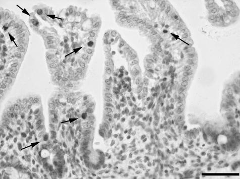
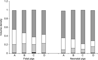
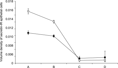
Stereological methods were used to quantify secretin and gastric inhibitory peptide (GIP)-immunoreactivity (GIP-IR) in paraffin sections of the duodenum, jejunum and ileum of fetal and neonatal piglets. In addition, sections were processed for GLP-1-immunohistochemistry. The volume density of the tunica mucosa increased after birth, giving rise to a decreased volume density of the tela submucosa and tunica muscularis. Generally known region-specific morphological distinctions were reflected in differing volume densities of the various layers. The highest volume density of GIP-IR epithelial cells was observed in the jejunum of the neonate. In contrast, the volume density of secretin-IR epithelial cells was highest in the duodenum of both fetal and neonatal piglets. The volume occupied by GIP-IR and secretin-IR epithelial cells increased in the jejunum after birth. Additionally, ileal secretin-IR epithelial cells were more numerous in the neonatal piglet. In conclusion, the quantitative and qualitative presence of GIP-IR and secretin-IR epithelial cells agree with earlier reports of their presence and co-localization between GIP-IR and GLP-1-IR, in the pig small intestine. Furthermore, the differences suggest that age- and region-related functional demands are temporally and probably causally related with the morphological diversification of the intestine and its endocrine cells.
Figures


Similar articles
-
The small intestine of the adult New Hampshire chicken: an immunohistochemical study.Anat Histol Embryol. 2011 Jun;40(3):163-8. doi: 10.1111/j.1439-0264.2010.01055.x. Epub 2010 Dec 6. Anat Histol Embryol. 2011. PMID: 21133986
-
A stereologic evaluation of glucagon-like peptide-1 (GLP-1) mucosal cells in the small intestine of the developing pig.Anat Embryol (Berl). 2002 May;205(2):153-7. doi: 10.1007/s00429-002-0235-z. Epub 2002 Apr 16. Anat Embryol (Berl). 2002. PMID: 12021917
-
Growth and morphological changes in the small and the large intestine in piglets during the first three days after birth.J Dev Physiol. 1992 Oct;18(4):161-72. J Dev Physiol. 1992. PMID: 1284564
-
Autoantibodies to duodenal gastric-inhibitory-peptide (GIP) cells and to secretin (S) cells in patients with coeliac disease, tropical sprue and maturity-onset diabetes.Clin Exp Immunol. 1980 Jul;41(1):33-42. Clin Exp Immunol. 1980. PMID: 7002390 Free PMC article.
-
On the physiology of GIP and GLP-1.Horm Metab Res. 2004 Nov-Dec;36(11-12):747-54. doi: 10.1055/s-2004-826158. Horm Metab Res. 2004. PMID: 15655703 Review.
Cited by
-
Pancreatic glucose-dependent insulinotropic polypeptide (GIP) (1-30) expression is upregulated in diabetes and PEGylated GIP(1-30) can suppress the progression of low-dose-STZ-induced hyperglycaemia in mice.Diabetologia. 2016 Mar;59(3):533-41. doi: 10.1007/s00125-015-3842-y. Epub 2015 Dec 22. Diabetologia. 2016. PMID: 26693710
-
Effects of secretin gene knockout on the diversity, composition, and function of gut microbiota in adult male mice.Front Cell Infect Microbiol. 2023 Dec 13;13:1257857. doi: 10.3389/fcimb.2023.1257857. eCollection 2023. Front Cell Infect Microbiol. 2023. PMID: 38156312 Free PMC article.
References
-
- Aiken KD, Roth KA. Temporal differentiation and migration of substance P, serotonin and secretin immunoreactive enteroendocrine cells in the mouse proximal small intestine. Dev. Dyn. 1992;194:303–310. - PubMed
-
- Aiken KD, Yu W, Wright JR, Roth KA. Adaptation of enteroendocrine cells in response to jejunal–ileal transposition in the rat. Gastroenterology. 1994;106:1576–1583. - PubMed
-
- Alumets J, Hakanson R, Sundler F. Ontogeny of endocrine cells in porcine gut and pancreas. Gastroenterology. 1983;85:1359–1372. - PubMed
-
- Argenzio RA. The pig as a model for studying the pathobiology of intestinal transport in infectious enteric disease. In: Tumbleson ME, Schook LB, editors. Advances in Swine in Biomedical Research. New York: Plenum Press; 1996. pp. 45–58.
-
- Burrin DG, Stoll B. Key nutrients and growth factors for the neonatal gastrointestinal tract. Clin. Perinatol. 2002;29:65–96. - PubMed
MeSH terms
Substances
LinkOut - more resources
Full Text Sources
