. 2004 Oct 5;101(40):14367-72.
doi: 10.1073/pnas.0405586101. Epub 2004 Sep 24.

A poxvirus protein forms a complex with left-handed Z-DNA: crystal structure of a Yatapoxvirus Zalpha bound to DNA

Affiliations

A poxvirus protein forms a complex with left-handed Z-DNA: crystal structure of a Yatapoxvirus Zalpha bound to DNA

Sung Chul Ha et al. Proc Natl Acad Sci U S A. .

Abstract

A conserved feature of poxviruses is a protein, well characterized as E3L in vaccinia virus, that confers IFN resistance on the virus. This protein comprises two domains, an N-terminal Z-DNA-binding protein domain (Zalpha) and a C-terminal double-stranded RNA-binding domain. Both are required for pathogenicity of vaccinia virus in mice infected by intracranial injection. Here, we describe the crystal structure of the Zalpha domain from the E3L-like protein of Yaba-like disease virus, a Yatapoxvirus, in a complex with Z-DNA, solved at a 2.0-A resolution. The DNA contacting surface of Yaba-like disease virus Zalpha(E3L) closely resembles that of other structurally defined members of the Zalpha family, although some variability exists in the beta-hairpin region. In contrast to the Z-DNA-contacting surface, the nonbinding surface of members of the Zalpha family are unrelated; this surface may effect protein-specific interactions. The presence of the conserved and tailored Z-DNA-binding surface, which interacts specifically with the zigzag backbone and syn base diagnostic of the Z-form, reinforces the importance to poxvirus infection of the ability of this protein to recognize the Z-conformation.


Figures

Fig. 1.
Fig. 1.
Sequence and secondary structural comparison of yabZαE3L with the other Z-DNA-binding domains and related poxvirus E3L domains. (A) Multiple sequence alignment of the Zα proteins. Numbering and secondary structure elements for the Zα domains of E3L from YLDV (yabZαE3L), YMTV (ymtvZαE3L), vaccinia virus (vvZαE3L), Orf virus (ovZαE3L), and Swinepox virus (svZαE3L), along with human ADAR1 (hZαADAR1) and mouse DLM1 (mZαDLM1) (accession numbers as in ref. , except YMTV NC 005179). Secondary structure is drawn on top of the sequence. The α-helix and β-sheet are represented by tubes and arrows, respectively. The conserved sequences of all Zα family are colored yellow. Residues from yabZαE3L that make contact with the DNA are marked with a blue triangle. (B) The overall structure of the yabZαE3L:Z-DNA complex. The N and C terminus and secondary structure elements are labeled. (C) Structural superposition of yabZαE3L:Z-DNA (sky blue), hZαADAR1-Z-DNA (red), mZαDLM1-Z-DNA (green), and vvcZαE3L (royal blue). All DNA atoms and structurally aligned Cα atoms were used for the superposition (64 Cα for hZαADAR1 and 57 Cα for ZαDLM).
Fig. 2.
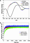
Fig. 2.
Interaction of yabZαE3L with Z-DNA. (A) Z-DNA binding of yabZαE3L as measured by CD. Unbound (dC-dG)6 is in the B-form under the buffer conditions used here (green curve). In the presence of a 2.5:1 (base pair:protein) molar ratio of DNA to protein (yabZαE3L in blue, hZαADAR1 in red and mZαDLM-1 in green), the DNA adopts the Z-conformation, as shown by the CD spectra. All curves are identical. (B) Kinetic measurement of DNA changing from B-DNA to Z-DNA in the presence of yabZαE3L, hZαADAR1, or mZαDLM1. The change in DNA conformation with time is measured by the change in ellipticity at 255 nm. hZαADAR1 and yabZαE3L show similar kinetics of conversion, whereas mZαDLM1 acts slightly more slowly.
Fig. 3.
Fig. 3.
Comparison of contacts between yabZαE3L or hZαADAR1 with Z-DNA. (A) Stereo view of the protein-DNA interface of the yabZαE3L:Z-DNA between chain A and chain C. (B) Stereo view of the protein-DNA interface of ZαADAR1:Z-DNA. Both diagrams are viewed from the C-terminal end of the recognition helix (α3). Water molecules are shown in green. Hydrogen bonds are drawn as dashed lines. The tyrosine on the recognition helix is oriented in an edge-on view. (C and D) Schematic diagrams of the protein-DNA interactions in the yabZαE3L:Z-DNA (C) and hZαADAR1:Z-DNA (D) complexes. Hydrogen bonds are represented by dashed lines and van der Waals contacts by open circles. The CH-π interaction between the conserved Tyr and a syn-guanosine is indicated by filled circles. Waters are shown by green ovals.
Fig. 4.
Fig. 4.
Surface charge distributions of yabZαE3L, hZαADAR1, mZαDLM1, and vvZαE3L (from left to right) viewed along the DNA-binding cleft (Upper) and rotated 180° on the back side (Lower). The red and blue areas represent the negatively and positively charged surfaces, respectively. Skeletal representations showing the bound Z-DNA strand are found in the first three diagrams.

Similar articles

Cited by

References

    1. Lee, H. J., Essani, K. & Smith, G. L. (2001) Virology 281, 170-192. - PubMed
    1. Whittaker, D. & Glaister, J. R. (1985) Lab. Anim. 19, 177-179. - PubMed
    1. Schielke, J. E., Kalishman, J., Liggitt, D. & Bielefeldt-Ohmann, H. (2002) Contemp. Top. Lab. Anim. Sci. 41, 27-29. - PubMed
    1. Rouhandeh, H., Vafai, A. & Kilpatrick, D. (1984) J. Ultrastruct. Res. 86, 100-105. - PubMed
    1. Knight, J. C., Novembre, F. J., Brown, D. R., Goldsmith, C. S. & Esposito, J. J. (1989) Virology 172, 116-124. - PubMed

Publication types

Associated data

LinkOut - more resources