Review
. 2004 Dec 1;384(Pt 2):201-32.
doi: 10.1042/BJ20041142.

The protein structures that shape caspase activity, specificity, activation and inhibition

Affiliations
Review

The protein structures that shape caspase activity, specificity, activation and inhibition

Pablo Fuentes-Prior et al. Biochem J. .

Abstract

The death morphology commonly known as apoptosis results from a post-translational pathway driven largely by specific limited proteolysis. In the last decade the structural basis for apoptosis regulation has moved from nothing to 'quite good', and we now know the fundamental structures of examples from the initiator phase, the pre-mitochondrial regulator phase, the executioner phase, inhibitors and their antagonists, and even the structures of some substrates. The field is as well advanced as the best known of proteolytic pathways, the coagulation cascade. Fundamentally new mechanisms in protease regulation have been disclosed. Structural evidence suggests that caspases have an unusual catalytic mechanism, and that they are activated by apparently unrelated events, depending on which position in the apoptotic pathway they occupy. Some naturally occurring caspase inhibitors have adopted classic inhibition strategies, but other have revealed completely novel mechanisms. All of the structural and mechanistic information can, and is, being applied to drive therapeutic strategies to combat overactivation of apoptosis in degenerative disease, and underactivation in neoplasia. We present a comprehensive review of the caspases, their regulators and inhibitors from a structural and mechanistic point of view, and with an aim to consolidate the many threads that define the rapid growth of this field.


Figures

Figure 1
Figure 1. Domain organization of human caspases
Human caspases have been grouped according to their sequence similarities. Notice that sequence identity divides caspases-1 to -10 into three subfamilies, in accordance with the physiological distinction between inflammatory, initiator and effector caspases. In contrast with the widespread distribution of these family members, caspase-14 is found mainly in the epidermis, may be involved in keratinocyte differentiation [–294], and is not activated in vivo at an Asp residue [295]. The positions of maturation cleavage sites are given, with the P1 aspartate residue highlighted in red (in italics in cases where the usage of the site has not been confirmed experimentally). Numberings correspond either to the Swiss-Prot entries (with exception of caspase-10, for which the sequence of the more commonly expressed isoform 10/a is given [296]) or to the caspase-1-based system used throughout this work (colour-coded).
Figure 2
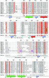
Figure 2. Structure-based sequence alignment of human caspase domains
Strictly conserved residues are shown in white with a red background; other conserved residues have a grey background. Single-nucleotide polymorphisms are indicated in bold italics. Residues involved in substrate recognition and catalysis are marked (*). Residues representing autolytic cleavage sites in the intersubunit linker are in red, and the cysteine residue that forms a dimer interface disulphide in caspase-2 is in orange. Caspase-14 is not activated by autolysis [295], and the cleavage site detected in vivo is presumably from another cellular protease [294,295]. Residues that have been mutated are in bold blue letters. The Ser residue reportedly phosphorylated in caspase-9 is shadowed yellow. The secondary structure representations (arrow, β-strand; cylinder, α-helix) follow the CATCH classification (http://www.biochem.ucl.ac.uk/bsm/cath/) for 1QTN [13].
Figure 3
Figure 3. The framework of apoptosis
Death is signalled by ligand-enforced clustering of receptors at the cell surface, which leads to the activation of initiator caspases-8 and -10 [297]. These caspases then directly cleave and activate the effector caspases-3 and -7 (and possibly caspase-6), which are predominantly responsible for the limited proteolysis that characterizes apoptotic morphology in a cell. On the other hand, genotoxic damage – transmitted by a mechanism thought to involve the release of cytochrome c from mitochondria – engages the same effector caspases [298]. The latter events progress through the initiator caspase-9 and its activator platform APAF-1 [55]. The common execution phase is regulated through direct caspase inhibition by XIAP, which can also regulate the active form of caspase-9. XIAP is under the influence of antagonist proteins such as Smac/DIABLO and HtrA2 that compete with caspases for IAPs [251]. Finally, these IAP antagonists may be sequestered by other IAPs, such as ML-IAP (melanoma IAP), acting as competitive sinks for the availability of the pro-apoptotic antagonists [299]. Although other modulators may regulate the apoptotic pathway in a cell-specific manner, this framework is considered to be common to most mammalian cells.
Figure 4
Figure 4. Structure of active caspases
(a) The crystal structure of human caspase-8 exemplifies the fundamental caspase fold, and is shown bound to the tetrapeptide aldehyde inhibitor acetyl-Ile-Glu-Thr-Asp-CHO ([13]; PDB entry 1QTN), which represents the highest-resolution structure of a caspase reported to date. Note the three-layer structure of a twisted, 12-stranded β-sheet that is sandwiched by α-helices. Most of the interdomain contact area is built by the central small subunits, with additional interactions (the characteristic ‘loop bundle’) tying together the C- and N-termini of large and small subunits from neighbouring domains. The bound inhibitor is represented with a ball-and-stick model, as are dithiane diol molecules trapped in the cleft between the two monomers (termed the central cavity, for obvious reasons). (b) Simplified topological diagram of the caspase structure, following the CATCH definition of secondary structure elements for 1QTN. An additional N-terminal α-helix of variable length (α0; not shown) is present in caspases-1 [6,7], -2 [75] and -9 [73], and closes the ‘bottom’ of the α/β barrel. Also not depicted is an additional α-helix found solely in the long 179-loop of caspase-8. The positions of catalytic dyad residues His-237 and Cys-285 (red), along with those of the specificity-determining arginine residues (Arg-179 and Arg-341), are indicated. The location of loops that contain important functional elements is indicated in blue text using the numbering convention designated throughout this review, along with an alternative designation [145].
Figure 5
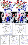
Figure 5. Caspase catalytic mechanism
Close-up of the active-site region in acetyl-Asp-Val-Ala-Asp-methyl ketone-inhibited caspase-3 (PDB code 1CP3; [10]) shown in standard orientation, i.e. with the active-site residues facing the viewer, and substrates running from left to right. The stereo plots display (a) a ribbon representation of the caspase (large subunit, blue; small subunit, red), and (b) the GRASP electrostatic surface potential of the caspase (contoured between −25 and +25 kBT/e) with stick inhibitor. Important residues are labelled in both panels. Hydrogen bonds were calculated using HBPLUS (http://www.biochem.ucl.ac.uk/bsm/hbplus/home.html) and are indicated with orange dotted lines in (a). Note that the inhibitor binds in an extended conformation, with backbone atoms of P3 and P1 residues hydrogen-bonded to strictly (Arg-341) and highly (Ser-339) conserved caspase residues. The guanidinium groups of Arg-179 and Arg-341 engage in strong salt bridges with the carboxylate of the P1 aspartate, which is further hydrogen-bonded to the side-chain carboxyamide of Gln-283. The combination of extended, β-sheet-like hydrogen bonding to the enzyme and of substrate recognition based mainly on interactions with the S1 and S4 pockets places caspases in a mechanistic sense closer to serine proteases, in particular those of the subtilisin clan. (c) Proposed substrate-hydrolysis mechanistic scheme. During the acylation step (1), the carbonyl oxygen of the non-covalently bound P1 residue is anchored through hydrogen bonds to the nitrogen atoms of Gly-238 and Cys-285 (the oxyanion hole). This increases the polarization of the C–O bond, and therefore facilitates nucleophilic attack of the sulphur atom of Cys-285 on the highly electrophilic carbonyl carbon. The result is a covalent enzyme–substrate adduct, the high-energy tetrahedral intermediate (2), as visualized in crystal structures of methyl ketone-inhibited caspases (see a). The imidazole moiety of His-237 acts as a general acid at this stage of catalysis by protonating the α-amino group of the leaving peptide product, thus avoiding re-formation of the peptide bond. Deacylation of the acyl-enzyme complex occurs then in a similar manner: the deprotonated His-237 side chain abstracts a proton from a water molecule, the hydrolytic water (3), which is thus activated to attack the thioester bond. Deacylation proceeds through a second tetrahedral intermediate (4), formed upon nucleophilic attack of the hydroxy group on the carbonyl carbon. (A putative, neutral gem-diol intermediate found in a recent quantum mechanics/molecular mechanics simulation of the hydrolysis of the acyl-enzyme complex in caspase-3 [79] is shown by grey atoms in parentheses. These authors also predicted that the catalytic histidine is activated by the hydroxy group of Ser-178, but this residue is not conserved in other caspases.) Rupture of the Sγ–C bond regenerates the enzyme in a non-covalent complex with the N-terminal peptide product (5). By analogy with serine proteases, it is conceivable that movements of the 341- and/or 381- substrate-binding loops are coupled to the latter reaction, thus allowing disruption of the main-chain–main-chain hydrogen bonds with the P1/P3 residues, and of the P1 carbonyl oxygen atom with the oxyanion hole. In other words, thioester hydrolysis and product release may be synchronized to ensure a high efficiency of catalysis.
Figure 6
Figure 6. Mechanisms of procaspase-7 activation
(a) Ribbon plots showing the crystal structures of human procaspase-7 ([124]; PDB code 1GQF), and both free ([11]; 1K86) and inhibitor-bound ([63]; 1F1J) active caspase-7. Loops that display significant changes during activation are coloured red for the 341-loop, blue for the 381-loop, and green for the intersubunit linker. Notice the turn of almost 180° at the Val-323–Glu-324 peptide bond in the intersubunit linker in the left and middle structures compared with the right (inhibitor-bound) form, leading to the insertion of residues N-terminal of Val-323 into the central cavity of the zymogen. Residues Thr-288 to Arg-318 were only poorly or not defined at all by electron density, suggesting enhanced flexibility. However, the distances between the defined N- and C-termini in the zymogen (left) suggest that the spatially adjacent large and small subunits derive from the same domain. (b) Cartoon version of the activation, showing the critical loop transitions. In this context, the unliganded active form is omitted as an intermediate in the generation of a fully functional active site.
Figure 7
Figure 7. Details of interdomain regions of an effector and an initiator caspase
Close-ups of the interdomain interfaces (left), surface (middle) and cavity depth (right) in caspase-7 (a) and caspase-8 (b). Caspase-7 is the acetyl-Asp-Glu-Val-Asp-CHO-inhibited form (PDB 1F1J; [63]) and caspase-8 is the acetyl-Ile-Glu-Thr-Asp-CHO-inhibited form (PDB 1QTN; [13]). Several interdomain residues are shown with all of their non-hydrogen atoms (colour-coded). Hydrogen bonds are denoted by orange dotted lines. The central cavity, formed at the dimer interface, is significantly larger in caspase-8, with the attendant possibility that the intersubunit linker that would be in the zymogen, and therefore not visible here, may partially occupy it without producing the steric clashes seen in the smaller caspase-7 central cavity.
Figure 8
Figure 8. Details of interdomain regions of caspase-9 reveal its activation mechanism
The crystal structure of caspase-9 is unusual in that it contains one catalytic domain in the active conformation (a) and one in a zymogen-like conformation (b) ([73]; PDB code 1JXQ). Loops that display significant changes between the two forms are coloured red for the 341-loop, blue for the 381-loop and green for the intersubunit linker, as in Figure 6(a). (c) Ribbon plot showing a close-up of the central cavity in human caspase-9. The unique insertion 240-loops (see also Figure 2) from both caspase-9 monomers intrude into the central cavity stabilized through important contacts with each other. In addition, two aliphatic residues that line the central cavity in procaspase-7, Val-390 and Met-393, are replaced by the bulkier aromatic side chains of Phe-390 and Phe-393 in the initiator caspase, thus effectively ‘sealing’ its central cavity. The well-conserved Arg-286, whose guanidinium group is sandwiched between Tyr-331 and Val-334 in active caspases-3 and -7, is substituted in caspase-9 by a glycine, leaving additional empty space for the elbow loop. Most notably, a hydrophobic pocket formed by some of the very same residues that seal the central cavity, i.e. Phe-240f′ and Phe-393′, together with Pro-324′ accepts the phenyl moiety of the elbow loop residue Phe-334 from the neighbouring monomer, which is thus anchored into the central cavity. In this manner, residues from one monomer indirectly prime the 341-loop of a neighbouring procaspase-9 molecule to adopt its active, substrate-binding conformation, without the need for intersubunit cleavage.
Figure 9
Figure 9. Caspase-9 packing and the apoptosome
(a) Tetrameric arrangement observed in the crystal structure of caspase-9 ([73]; PDB code 1JXQ), colour-coded to show the active (red tones) and inactive (green tones) domain of each standard caspase dimer. (b) Average map of the APAF-1·procaspase-9 complex [165]. The arrowed region of central density represents bound procaspase-9 molecules sandwiched between two APAF-1 rings. The scale bar corresponds to 100 Å. Reprinted from Molecular Cell, Vol. 9, D. Acehan, X. Jiang, D. G. Morgan, J. E. Heuser, X. Wang and C. W. Akey, Three-dimensional structure of the apoptosome: implications for assembly, procaspase-9 binding, and activation, pp. 423–432, Copyright (2002), with permission from Elsevier.
Figure 10
Figure 10. Mechanisms of caspase inhibition by XIAP
(a) Schematic diagram of the domain organization of human XIAP. The sequence critical for the inhibition of effector caspases by XIAP is given [254,256]; the arrow above the sequence indicates that it binds in a ‘reverse mode’ to effector caspases (see text and Figure 11b). A caspase cleavage site in XIAP [253,263] is indicated; other cleavages are also detected [166]. (b) Cartoon depicting the mode of binding of BIR2 to caspase-3 and of BIR3 to caspase-9 (caspase large subunits in blue, and small subunits in red) bound to the respective BIR (orange). The shallow groove on each BIR domain symbolizes the Smac pocket into which the four N-terminal residues constituting an IAP-binding motif fit. Although important for caspase-9 inhibition [122], the significance of this groove in BIR2 is less well understood in terms of caspase-3 or -7 inhibition [255]. (c) Ribbon diagram of the inhibitory interactions, with the same colour coding. The N- and C-termini of the subunits of the complex are labelled, and important side chains in the interactions are shown, but for simplicity are not labelled.
Figure 11
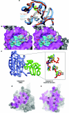
Figure 11. Details of caspase inhibition by XIAP
(a) The catalytic cleft of caspase-3 in three different forms, i.e. inhibitor [CMK (chloromethyl ketone)]-bound (blue; PDB code 1CP3; [10]), active with free catalytic site (red; PDB 1QX3; [67]) or bound to BIR2 (grey; PDB 1I30; [255]), is highly conserved. There are no substantial alterations in main-chain or side-chain conformations with the docking of BIR2 to the cleft, and it is only when the tetrapeptide CMK inhibitor binds that Tyr-338 must rotate to accept the P2 side chain (green arrow). Consequently, the linker region of BIR2 appears perfectly adapted to the unliganded active form of caspase-7. (b) Superposition of the XIAP inhibitory region (pink sticks) and acetyl-DVAD-CMK (blue sticks) on the active-site cleft of caspase-3 from PDB file 1I30 [255]. The orientation is as for Figure 5 and shows the small 4 Å footprint made by acetyl-DVAD-CMK side-chains (cyan) compared with that made by XIAP side chains (magenta). Note the manifold interactions of the two residues in the BIR1–BIR2 linker that are critical for activity, i.e. Leu-141 and Asp-148 [256]. The former engages in van der Waals contacts with Tyr-338 and Phe-381h, while Asp-148 occupies the S4 pocket of the caspase, hydrogen-bonding in particular residues of the 341 substrate-binding loop (Arg-341, Ser-343 and Trp-348). In addition, the carboxylate of Asp-148 is clamped to the C-terminal residue of XIAP (BIR2), Arg-233 (not shown). (c) Crystal structure of BIR3 (green)-bound caspase-9 (blue) taken from PDB 1NW9 [122]. The BIR domain takes the position of the dimeric partner domain of caspase-9, essentially monomerizing the caspase, thus reversing the process of zymogen activation. The blow-up depicts a conformational relay upon BIR2 binding (green residues) that forces Phe-390 from the active conformation (red) to the inactive one (blue). A rotation of Phe-390 requires a compensatory rotation in Tyr-331 and a concomitant expulsion of the catalytic Cys-285 into a non-catalytic location. (d, e) The 4 Å footprint of caspase-9 residues (shaded magenta) in contact with either its dimeric partner caspase-9 (d) or BIR3 (e) at the dimer interface. Note that although the contacts are more extensive in the caspase-9 dimer, most of the crucial contacts are the same. An exception is the segment Ala-298–Phe-301, which forms the IBM interacting with the Smac pocket on BIR3. Rendered with PyMol.
Figure 12
Figure 12. IAP domains bound to Smac/DIABLO and caspase neoepitopes
The GRASP electrostatic surface potential of XIAP BIR3 (contoured between −15 and +15 kBT/e) with stick representation of the neoepitope generated following cleavage of caspase-9 at Asp-297 (blue; PDB 1NW9; [122]) superimposed on the N-terminal tetrapeptide of mature Smac/DIABLO (orange; PDB 1G73; [278]). Residues are identified by the corresponding colour. Notice the highly negative end of the Smac pocket, which helps to counter the energetic penalty of burying the positive charge on the N-terminal amine of IBM peptides.

Similar articles

Cited by

References

    1. Schechter I., Berger A. On the size of the active site in proteases. I. Papain. Biochem. Biophys. Res. Commun. 1967;27:157–162. - PubMed
    1. Thornberry N. A., Bull H. G., Calaycay J. R., Chapman K. T., Howard A. D., Kostura M. J., Miller D. K., Molineaux S. M., Weidner J. R., Aunins J. A novel heterodimeric cysteine protease is required for interleukin-1β processing in monocytes. Nature (London) 1992;356:768–774. - PubMed
    1. Cerretti D. P., Kozlosky C. J., Mosley B., Nelson N., Van Ness K., Greenstreet T. A., March C. J., Kronheim S. R., Druck T., Cannizzaro L. A., et al. Molecular cloning of the interleukin-1β converting enzyme. Science. 1992;256:97–100. - PubMed
    1. Yuan J., Shaham S., Ledoux S., Ellis H. M., Horvitz H. M. The C. elegans cell death gene ced-3 encodes a protein similar to mammalian interleukin-1β-converting enzyme. Cell. 1993;75:641–652. - PubMed
    1. Alnemri E. S., Livingston D. J., Nicholson D. W., Salvesen G., Thornberry N. A., Wong W. W., Yuan J. Human ICE/CED-3 protease nomenclature. Cell. 1996;87:171. - PubMed

Publication types