

Reversal of glandular polarity in the lymphovascular compartment of breast cancer
- PMID: 15452174
- PMCID: PMC1770446
- DOI: 10.1136/jcp.2004.016980
Reversal of glandular polarity in the lymphovascular compartment of breast cancer
Abstract
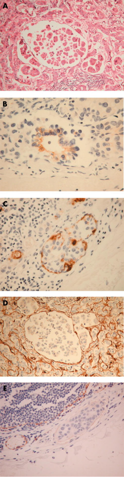
Aim: To investigate the polarity of breast invasive ductal carcinoma cells by comparing the polarity of the tumour located within lymphovascular spaces with that located in the extravascular compartment.
Methods: An immunohistochemical study identifying the apical HMFG-1, basolateral AUA-1, and basal laminin polarity markers of 11 cases of invasive ductal carcinoma (grades 1 or 2) metastatic to lymph nodes, all of which contained areas of tumour within and outside of lymphovascular spaces.
Results: Only one of 11 tumours had a focus of apparent reversed glandular polarity in the larger extravascular tumour compartment (with AUA-1 present internally and HMFG-1 expressed externally on tumour clumps), but six of the 11 tumours showed reversed glandular polarity (either with AUA-1, or HMFG-1, or both) within the very much smaller lymphovascular space tumour compartment. Laminin was not identified in association with lymphovascular tumour.
Conclusions: Reversed glandular polarity in invasive ductal breast carcinomas was identified and was significantly more frequent within vessels than outside of them. Reversal of tumour glandular polarity within lymphovascular spaces allows direct interaction between apical domain-type molecules-which are then aberrantly expressed on the external surface of tumour clumps-and lymphovascular endothelium. Such interactions may affect the establishment of metastatic disease.
Figures
Similar articles
-
Invasive ductal carcinomas of the breast showing partial reversed cell polarity are associated with lymphatic tumor spread and may represent part of a spectrum of invasive micropapillary carcinoma.Am J Surg Pathol. 2010 Nov;34(11):1637-46. doi: 10.1097/PAS.0b013e3181f5539c. Am J Surg Pathol. 2010. PMID: 20975342
-
Expression of CD31 by cells of extensive ductal in situ and invasive carcinomas of the breast.J Pathol. 2001 Jun;194(2):254-61. doi: 10.1002/1096-9896(200106)194:2<254::AID-PATH880>3.0.CO;2-2. J Pathol. 2001. PMID: 11400156
-
The biological and prognostic significance of cell polarity and E-cadherin in grade I infiltrating ductal carcinoma of the breast.J Pathol. 1999 Sep;189(1):20-7. doi: 10.1002/(SICI)1096-9896(199909)189:1<20::AID-PATH394>3.0.CO;2-2. J Pathol. 1999. PMID: 10451483
-
A comparison of the patterns of laminin expression in fibroadenoma, fibrocystic diseases, pre-invasive and invasive ductal breast carcinoma.Pathology. 2001 Aug;33(3):303-6. Pathology. 2001. PMID: 11523929
-
[Invasive ductal carcinomas of breast showing partial reversed cell polarity are associated with lymphatic tumor spread].Zhonghua Bing Li Xue Za Zhi. 2012 May;41(5):305-8. doi: 10.3760/cma.j.issn.0529-5807.2012.05.005. Zhonghua Bing Li Xue Za Zhi. 2012. PMID: 22883668 Chinese.
Cited by
-
Inverted apicobasal polarity in health and disease.J Cell Sci. 2024 Mar 1;137(5):jcs261659. doi: 10.1242/jcs.261659. Epub 2024 Mar 11. J Cell Sci. 2024. PMID: 38465512 Free PMC article. Review.
-
Normal morphogenesis of epithelial tissues and progression of epithelial tumors.Wiley Interdiscip Rev Syst Biol Med. 2012 Jan-Feb;4(1):51-78. doi: 10.1002/wsbm.159. Epub 2011 Sep 2. Wiley Interdiscip Rev Syst Biol Med. 2012. PMID: 21898857 Free PMC article. Review.
-
Phosphoinositides in cell architecture.Cold Spring Harb Perspect Biol. 2011 Aug 1;3(8):a004796. doi: 10.1101/cshperspect.a004796. Cold Spring Harb Perspect Biol. 2011. PMID: 21576256 Free PMC article. Review.
-
From cells to organs: building polarized tissue.Nat Rev Mol Cell Biol. 2008 Nov;9(11):887-901. doi: 10.1038/nrm2523. Nat Rev Mol Cell Biol. 2008. PMID: 18946477 Free PMC article. Review.
-
Establishing epithelial glandular polarity: interlinked roles for ARF6, Rac1, and the matrix microenvironment.Mol Biol Cell. 2012 Dec;23(23):4495-505. doi: 10.1091/mbc.E12-03-0246. Epub 2012 Oct 10. Mol Biol Cell. 2012. PMID: 23051733 Free PMC article.
References
-
- Cowley GP, Smith MEF. Modulation of E-cadherin expression and morphological phenotype in the intravascular component of adenocarcinomas. Int J Cancer 1995;60:325–9. - PubMed
-
- Spurr NK, Durbin H, Sheer D, et al. Characterization and chromosomal assignment of a human cell surface antigen defined by the monoclonal antibody AUA-1. Int J Cancer 1986;38:631–6. - PubMed
-
- Adams SA, Sherwood AJ, Smith MEF. Malignant mesothelioma: PAS/D positivity and inversion of polarity in intravascular tumour. Histopathology 2002;41:1–3. - PubMed
-
- Kirkland S . Polarity and differentiation of human rectal adenocarcinoma cells in suspension and collagen gel cultures. J Cell Sci 1988;91:615–21. - PubMed
Publication types
MeSH terms
Substances
LinkOut - more resources
Full Text Sources
Medical