Comparative Study
. 2004 Oct 4;200(7):825-34.
doi: 10.1084/jem.20041389. Epub 2004 Sep 27.

Coordinate expression and trans presentation of interleukin (IL)-15Ralpha and IL-15 supports natural killer cell and memory CD8+ T cell homeostasis

Affiliations
Comparative Study

Coordinate expression and trans presentation of interleukin (IL)-15Ralpha and IL-15 supports natural killer cell and memory CD8+ T cell homeostasis

Patrick R Burkett et al. J Exp Med. .

Abstract

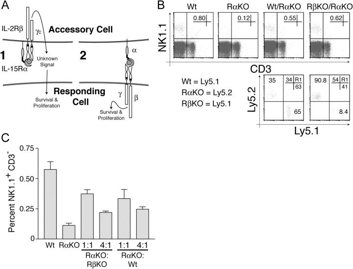
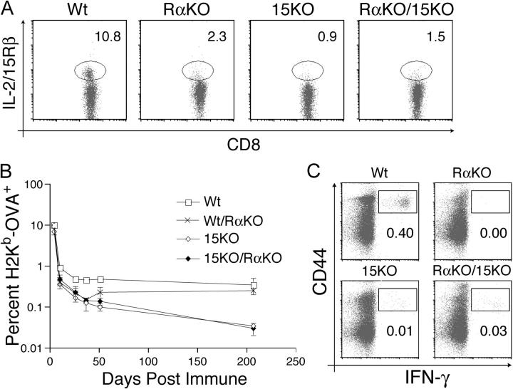
The high affinity interleukin (IL)-15 receptor, IL-15Ralpha, is essential for supporting lymphoid homeostasis. To assess whether IL-15Ralpha's role in vivo is to trans present IL-15, we generated mixed bone marrow chimera from IL-15Ralpha- and IL-2/15Rbeta-deficient mice. We find that IL-15Ralpha-competent, IL-2/15Rbeta-deficient cells are able to support IL-15Ralpha-deficient natural killer (NK) and memory CD8+ T cells, thus ruling out secondary signals on these cells and demonstrating that IL-15Ralpha-mediated presentation of IL-15 in trans is the primary mechanism by which IL-15Ralpha functions in vivo. Surprisingly, using IL-15- and IL-15Ralpha-deficient mixed chimera, we also find that IL-15 and IL-15Ralpha must be expressed by the same cells to present IL-15 in trans, indicating that IL-15Ralpha is required on a cellular level for the elaboration of IL-15. These studies indicate that IL-15Ralpha defines homeostatic niches for NK and memory CD8+ T cells by controlling both the production and the presentation of IL-15 in trans to NK and CD8+ memory T cells.


Figures

Figure 1.
Figure 1.
IL-15Rα expression by RAG-1–independent hematopoietic cells is sufficient to maintain NK and memory CD8+ T cells. IL-15Rα−/− mice were lethally irradiated and reconstituted with bone marrow from either IL-15Rα+/− (Wt) or IL-15Rα−/− (RαKO) mice, or a 1:1 mixture of IL-15Rα+/− and IL-15Rα−/− bone marrow (Wt/RαKO), or a 1:1 mixture of RAG-1−/− and IL-15Rα−/− bone marrow (RAG/RαKO). (A) Flow cytometric analyses of NK cell reconstitution in chimeric mice. The percentage of total lymphocytes (defined by forward and side scatter as the R1 gate) that are NK1.1+ CD3 cells is indicated in the top plots. The bottom plots are gated on NK1.1+ CD3 cells. The percentage of total lymphocytes expressing either Ly5.1 or Ly5.2 is indicated in the top right corner of the bottom plots. Note that RAG-1−/− hematopoietic cells support peripheral NK cell development. (B) Graphic representation of percentages of H2Kb-OVA+ CD8+ T cells in immunized chimeric mice. 8 wk after irradiation and reconstitution, OT-1+ CD8+ T cells were adoptively transferred into the indicated chimeric mice, after which mice were immunized with OVA and poly I:C. The percentage of total lymphocytes that are H2Kb-OVA+ CD8+ T cells after immunization was tracked via serial peripheral blood analyses. Note that RAG-1−/− hematopoietic cells support memory CD8+ T cell homeostasis. Data represent mean ± SEM of at least three mice per group.
Figure 2.
Figure 2.
IL-15Rα, but not IL-2/15Rβ, expression is required by hematopoietic cells to support NK cell development and survival. (A) Model illustrating two distinct mechanisms by which non-cell–autonomous IL-15Rα expression is required to support IL-15–dependent cell types. Mechanism 1 requires IL-2Rβ expression on accessory cells to mediate signal transduction, whereas mechanism 2 (trans presentation) does not. (B) Flow cytometric analysis of NK cell reconstitution in indicated chimeric mice depends upon IL-15Rα–, but not IL-2/15Rβ–, competent cells. IL-15Rα−/− mice were lethally irradiated and reconstituted with either IL-15Rα+/− (Wt) or IL-15Rα−/− (RαKO) bone marrow cells, or a mixture of IL-15Rα+/− and IL-15Rα−/− bone marrow cells (Wt/RαKO), or a mixture of IL-2/15Rβ−/− and IL15Rα−/− bone marrow cells (RβKO/RαKO). NK cell populations in chimeric mice were assessed 8 wk after reconstitution. The percentages of total lymphocytes that are NK cells (NK1.1+ CD3 cells) are indicated in each of the top panels. The bottom plots are gated on NK1.1+ CD3 cells in WT/RαKO and RβKO/RαKO chimera. The percentages of total lymphocytes expressing either Ly5.1 or Ly5.2 in WT/RαKO and RβKO/RαKO chimera are shown in the top right corner of the bottom panels. Note that NK cell reconstitution in chimeric mice depends upon IL-15Rα–, but not IL-2/15Rβ–, competent cells. (C) The frequency of IL-15Rα–competent cells controls the frequency of peripheral NK cells. IL-15Rα−/− mice were lethally irradiated and reconstituted with bone marrow from either IL-15Rα+/− or IL-15Rα−/− mice, or with various ratios of IL-15Rα+/− and IL-15Rα−/− bone marrow, or assorted ratios of IL-2/15Rβ−/− and IL-15Rα−/− bone marrow. Mice were bled and NK cell populations in chimeric mice were assessed 8 wk after reconstitution. The percentage of total lymphocytes that are NK1.1+ CD3 cells is shown. Plots are representative of at least two mice per condition, and all experiments were performed three times with similar results.
Figure 3.
Figure 3.
IL-15Rα, but not IL-2/15Rβ, expression is required by hematopoietic cells to support CD8+ memory T cell homeostasis. IL-15Rα−/− mice were lethally irradiated and reconstituted with bone marrow from either IL-15Rα+/− (Wt) or IL-15Rα−/− (RαKO) mice, or a mixture of IL-2/15Rβ−/− and IL-15Rα−/− (RβKO/RαKO) bone marrow. (A) Flow cytometric analyses of endogenous CD44hi IL-2/15Rβhi (memory phenotype) CD8+ T cells in spleens of chimeric mice. The percentages of total CD8+ T cells that are memory phenotype CD8+ T cells in the indicated chimeric mice were assessed 8 wk after reconstitution. The percentage of CD8+ T cells in each quadrant is shown. Plots are gated on CD8+ T cells and are representative of at least three mice per condition. (B) Graphic representation of peripheral blood H2Kb-OVA+ CD8+ T cells after immunization of chimeric mice. 8 wk after reconstitution, mice received OT-1+ CD8+ T cells and were immunized with OVA and poly I:C. The percentage of total lymphocytes that were H2Kb-OVA+ CD8+ T cells was quantitated by flow cytometric analyses of serial peripheral blood samples after immunization. Data represent mean ± SEM of at least two mice per group.
Figure 4.
Figure 4.
Two models for the relationship of IL-15–producing and –presenting cells. In model 1, IL-15 is secreted by a distinct IL-15–producing cell, and subsequently bound and presented by an IL-15Rα–competent cell. In model 2, coordinate expression of IL-15 and IL-15Rα is required for efficient presentation of IL-15 to responding cells.
Figure 5.
Figure 5.
Coordinate expression of IL-15 and IL-15Rα is required for NK cell development and maintenance. IL-15Rα−/− mice were lethally irradiated and reconstituted with bone marrow cells from either IL-15Rα+/− (Wt), IL-15Rα−/− (RαKO), or IL-15−/− (15KO), mice, or with 1:1 mixtures of either IL-15Rα+/− and IL-15Rα−/− bone marrow cells (Wt/RαKO), or IL-15Rα−/− and IL-15−/− bone marrow cells (15KO/RαKO). (A) Flow cytometric analyses of endogenous NK cells in bone marrow (BM) and spleens (SPL) of chimeric mice 8 wk after reconstitution with the indicated bone marrow genotypes. Note that endogenous NK cells are not supported in chimera generated from a mixture of IL-15−/− and IL-15Rα2/2 bone marrows. Data are representative of at least four mice per group. (B) Flow cytometric analysis of IL-15Rα expression on LPS-stimulated, bone marrow–derived dendritic cells of the indicated genotypes. Bone marrow–derived dendritic cells were stimulated with LPS for 24 h in vitro and stained for IL-15Rα expression. (C) Graphic representation of survival of adoptively transferred NK cells after transfer into the indicated mixed chimera. The percentage of total lymphocytes that were adoptively transferred NK cells (i.e., CFSE+ NK1.1+ CD3 cells) is indicated at each time point. Note that adoptively transferred NK cells are not supported in chimera generated from a mixture of IL-15−/− and IL-15Rα2/2 bone marrows. Data represent mean ± SEM of at least two mice per group.
Figure 6.
Figure 6.
Homeostasis of memory phenotype and memory CD8+ T cells requires coordinate expression of IL-15 and IL-15Rα. IL-15Rα−/− mice were lethally irradiated and reconstituted with bone marrow from either IL-15Rα+/− (Wt), IL-15Rα−/− (RαKO), or IL-15−/− (15KO) mice, or with a 1:1 mixture of IL-15Rα−/− and IL-15−/− bone marrow cells (RαKO/15KO). (A) Flow cytometric analysis of IL-2/15Rβhi memory phenotype CD8+ T cells 8 wk after reconstitution. The percentage of total CD8+ T cells that are IL-2/15Rβhi memory phenotype CD8+ T cells is indicated. Data are representative of at least five mice per condition. (B) Graphic representation of the percentages of memory H2Kb-OVA+ CD8+ T cells after immunization of the indicated chimeric mice. 8 wk after reconstitution, mice received OT-1+ CD8+ T cells and were then immunized with OVA and poly I:C. The percentage of total lymphocytes that are H2Kb-OVA+ CD8+ T cells after immunization was quantitated by serial peripheral blood analyses. Data represent mean ± SEM of at least three mice per group. (C) Flow cytometric analysis of IFN-γ production by memory CD8+ T cells in chimeric mice. IL-15 and IL-15Rα must be expressed by the same cell for optimal memory CD8+ T cell function. 8 wk after reconstitution, chimeric mice received OT-1+ CD8+ T cells and were then immunized with OVA and poly I:C. 90 d after immunization, splenocytes were stimulated with SIINFEKL, and IFN-γ production was assessed by intracellular staining. The percentage of total lymphocytes that are CD8+ CD44hi IFN-γ+ is indicated. Plots are gated on CD8+ cells and are representative of at least three mice.

References

    1. Schluns, K.S., W.C. Kieper, S.C. Jameson, and L. Lefrancois. 2000. Interleukin-7 mediates the homeostasis of naive and memory CD8 T cells in vivo. Nat. Immunol. 1:426–432. - PubMed
    1. Becker, T.C., E.J. Wherry, D. Boone, K. Murali-Krishna, R. Antia, A. Ma, and R. Ahmed. 2002. Interleukin 15 is required for proliferative renewal of virus-specific memory CD8 T cells. J. Exp. Med. 195:1541–1548. - PMC - PubMed
    1. Goldrath, A.W., P.V. Sivakumar, M. Glaccum, M.K. Kennedy, M.J. Bevan, C. Benoist, D. Mathis, and E.A. Butz. 2002. Cytokine requirements for acute and basal homeostatic proliferation of naive and memory CD8+ T cells. J. Exp. Med. 195:1515–1522. - PMC - PubMed
    1. Burkett, P.R., R. Koka, M. Chien, S. Chai, F. Chan, A. Ma, and D.L. Boone. 2003. IL-15R alpha expression on CD8+ T cells is dispensable for T cell memory. Proc. Natl. Acad. Sci. USA. 100:4724–4729. - PMC - PubMed
    1. Koka, R., P.R. Burkett, M. Chien, S. Chai, F. Chan, J.P. Lodolce, D.L. Boone, and A. Ma. 2003. Interleukin (IL)-15Rα–deficient natural killer cells survive in normal but not IL-15Rα–deficient mice. J. Exp. Med. 197:977–984. - PMC - PubMed

Publication types