Comparative Study
. 2004 Oct 4;200(7):905-16.
doi: 10.1084/jem.20040402. Epub 2004 Sep 27.

Chlamydia inhibit host cell apoptosis by degradation of proapoptotic BH3-only proteins

Affiliations
Comparative Study

Chlamydia inhibit host cell apoptosis by degradation of proapoptotic BH3-only proteins

Silke F Fischer et al. J Exp Med. .

Abstract

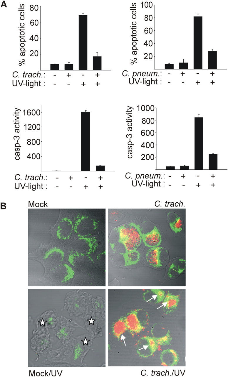
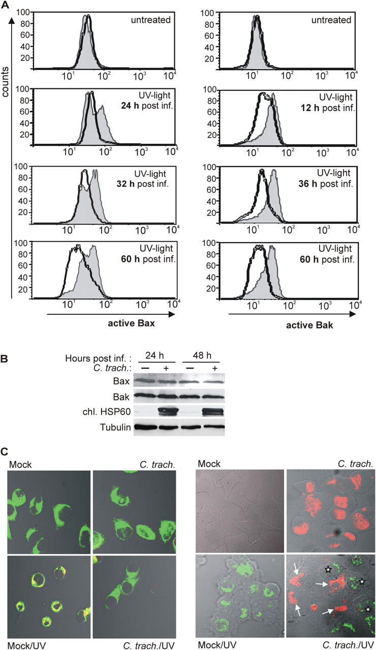
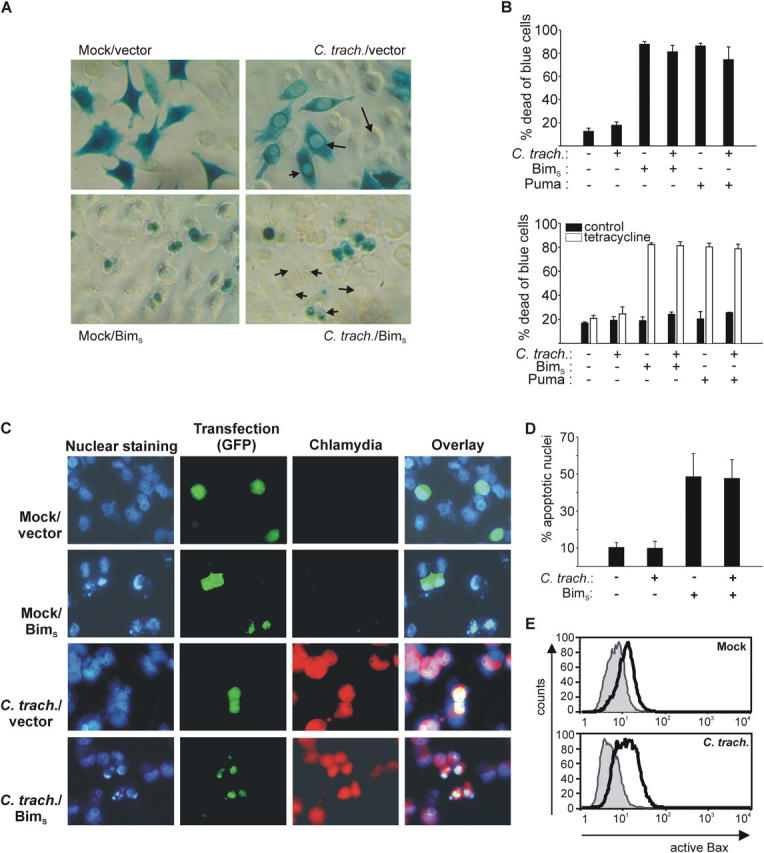
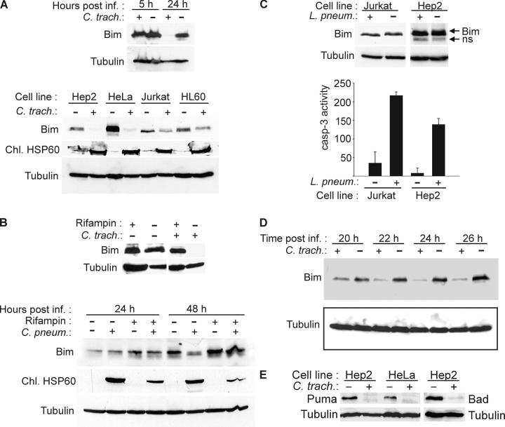
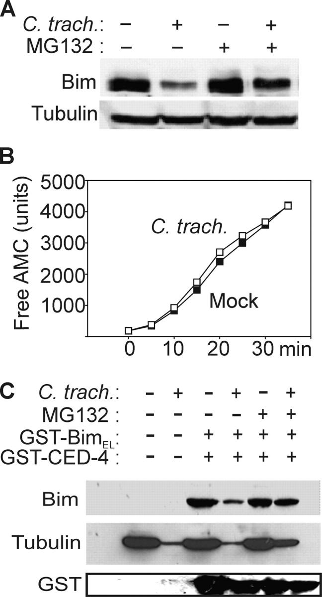
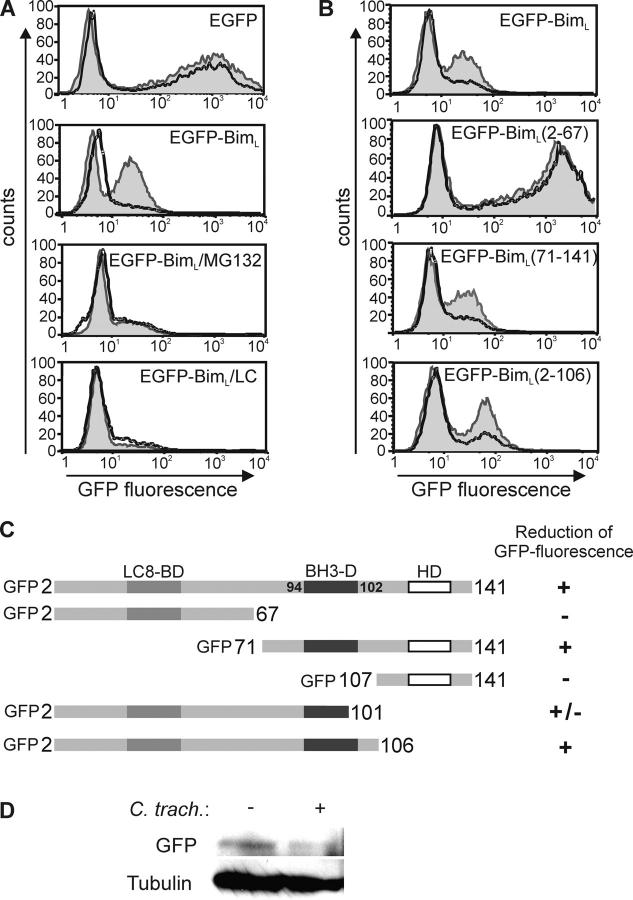
Chlamydia are obligate intracellular bacteria that replicate in a vacuole inside a host cell. Chlamydial infection has been shown to protect the host cell against apoptotic stimuli. This is likely important for the ability of Chlamydia to reproduce in human cells. Here we show that resistance to apoptosis is conveyed by the destruction of the proapoptotic BH3-only proteins Bim/Bod, Puma, and Bad during infection. Apoptotic stimuli were blocked upstream of the mitochondrial activation of Bax/Bak. During infection with both species, Chlamydia trachomatis and Chlamydia pneumoniae, Bim protein gradually disappeared without noticeable changes in Bim mRNA. The disappearance was blocked by inhibitors of the proteasome. Infected cells retained sensitivity to Bim expressed by transfection, indicating functional relevance of the Bim disappearance. Fusion to Bim targeted the green fluorescent protein for destruction during infection. Analysis of truncation mutants showed that a short region of Bim containing the BH3 domain was sufficient for destruction during chlamydial infection. Like Bim, Puma and Bad proteins disappeared during infection. These results reveal a novel way by which microbes can interfere with the host cell's apoptotic machinery, and provide a molecular explanation of the cellular resistance to apoptosis during infection with Chlamydia.


Figures

Figure 1.
Figure 1.
Infection with C. trachomatis or C. pneumoniae inhibits UV light–induced apoptosis. Hep2 cells were infected with Chlamydia or not (mock). After 24 (C. trachomatis) or 48 h (C. pneumoniae), apoptosis was induced by UV irradiation (1,000 J/m2). 6 h later, cells were analyzed. (A) Nuclear apoptosis and caspase 3–like activity. Cells were stained with Hoechst dye and nuclear apoptosis was scored under a fluorescence microscope (top; at least 300 nuclei were counted per sample and values are mean/SD of triplicates) or lysed, and DEVD cleaving activity was measured in cell extracts (bottom; mean/SD of triplicate measurements). (B) Cytochrome c release from mitochondria. Cells were stained for cytochrome c (green) and chlamydial LPS (red), and analyzed by laser-scanning microscopy. Arrows point at chlamydial inclusions in cells that have retained cytochrome c in their mitochondria. Asterisks indicate noninfected cells with typical apoptotic morphology and release of cytochrome c from mitochondria (the cytochrome c signal is diminished upon release). The figure shows results typical of three independent experiments.
Figure 2.
Figure 2.
Activation of Bax and Bak by UV light is inhibited in C. trachomatis–infected cells. (A) On the left, detection of active Bax by flow cytometry is shown. HeLa cells were either infected or left uninfected. At the time points indicated, cells were treated with UV light (1,000 J/m2; bottom) or not (top). Cells were collected 6 h later and stained for active Bax. Normal line/shaded area, uninfected cells; bold line, C. trachomatis–infected cells. On the right, detection of active Bak is shown. Data are representative of three experiments. (B) Detection of Bax and Bak by Western blot. HeLa cells were infected or left uninfected. 24 or 48 h later, cells were collected and subjected to Western blotting with antibodies specific for total Bax, total Bak, chlamydial HSP60 (as a control of infection), and tubulin (as a loading control). Similar results were obtained in three independent experiments. (C) Active Bax localizes to mitochondria and is only detectable in noninfected cells. In Hep2 cells with or without (mock) chlamydial infection, apoptosis was induced by UV irradiation (1,000 J/m2). After 6 h, cells were doubly labeled with anti-active Bax (red) and the mitochondrial marker MitoTracker Green FM (left, green; colocalization results in yellow color) or anti-active Bax (green) and anti-chlamydial LPS (right, red). Active Bax was not detected in infected cells (arrows). Asterisks indicate noninfected cells positive for active Bax. For the results shown in the right panel, infectious dose was reduced (MOI: 0.5). Similar results were obtained in three independent experiments.
Figure 3.
Figure 3.
Chlamydia-infected cells retain sensitivity to cell death induced by overexpression of BimS and Puma. (A) HeLa cells were infected with C. trachomatis or not (mock). 4 h after infection, cells were transfected by lipofection with BimS expression vector or control vector together with CMV-LacZ. 3 h later, medium was changed and 12 h later, cells were stained for β-galactosidase activity. Blue cells were viewed under a microscope and scored alive or dead. Examples of chlamydial inclusions are illustrated with arrows. (B) Quantification of dead and blue cells (graphs show percentages of dead cells; mean/SD of triplicate wells). At least 300 cells per sample were counted. On the top, HeLa cells were infected with C. trachomatis and transfected 8 h later. 20 h later, cells were stained for β-galactosidase expression and counted. This experiment was performed three times with similar results. On the bottom, HeLa cells stably expressing the tet repressor were transfected with reporter and a vector where the expression of either BimS or Puma is placed under the control of a tetracycline-inducible promoter. 3 h later, some wells were infected with C. trachomatis. 24 h later, BimS/Puma expression was induced by the addition of tetracycline and cells were fixed and stained as described above after an additional 10 h. This experiment was performed twice with similar results. Although tetracycline may inhibit the growth of Chlamydia, our earlier results indicate that the antiapoptotic activity is long-lived (it was still detectable after 48 h of rifampin treatment; reference 11). (C) BimS induces nuclear condensation in cells infected with C. trachomatis. HeLa cells were infected with C. trachomatis at an MOI of 3 or mock infected as indicated. After 8 h, cultures were cotransfected with EGFP and either empty vector or BimS expression constructs. 16 h later, cells were fixed and stained with antichlamydial LPS antibody to visualize chlamydial inclusions and Hoechst dye to visualize nuclear morphology. The micrographs show EGFP fluorescence (green, as a marker of transfection), Chlamydia (red), and nuclei/DNA (blue). Note the apoptotic morphology in both uninfected and infected cells transfected with BimS. Similar results were obtained in three separate experiments. (D) Quantification of the results illustrated in C. At least 200 GFP+ cells per transfection were counted, and nuclei were scored as normal or apoptotic. For infected cultures, only cells staining positive for chlamydial antigen were counted. Data are mean/SD of three independent experiments. (E) Normal Bax activation by BimS in infected cells. Hep2 cells were infected or not and were cotransfected by electroporation with an EGFP expression plasmid as a marker, and either empty vector or a BimS expression vector 10 h later. 16 h later, cells were stained for active Bax and analyzed by flow cytometry. The histograms show expression of active Bax in EGFP+ cells. Normal line/shaded area, empty vector; bold line, BimS expression vector.
Figure 4.
Figure 4.
Disappearance of Bim, Puma, and Bad in Chlamydia-infected cells. (A) Analysis of SDS and Triton X-100 extracts of various cell lines. Cells were infected with C. trachomatis or left uninfected and harvested after 24 h of infection or as indicated. SDS extracts from MCF-7 cells (top) or Triton X-100 extracts (bottom) were subjected to Western blotting with antibodies against Bim (isoform BimEL is detected) and chlamydial HSP60 (as control for chlamydial infection). Detection of tubulin served as a loading control. SDS extracts were used in the initial experiments to exclude the possibility of Bim degradation during extraction. (B) Rifampin treatment prevents degradation of Bim by both chlamydial species. Hep2 cells were infected with C. trachomatis (24 h before analysis) or C. pneumoniae (as indicated) in the presence or absence of 10 μg/ml rifampin. Triton X-100 extracts were analyzed by Western blotting. (C) No disappearance of Bim is seen in Legionella-infected cells. Jurkat and Hep2 cells were infected with L. pneumophila for 8 h. Triton X-100 extracts were analyzed by Western blotting (ns, nonspecific band). Caspase 3–like activity was measured in cell extracts as described above to confirm infection, which is known to cause caspase activation. (D) Kinetics of Bim disappearance. At the indicated time points after infection, Triton X-100 extracts of Hep2 cell samples were taken and analyzed by anti-Bim Western blotting. The figure shows typical results from at least three independent experiments. (E) Disappearance of Puma and Bad in infected cells. Hep2 or HeLa cells were infected with C. trachomatis. 24 h later, the expression of Puma or Bad was assessed by Western blotting.
Figure 5.
Figure 5.
Bim disappearance requires proteasomal activity. (A) Disappearance of Bim is prevented by a proteasome inhibitor. Hep2 cells were infected or not with C. trachomatis and treated with 500 nM MG132 and analyzed by Bim-specific Western blotting. (B) Infection with C. trachomatis does not increase proteasomal activity. As a measure of proteasomal activity, cleavage of Suc-LLVY-AMC was analyzed in cytosolic extracts from C. trachomatis and noninfected cells. ▪, uninfected; □, C. trachomatis–infected. The appearance of free AMC over time is shown. (C) Nonspecific proteasomal degradation of proteins in cytosolic extracts from infected cells. Cytosolic extracts of cells infected with C. trachomatis or not were incubated for 1.5 h at 37°C with GST-BimEL, GST-CED-4, and 500 nM proteasome inhibitor MG132 as indicated. Western blotting was performed using antibodies against Bim, tubulin, or GST.
Figure 6.
Figure 6.
The BH3 domain is required for degradation of EGFP–BimL mutants by chlamydial infection. Hep2 cells were transfected with EGFP or EGFP–BimL, or its mutants by electroporation. After 4 h, cells were left uninfected or infected with C. trachomatis. 20 h after infection, cells were collected for the detection of EGFP fluorescence by flow cytometry. (A) Chlamydial infection leads to the proteasome inhibitor–sensitive disappearance of fluorescence of EGFP–BimL. 500 nM MG132 or 10 μM lactacystin (LC) were added at the time of transfection. Normal line/shaded area, uninfected cells; bold line, C. trachomatis–infected cells. (B) EGFP–BimL mutants require the BH3 domain for degradation during chlamydial infection. Normal line/shaded area, uninfected cells; bold line, C. trachomatis–infected cells. The figure shows typical results from five independent experiments. (C) Schematic representation of the constructs used and the susceptibility to Chlamydia-mediated loss of fluorescence. EGFP was fused to the NH2 terminus of all Bim mutants. Numbers represent amino acids in the BimL sequence. Localization of the domains is given at the top. LC8-BD, dynein light chain 8 binding domain; BH3-D, BH3 domain; HD, hydrophobic domain. (D) Disappearance of the EGFP–BimL Western blot band upon infection with C. trachomatis. Hep2 cells were transfected with EGFP–BimL, and one aliquot was infected with C. trachomatis (MOI: 3). 20 h later, cells were lysed and EGFP–BimL was detected by anti-GFP Western blotting. Tubulin served as a loading control. Similar results were obtained in three experiments.

References

    1. Grayston, J.T., and S. Wang. 1975. New knowledge of chlamydiae and the diseases they cause. J. Infect. Dis. 132:87–105. - PubMed
    1. Grayston, J.T. 1992. Chlamydia pneumoniae, strain TWAR pneumonia. Annu. Rev. Med. 43:317–323. - PubMed
    1. Vaux, D.L., G. Haecker, and A. Strasser. 1994. An evolutionary perspective on apoptosis. Cell. 76:777–779. - PubMed
    1. Vaux, D.L., and A. Strasser. 1996. The molecular biology of apoptosis. Proc. Natl. Acad. Sci. USA. 93:2239–2244. - PMC - PubMed
    1. Roulston, A., R.C. Marcellus, and P.E. Branton. 1999. Viruses and apoptosis. Annu. Rev. Microbiol. 53:577–628. - PubMed

Publication types

MeSH terms