. 2004 Oct;78(20):10839-47.
doi: 10.1128/JVI.78.20.10839-10847.2004.

VP7 mediates the interaction of rotaviruses with integrin alphavbeta3 through a novel integrin-binding site

Affiliations

VP7 mediates the interaction of rotaviruses with integrin alphavbeta3 through a novel integrin-binding site

Selene Zárate et al. J Virol. 2004 Oct.

Abstract

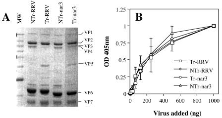
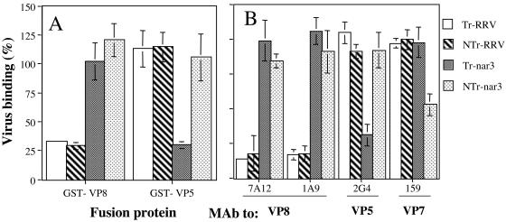
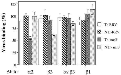
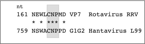
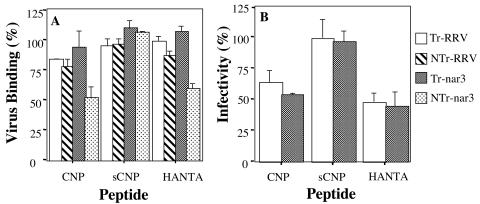
Rotavirus entry is a complex multistep process that depends on the trypsin cleavage of the virus spike protein VP4 into polypeptides VP5 and VP8 and on the interaction of these polypeptides and of VP7, the second viral surface protein, with several cell surface molecules, including integrin alphavbeta3. We characterized the effect of the trypsin cleavage of VP4 on the binding to MA104 cells of the sialic acid-dependent virus strain RRV and its sialic acid-independent variant, nar3. We found that, although the trypsin treatment did not affect the attachment of these viruses to the cell surface, their binding was qualitatively different. In contrast to the trypsin-treated viruses, which initially bound to the cell surface through VP4, the non-trypsin-treated variant nar3 bound to the cell through VP7. Amino acid sequence comparison of the surface proteins of rotavirus and hantavirus, both of which interact with integrin alphavbeta3 in an RGD-independent manner, identified a region shared by rotavirus VP7 and hantavirus G1G2 protein in which six of nine amino acids are identical. This region, which is highly conserved among the VP7 proteins of different rotavirus strains, mediates the binding of rotaviruses to integrin alphavbeta3 and probably represents a novel binding motif for this integrin.


Figures

FIG. 1.
FIG. 1.
Binding of trypsin-treated or untreated viral particles to MA104 cells. (A) SDS-PAGE analysis of Tr- or NTr-RRV and -nar3 viruses. Rotaviruses were propagated in MA104 cells in the absence of trypsin; the resultant viral cell lysates were then treated with 100 μg of trypsin/ml for 30 min at 37°C or mock treated, and the virus particles were purified by isopycnic centrifugation. The viral proteins were separated by SDS-10% PAGE and were Coomassie blue stained. The location of the individual proteins is indicated. (B) Binding of Tr- or NTr-RRV and -nar3 viruses to the surface of MA104 cells. The indicated amounts of purified TLPs were added to cells grown in 48-well plates. The cells were incubated with the viruses for 1 h at 4°C, and the amount of cell-bound virus was determined by an ELISA. The total amount of viral particles added in each assay (in nanograms) is plotted against the OD at 405 nm (OD405) reading obtained in the ELISA. A representative result of at least two independent experiments performed in duplicate is shown.
FIG. 2.
FIG. 2.
Cell binding of Tr- or NTr-RRV and -nar3 viruses in the presence of the recombinant proteins GST-VP8 and GST-VP5 or in the presence of MAbs to the viral proteins. (A) MA104 cells grown in 48-well plates were preincubated with 1.5 μg of affinity-purified GST-VP8 or GST-VP5 proteins. After removal of the excess of protein, 500 ng of the indicated purified virus was added. The cells were further incubated for 1 h at 4°C, and the amount of cell-bound virus was determined as described in Materials and Methods. (B) Purified viral particles (500 ng) were preincubated for 1 h at room temperature with 50 μg of the indicated MAbs to rotavirus proteins/ml. After this incubation, the virus-antibody mixture was added to a monolayer of MA104 cells, followed by incubation for 1 h at 4°C, and the amount of cell-bound virus determined. The data are expressed as the percentage of the virus bound when the cells were preincubated with PBS as a control. The arithmetic means and standard deviations for two independent experiments performed in duplicate are shown.
FIG. 3.
FIG. 3.
Cell binding of Tr- or NTr-RRV and -nar3 viruses in the presence of antibodies to different integrin subunits. MA104 cells grown in 48-well plates were preincubated with antibodies to the indicated integrin subunits for 1 h at 4°C, the excess of antibody was washed, and a fixed amount of purified virus particles (500 ng) was added to the cells, followed by further incubation for 1 h at 4°C. The cell-bound virus was determined as indicated in Materials and Methods. The antibodies used included polyclonal goat antibody to β3 (20 μg/ml), MAb P4G11 to β1 (10 μg/ml), MAb LM609 to αvβ3 (10 μg/ml), and MAb P1E6 to α2 (10 μg/ml). The data are expressed as the percentage of the virus bound when the cells were preincubated with PBS as a control. The arithmetic means and standard deviations for two independent experiments performed in duplicate are shown.
FIG. 4.
FIG. 4.
Alignment of rotavirus RRV protein VP7 (aa 161 to 169) with the G1G2 protein of hantavirus L99 (aa 759 to 767).
FIG. 5.
FIG. 5.
Effect of the synthetic peptides on the binding and infectivity of RRV and nar3 rotaviruses. (A) MA104 cells in 48-well plates were preincubated with 4 mg of each peptide/ml for 1 h at 4°C, the excess of peptide was removed, and 500 ng of Tr- or NTr-RRV and -nar3 viruses was then added, followed by incubation for 1 h at 4°C. The amount of bound virus was determined by an ELISA. The data are expressed as the percentage of the virus bound when the cells were preincubated with PBS as a control. The arithmetic means and standard deviations for two independent experiments performed in duplicate are shown. (B) MA104 cells grown in 96-well plates were preincubated for 1 h at 37°C with 4 mg of peptides CNP, sCNP, or HANTA/ml; the excess peptide was removed, and 2,000 FFU per well of either RRV or nar3 virus were then added and adsorbed for 1 h at 37°C. The excess inoculum was removed, and the infection was allowed to proceed for 14 h at 37°C. Finally, the cells were fixed and immunostained as described in Materials and Methods. The data are expressed as the percentage of virus infectivity obtained when the cells were preincubated with MEM as a control. The average numbers of foci representing 100% infectivity were 157 and 207 for RRV and nar3, respectively. The arithmetic means and standard deviations of three independent experiments performed in duplicate are shown.
FIG. 6.
FIG. 6.
Direct binding of peptide CNP to purified αvβ3 integrin. (A) ELISA plates coated with αvβ3 or α5β1 integrins (100 ng per well) were incubated for 1 h at 37°C with increasing amounts of biotinylated peptides. The amount of peptide bound to the plate was detected by a streptavidin-peroxidase assay. The amount of peptide added is plotted against the OD490 reading obtained in the ELISA. (B) The indicated amounts of nonbiotinylated CNP or sCNP peptide were added to a 96-well plate coated with integrin αvβ3 for 1 h at 37°C. After this incubation, 2 mg of biotinylated-CNP peptide/ml was added, and the plates were further incubated for 1 h at 37°C. The biotinylated peptide bound to the well was determined with streptavidin-peroxidase as described above. The data are expressed as the percentage of peptide bound when the wells were preincubated with PBS as a control. The arithmetic means and standard deviations for two independent experiments performed in duplicate are shown.
FIG. 7.
FIG. 7.
Binding of NTr-nar3 rotavirus to purified integrin αvβ3. (A) ELISA plates coated with αvβ3 or α5β1 integrins (100 ng per well) were incubated for 1 h at 37°C with increasing amounts of Tr- or NTr-nar3 viral particles. The virus bound to the plates was detected with a rabbit serum against rotavirus. The amount of virus added to the plate is plotted against the OD490 reading obtained for the virus bound. (B) Different amounts of nonbiotinylated CNP or sCNP peptide were added to a 96-well plate coated with αvβ3 integrin for 1 h at 37°C. After this incubation, NTr-nar3 virus (300 ng/well) was added, followed by further incubation for 1 h at 37°C. The amount of virus bound to the plate was detected with specific antibodies to rotavirus. The data are expressed as the percentage of virus bound when the αvβ3 integrin was preincubated with PBS as a control. The arithmetic means and standard deviations of two independent experiments performed in duplicate are shown.
FIG. 8.
FIG. 8.
The binding of CNP to αvβ3 integrin is independent of the RGD-binding site. Left panel, Different amounts of RGD or HANTA peptides were added to a 96-well plate coated with αvβ3 integrin for 1 h at 37°C. After incubation, a fixed amount (1 mg/ml) of biotinylated CNP peptide was added, followed by incubation for 1 h at 37°C. The biotinylated peptide bound to the well was determined by a streptavidin-peroxidase assay. Right panel, An αvβ3 integrin coated plate was preincubated with the indicated amounts of CNP or RGD peptides, and a fixed amount (1 mg/ml) of biotinylated RGD peptide was then added, followed by incubation for 1 h at 37°C. The amount of biotinylated RGD peptide was determined by a streptavidin-peroxidase assay. The data are expressed as the percentage of the peptide bound when the wells were preincubated with PBS as a control. The arithmetic means and standard deviations of two independent experiments performed in duplicate are shown.

Similar articles

Cited by

References

    1. Arias, C. F., C. A. Guerrero, E. Mendez, S. Zárate, P. Isa, R. Espinosa, P. Romero, and S. Lopez. 2001. Early events of rotavirus infection: the search for the receptor(s). John Wiley & Sons, Inc., New York, N.Y. - PubMed
    1. Arias, C. F., M. Lizano, and S. López. 1987. Synthesis in Escherichia coli and immunological characterization of a polypeptide containing the cleavage site associated with trypsin enhancement of rotavirus SA11 infectivity. J. Gen. Virol. 68:633-642. - PubMed
    1. Arias, C. F., P. Romero, V. Alvarez, and S. López. 1996. Trypsin activation pathway of rotavirus infectivity. J. Virol. 70:5832-5839. - PMC - PubMed
    1. Ciarlet, M., and M. K. Estes. 1999. Human and most animal rotavirus strains do not require the presence of sialic acid on the cell surface for efficient infectivity. J. Gen. Virol. 80:943-948. - PubMed
    1. Clark, S. M., J. R. Roth, M. L. Clark, B. B. Barnett, and R. S. Spendlove. 1981. Trypsin enhancement of rotavirus infectivity: mechanism of enhancement. J. Virol. 39:816-822. - PMC - PubMed

Publication types

LinkOut - more resources