. 2004 Oct;78(20):10848-55.
doi: 10.1128/JVI.78.20.10848-10855.2004.

Lentivirus-mediated RNA interference of DC-SIGN expression inhibits human immunodeficiency virus transmission from dendritic cells to T cells

Affiliations

Lentivirus-mediated RNA interference of DC-SIGN expression inhibits human immunodeficiency virus transmission from dendritic cells to T cells

Jean-François Arrighi et al. J Virol. 2004 Oct.

Abstract

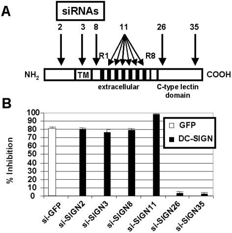
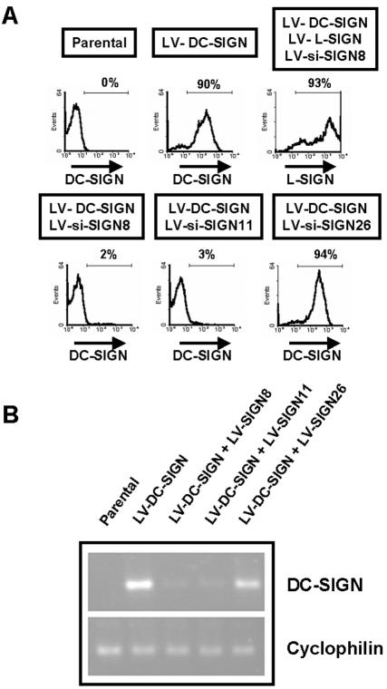
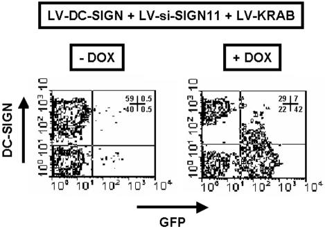
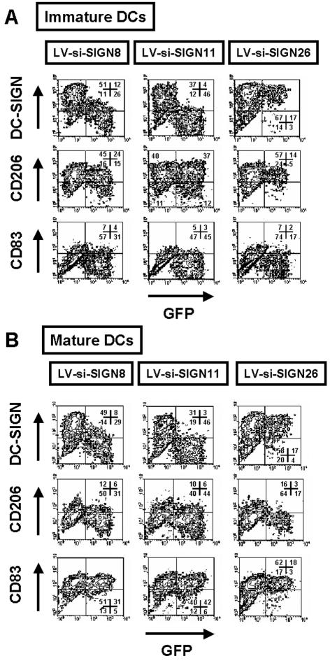
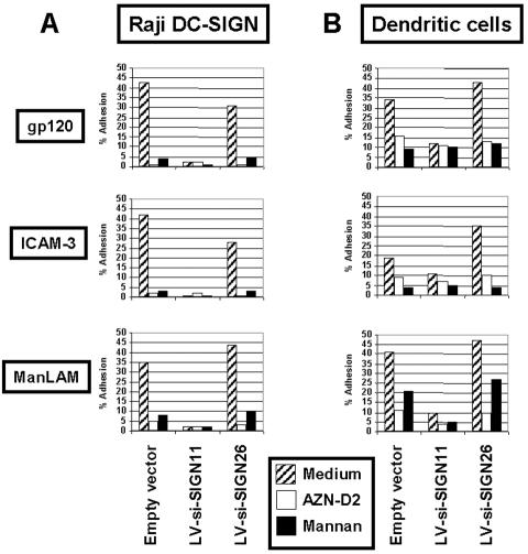
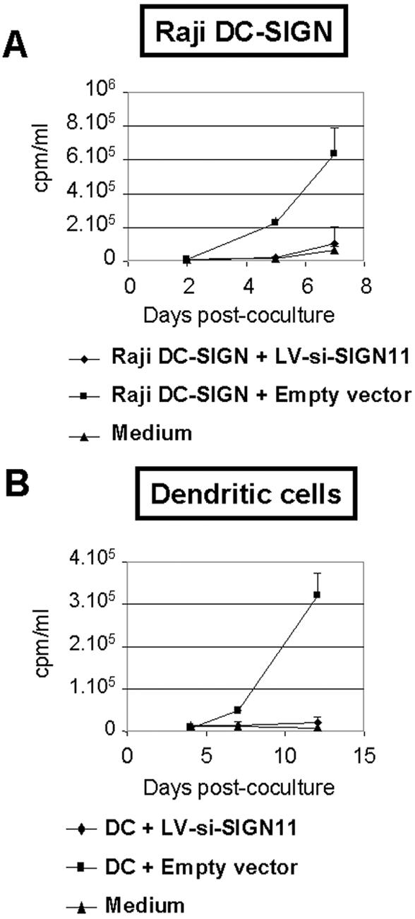
In the early events of human immunodeficiency virus type 1 (HIV-1) infection, immature dendritic cells (DCs) expressing the DC-specific intercellular adhesion molecule 3-grabbing nonintegrin (DC-SIGN) receptor capture small amounts of HIV-1 on mucosal surfaces and spread viral infection to CD4(+) T cells in lymph nodes (22, 34, 45). RNA interference has emerged as a powerful tool to gain insight into gene function. For this purpose, lentiviral vectors that express short hairpin RNA (shRNA) for the delivery of small interfering RNA (siRNA) into mammalian cells represent a powerful tool to achieve stable gene silencing. In order to interfere with DC-SIGN function, we developed shRNA-expressing lentiviral vectors capable of conditionally suppressing DC-SIGN expression. Selectivity of inhibition of human DC-SIGN and L-SIGN and chimpanzee and rhesus macaque DC-SIGN was obtained by using distinct siRNAs. Suppression of DC-SIGN expression inhibited the attachment of the gp120 envelope glycoprotein of HIV-1 to DC-SIGN transfectants, as well as transfer of HIV-1 to target cells in trans. Furthermore, shRNA-expressing lentiviral vectors were capable of efficiently suppressing DC-SIGN expression in primary human DCs. DC-SIGN-negative DCs were unable to enhance transfer of HIV-1 infectivity to T cells in trans, demonstrating an essential role for the DC-SIGN receptor in transferring infectious viral particles from DCs to T cells. The present system should have broad applications for studying the function of DC-SIGN in the pathogenesis of HIV as well as other pathogens also recognized by this receptor.


Figures

FIG. 1.
FIG. 1.
Design and screening of siRNAs targeting human DC-SIGN expression. (A) Design of siRNAs. Six different siRNAs against DC-SIGN were designed to target distinct parts of the DC-SIGN mRNA sequence. siRNA11 targeted tandem repeats (R1 to R8) present in exon III. TM, transmembrane domain. (B) Screening of siRNA-expressing vectors. Subconfluent 293T cells were cotransfected with pSUPER control plasmid, si-GFP, or si-SIGN constructs along with human DC-SIGN and pEGFP-N1 expression plasmids. The mean fluorescence intensity of DC-SIGN and GFP expression was measured in the GFP-positive and the DC-SIGN-positive cell populations, respectively. Histograms show the percentages of inhibition relative to that under the pSUPER control experimental condition. Means ± standard errors of results from four independent experiments are shown.
FIG. 2.
FIG. 2.
Suppression of DC-SIGN expression in cell lines through lentivirus-mediated delivery of siRNAs. (A) Generation of cell lines stably expressing siRNAs. Stable Raji transfectants were generated by transduction with the indicated lentiviral vectors. While both LV-si-SIGN8 and LV-si-SIGN11 suppressed DC-SIGN expression, only LV-si-SIGN11 suppressed L-SIGN expression. One month posttransduction, flow cytometric analysis of GFP, DC-SIGN, and L-SIGN expression was performed. The percentages of cells positive for the indicated cell surface markers are shown. (B) Effect of siRNAs on DC-SIGN mRNA. Total RNA extracted from Raji transfectants was subjected to RT-PCR to assess levels of mRNA corresponding to DC-SIGN and cyclophilin. Both LV-si-SIGN8 and LV-si-SIGN11 downregulated DC-SIGN mRNA expression but not cyclophilin mRNA expression.
FIG. 3.
FIG. 3.
Conditional suppression of DC-SIGN expression was evaluated in HeLa cell lines transduced with LV-DC-SIGN, LV-si-SIGN11, and LV-KRAB in the presence (+) or absence (−) of doxycycline (DOX). One week posttransduction, flow cytometric analysis of GFP and DC-SIGN expression was performed. Cell percentages corresponding to each quadrant of two-dimensional plots are shown.
FIG. 4.
FIG. 4.
Generation of DCs with DC-SIGN expression knocked down. DC progenitors were transduced with the indicated lentiviral vectors and differentiated either into immature DCs for 6 days with GM-CSF and IL-4 or into mature DCs by addition of LPS for the last 2 days of culture. Immature or mature DCs harvested at day 6 were analyzed by flow cytometry for the indicated cell surface antigens. LV-si-SIGN8 and LV-si-SIGN11 specifically inhibited DC-SIGN expression in DCs, and control vectors did not. Cell percentages corresponding to each quadrant of two-dimensional plots are shown. Representative results from one experiment out of four are presented.
FIG. 5.
FIG. 5.
Effect of DC-SIGN knockdown on HIV-1 gp120 adhesion. Raji transfectants (A) or transduced immature DCs (B) were resuspended in adhesion buffer and preincubated with anti-DC-SIGN neutralizing MAb (AZN-D2), mannan, or medium alone for 45 min at 37°C. Carboxylate-modified TransFluorSpheres coated with ICAM-3, gp120, or ManLAM were added, and the suspension was incubated for 30 min at 37°C. Adhesion was determined by measuring the percentages of cells with bound fluorescent beads within the GFP-positive cell population by flow cytometric analysis. The results shown are representative of those from three independent experiments giving similar results.
FIG. 6.
FIG. 6.
DCs with knocked-down DC-SIGN expression are unable to confer trans-enhancement of HIV-1 infection on T cells. Raji DC-SIGN transfectants (A) or sorted transduced immature DCs (B) were incubated with HIV-1 at a MOI of 0.001 and directly cocultured with activated PBLs. As a control, virus alone was added to cell culture medium (in the absence of Raji DC-SIGN transfectants or sorted DCs) but with addition of activated PBLs (medium). Viral production, expressed in counts per minute per milliliter, was monitored by reverse transcriptase assay of coculture supernatants. Data are means ± standard errors of results from one of three (A) or two (B) sets of duplicate experiments giving similar results.

Similar articles

Cited by

References

    1. Abbas-Terki, T., W. Blanco-Bose, N. Deglon, W. Pralong, and P. Aebischer. 2002. Lentiviral-mediated RNA interference. Hum. Gene Ther. 13:2197-2201. - PubMed
    1. Alvarez, C. P., F. Lasala, J. Carrillo, O. Muniz, A. L. Corbi, and R. Delgado. 2002. C-type lectins DC-SIGN and L-SIGN mediate cellular entry by Ebola virus in cis and in trans. J. Virol. 76:6841-6844. - PMC - PubMed
    1. An, D. S., Y. Xie, S. H. Mao, K. Morizono, S. K. Kung, and I. S. Chen. 2003. Efficient lentiviral vectors for short hairpin RNA delivery into human cells. Hum. Gene Ther. 14:1207-1212. - PubMed
    1. Arrighi, J. F., C. Hauser, B. Chapuis, R. H. Zubler, and V. Kindler. 1999. Long-term culture of human CD34(+) progenitors with FLT3-ligand, thrombopoietin, and stem cell factor induces extensive amplification of a CD34(−)CD14(−) and a CD34(−)CD14(+) dendritic cell precursor. Blood 93:2244-2252. - PubMed
    1. Arrighi, J. F., C. Soulas, C. Hauser, S. Saeland, B. Chapuis, R. H. Zubler, and V. Kindler. 2003. TNF-alpha induces the generation of Langerin/(CD207)+ immature Langerhans-type dendritic cells from both CD14-CD1a and CD14+CD1a− precursors derived from CD34+ cord blood cells. Eur. J. Immunol. 33:2053-2063. - PubMed

Publication types

MeSH terms