. 2004 Oct;78(20):10888-905.
doi: 10.1128/JVI.78.20.10888-10905.2004.

Identification of an hnRNP A1-dependent splicing silencer in the human papillomavirus type 16 L1 coding region that prevents premature expression of the late L1 gene

Affiliations

Identification of an hnRNP A1-dependent splicing silencer in the human papillomavirus type 16 L1 coding region that prevents premature expression of the late L1 gene

Xiaomin Zhao et al. J Virol. 2004 Oct.

Abstract

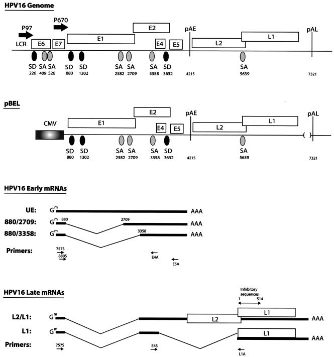
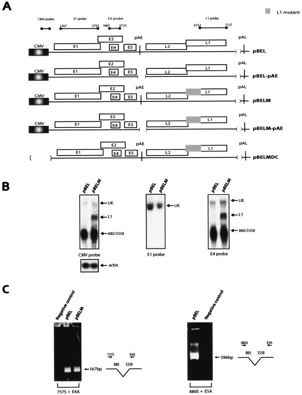
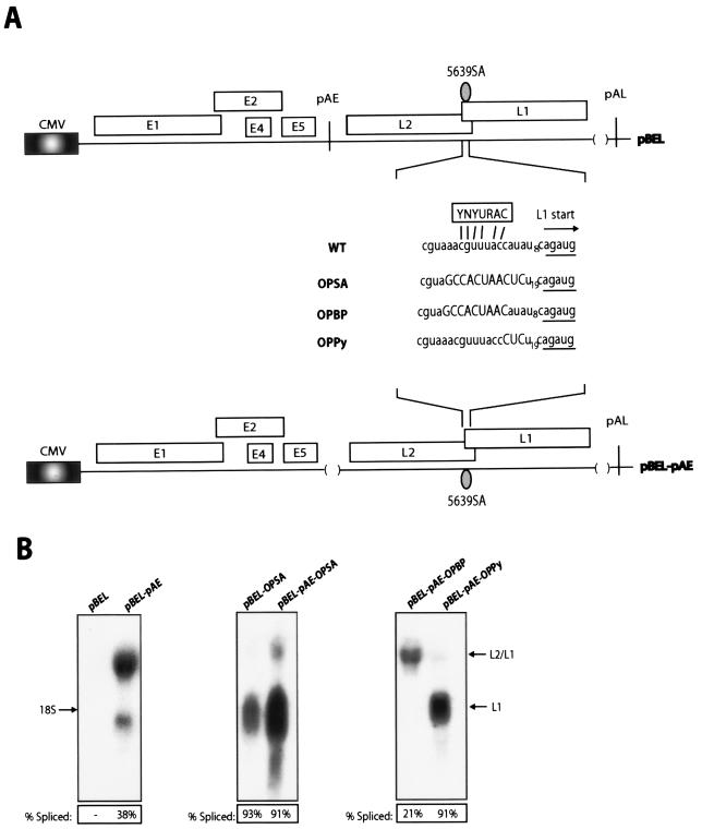
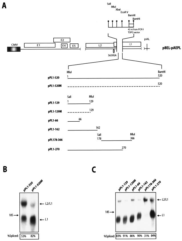
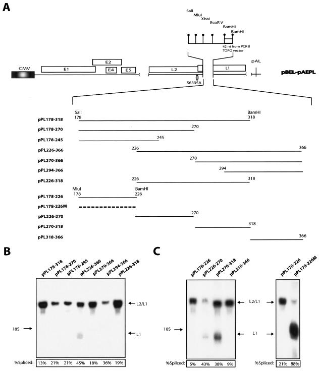
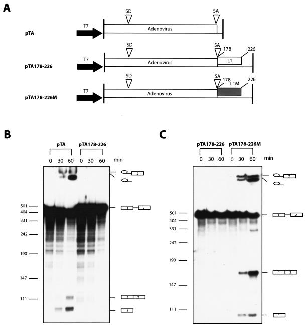
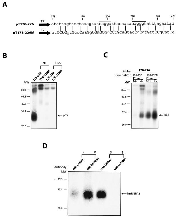
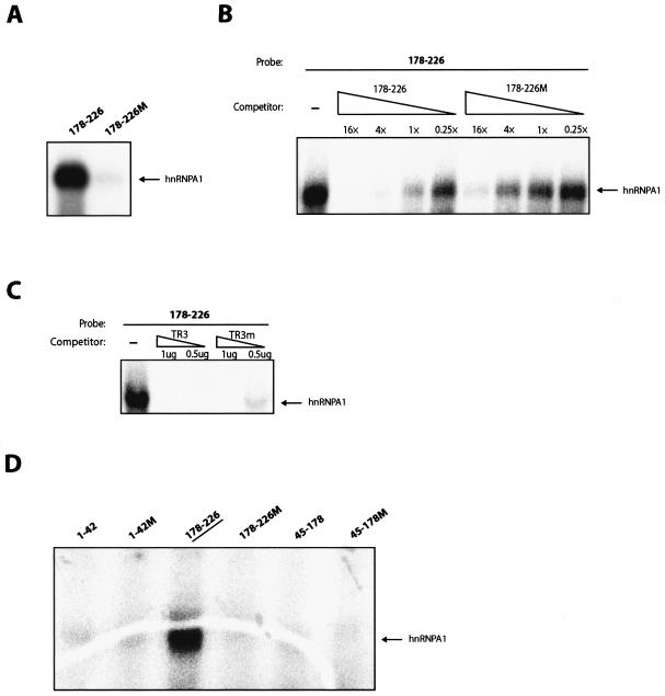
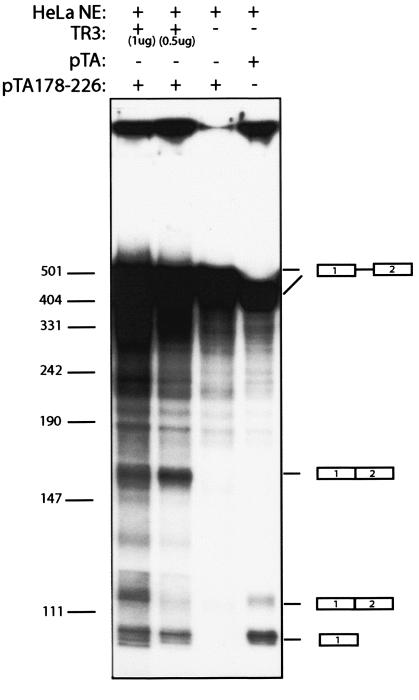
We have previously identified cis-acting RNA sequences in the human papillomavirus type 16 (HPV-16) L1 coding region which inhibit expression of L1 from eukaryotic expression plasmids. Here we have determined the function of one of these RNA elements, and we provide evidence that this RNA element is a splicing silencer which suppresses the use of the 3' splice site located immediately upstream of the L1 AUG. We also show that this splice site is inefficiently utilized as a result of a suboptimal polypyrimidine tract. Introduction of point mutations in the L1 coding region that altered the RNA sequence without affecting the L1 protein sequence resulted in the inactivation of the splicing silencer and induced splicing to the L1 3' splice site. These mutations also prevented the interaction of the RNA silencer with a 35-kDa cellular protein identified here as hnRNP A1. The splicing silencer in L1 inhibits splicing in vitro, and splicing can be restored by the addition of RNAs containing an hnRNP A1 binding site to the reaction, demonstrating that hnRNP A1 inhibits splicing of the late HPV-16 mRNAs through the splicing silencer sequence. While we show that one role of the splicing silencer is to determine the ratio between partially spliced L2/L1 mRNAs and spliced L1 mRNAs, we also demonstrate that it inhibits splicing from the major 5' splice site in the early region to the L1 3' splice site, thereby playing an essential role in preventing late gene expression at an early stage of the viral life cycle. We speculate that the activity of the splicing silencer and possibly the concentration of hnRNP A1 in the HPV-16-infected cell determines the ability of the virus to establish a persistent infection which remains undetected by the host immune surveillance.


Figures

FIG. 1.
FIG. 1.
Schematic representation of the HPV-16 genome. Boxes indicate the protein coding regions. Numbers refer to nucleotide positions in the HPV-16R sequence. The major p97 promoter and the differentiation-dependent promoter p670 (19) are indicated. Splice sites and polyadenylation signals are shown. The late UTR which contains RNA instability elements was originally deleted to increases the chances of obtaining detectable levels of late mRNAs. The structure of the pBEL expression plasmid is shown, and the predicted major mRNAs are displayed. The RT-PCR primers used here are shown as arrows under the schematic structures of the mRNAs. The previously identified inhibitory element in the first 514 nucleotides of the L1 coding sequence is indicated above the L1 gene (9, 51). pAE, early poly(A) signal; pAL, late poly(A) signal, CMV, human cytomegalovirus immediate-early promoter; SD, 5′ ss; SA, 3′ ss.
FIG.2.
FIG.2.
(A) Schematic representation of pBEL and the pBEL-derived plasmids. Boxes indicate the protein coding regions. The CMV promoter is indicated. The probes used in Northern blots are indicated above the diagram. The CMV probe detects all mRNAs produced from the CMV-driven plasmids and has been described previously (9). Numbers refer to nucleotide positions in the HPV-16R sequence. (B) Northern blots of total RNA extracted from HeLa cells transfected with pBEL and pBELM are shown hybridized to the indicated radiolabeled probes. UE, unspliced early mRNAs; L1, the spliced late mRNA; 880/3358, early mRNA spliced from the 5′ ss at position 880 to the 3′ss at position 3358 followed by polyadenylation at pAE. The same samples were hybridized to a human β-actin probe to control for loading. The data variation in each transfection experiment was less than 20%. (C) RT-PCR on the RNA samples shown in Fig. 2B using primers 757S or 880S in combination with either E4A or E5A (Fig. 1). Negative control, RNA sample from cells transfected with unrelated plasmid. (D) Northern blot of total RNA extracted from HeLa cells transfected with pBEL, pBELM, pBEL-pAE, pBELM-pAE, ors pBELMDC hybridized to the L1 probe. Both L2/L1 and L1 mRNAs are spliced from the 5′ ss at position 880 to the 3′ splice site at position 3358. The L2/L1 mRNA then remains unspliced until polyadenylation at pAL, whereas the L1 mRNA is spliced also between the 5′ss at position 3632 and the 3′ ss at position 5639. The truncated L1 mRNA is polyadenylated at a previously identified cryptic poly(A) signal at position 5170 in the HPV-16R genome (33). Spliced mRNA as a percentage of total late RNA in each lane is indicated at the bottom of the gel. The same samples were hybridized to a human β-actin probe to control for loading. The data variation in each transfection experiment was less than 20%. (E) RT-PCR of the RNA samples in Fig. 2D using primers 757S and L1A (left panel) or primers E4S and L1A (right panel). All RT-PCR products were cloned and sequenced. (F) Total, cytoplasmic, and nuclear RNAs were extracted from HeLa cells transfected with pBEL or pBELM. The blotted RNA was probed with the L1 probe.
FIG.2.
FIG.2.
(A) Schematic representation of pBEL and the pBEL-derived plasmids. Boxes indicate the protein coding regions. The CMV promoter is indicated. The probes used in Northern blots are indicated above the diagram. The CMV probe detects all mRNAs produced from the CMV-driven plasmids and has been described previously (9). Numbers refer to nucleotide positions in the HPV-16R sequence. (B) Northern blots of total RNA extracted from HeLa cells transfected with pBEL and pBELM are shown hybridized to the indicated radiolabeled probes. UE, unspliced early mRNAs; L1, the spliced late mRNA; 880/3358, early mRNA spliced from the 5′ ss at position 880 to the 3′ss at position 3358 followed by polyadenylation at pAE. The same samples were hybridized to a human β-actin probe to control for loading. The data variation in each transfection experiment was less than 20%. (C) RT-PCR on the RNA samples shown in Fig. 2B using primers 757S or 880S in combination with either E4A or E5A (Fig. 1). Negative control, RNA sample from cells transfected with unrelated plasmid. (D) Northern blot of total RNA extracted from HeLa cells transfected with pBEL, pBELM, pBEL-pAE, pBELM-pAE, ors pBELMDC hybridized to the L1 probe. Both L2/L1 and L1 mRNAs are spliced from the 5′ ss at position 880 to the 3′ splice site at position 3358. The L2/L1 mRNA then remains unspliced until polyadenylation at pAL, whereas the L1 mRNA is spliced also between the 5′ss at position 3632 and the 3′ ss at position 5639. The truncated L1 mRNA is polyadenylated at a previously identified cryptic poly(A) signal at position 5170 in the HPV-16R genome (33). Spliced mRNA as a percentage of total late RNA in each lane is indicated at the bottom of the gel. The same samples were hybridized to a human β-actin probe to control for loading. The data variation in each transfection experiment was less than 20%. (E) RT-PCR of the RNA samples in Fig. 2D using primers 757S and L1A (left panel) or primers E4S and L1A (right panel). All RT-PCR products were cloned and sequenced. (F) Total, cytoplasmic, and nuclear RNAs were extracted from HeLa cells transfected with pBEL or pBELM. The blotted RNA was probed with the L1 probe.
FIG. 3.
FIG. 3.
(A) Schematic representation of the pBEL and pBEL-pAE plasmids. The wt and the various mutant branch-point and polypyrimidine sequences are shown. The optimal branch point is displayed above the wt sequence. The OPSA sequence contains optimized branch point and polypyrimidine tract, while OPBP contains only the optimized branch point, and OPPy contains the optimized polypyrimidine tract only. (B) Northern blots on total RNA extracted from HeLa cells transfected with the indicated plasmids. All blots were probed with the L1 probe (Fig. 2A). Spliced mRNA as a percentage of total late RNA in each lane is indicated at the bottom of each gel. The data variation in each transfection experiment was less than 20%.
FIG. 4.
FIG. 4.
(A) Schematic diagram of the pBEL-pAEPL plasmid. A polylinker including a small sequence from the pCRII-TOPO cloning vector (Invitrogen) was inserted into L1, thereby replacing nucleotides 23 to 513 of L1 (numbering starts at A in the ATG of L1). The indicated wt and mutant L1 sequences of various lengths were inserted into the polylinker as MluI-BamHI or SalI-MluI fragments, as indicated. The numbering of the fragments starts at A in the L1 ATG. (B) Northern blot on total RNA extracted from HeLa cells transfected with pPL1-520 or pPL1-520 M. The blot is probed with the L1 probe (Fig. 2A). Spliced mRNA as a percentage of total late RNA in each lane is indicated at the bottom of each gel. (C) Northern blot on total RNA extracted from HeLa cells transfected with the indicated plasmids. The blot was probed with the L1 probe (Fig. 2A). Spliced mRNA as a percentage of total late RNA in each lane is indicated at the bottom of each gel. The data variation in each transfection experiment was less than 20%.
FIG. 5.
FIG. 5.
(A) Schematic representation of the pBEL-pAEPL plasmid. A polylinker including a small sequence from the pCRII-TOPO cloning vector (Invitrogen) was inserted into L1, thereby replacing nucleotides 23 to 513 of L1 (numbering starts at A in the ATG of L1). The indicated L1 sequences of various length were inserted into the polylinker as SalI-BamHI or MluI-BamHI fragments. The numbering of the fragments start at the A in the L1 ATG. (B and C) Northern blot on total RNA extracted from HeLa cells transfected with the indicated plasmids. The blots were probed with the L1 probe (see Fig. 2A). Spliced mRNA as a percentage of total late RNA in each lane is indicated at the bottom of each gel. The data variation in each transfection experiment was less than 20%.
FIG. 6.
FIG. 6.
(A) Schematic representation of the pTA, pTA178-226, and pTA178-226 M plasmids used for in vitro synthesis of RNA substrates for the in vitro splicing reactions. The numbering of the inserted HPV-16 L1 fragments start at the A in the L1 ATG. L1, L1 wt sequence from position 178 to 226; L1 M, L1 mutant sequence from position 178 to 226; T7, T7 RNA polymerase promoter; SD, adenovirus 5′ ss; SA, adenovirus 3′ ss. (B) In vitro splicing using RNA derived from pTA or pTA178-226. The splicing products are indicated. (C) In vitro splicing using RNA derived from pTA178-226 or pTA178-226 M. The splicing products are indicated.
FIG. 7.
FIG. 7.
(A) The sequences of the in vitro-transcribed inserts in plasmids pT178-228 and pT178-226 M are shown. The mutant positions are indicated with lines, and altered nucleotides are capitalized. These mutations were originally inserted in the L1 coding sequence in a way which did not affect the protein sequence of L1 while the L1 RNA sequence was altered (9). The numbering of the fragments start at the A in the L1 ATG. The two lines above the HPV-16 L1 sequence indicate two potential hnRNP A1 binding sites (5). (B) UV cross-linking of nuclear or cytoplasmic S100 extract to radiolabeled RNAs from the indicated plasmids. The p35 protein binding and cross-linking specifically to the wt HPV-16 L1 sequence is indicated. (C) UV cross-linking of nuclear extract to radiolabeled RNAs from plasmid pT178-226 in the presence of cold competitor RNA derived from the wt L1 sequence in pT178-226 or the mutant L1 sequence in pT178-226 M. Fold excess of the cold RNA is indicated. The p35 protein binding and cross-linking specifically to the wt HPV-16 L1 sequence is indicated. (D) Immunoprecipitation of the 35-kDa protein which UV cross-links specifically to the wt 178-226 sequence with a monoclonal antibody against hnRNP A1 (mAb hnRNP A1), but not with a monoclonal antibody against HPV-16 L1 capsid protein (mAb CAMvir).
FIG. 8.
FIG. 8.
(A) UV cross-linking of recombinant His-tagged hnRNP A1 to radiolabeled RNAs from plasmid pT178-226 or pT178-226 M. (B) UV cross-linking of recombinant His-tagged hnRNP A1 to radiolabeled RNAs from plasmid pT178-226 in the presence of cold competitor RNA derived from the wt L1 sequence in pT178-226 or the mutant L1 sequence in pT178-226 M. n-fold excess of cold competitor is indicated. (C) UV cross-linking of recombinant His-tagged hnRNP A1 to radiolabeled RNA from plasmid pT178-226 in the absence or presence of wt or mutant single-stranded telomeric DNA repeats named TR3 and TR3m, respectively (60). hnRNP A1 has been shown previously to bind specifically to TR3 but less efficiently to the mutant named TR3m. (D) UV cross-linking of recombinant His-tagged hnRNP A1 to radiolabeled RNAs, which encode wt and mutant HPV-16 sequences derived from various parts of L1. The numbering of the fragments start at the A in the L1 ATG.
FIG. 9.
FIG. 9.
In vitro splicing using RNA derived from pTA or pTA178-226, in the absence or presence of the single-stranded telomeric DNA repeat competitor named TR3 (60). It has been shown previously that hnRNP A1 binds specifically to TR3 (60). The splicing products are indicated.

Similar articles

Cited by

References

    1. Baker, C. C. 1997. Posttranscriptional regulation of papillomavirus gene expression, p. 11-16. In S. R. Billakanti, C. E. Calef, A. D. Farmer, A. L. Halpern, and G. L. Myres (ed.), Human papillomaviruses: a compilation and analysis of nucleic acid and amino acid sequences. Theoretical Biology and Biophysics, Los Alamos National Laboratory, Los Alamos, N.Mex.
    1. Barksdale, S. K., and C. C. Baker. 1995. The human immunodeficiency virus type 1 Rev protein and the Rev-responsive element counteract the effect of an inhibitory 5′ splice site in a 3′ untranslated region. Mol. Cell. Biol. 15:2962-2971. - PMC - PubMed
    1. Bilodeau, P. S., J. K. Domsic, A. Mayeda, A. R. Krainer, and C. M. Stoltzfus. 2001. RNA splicing at human immunodeficiency virus type 1 3′ splice site A2 is regulated by binding of hnRNP A/B proteins to an exonic splicing silencer element. J. Virol. 75:8487-8497. - PMC - PubMed
    1. Blanchette, M., and B. Chabot. 1999. Modulation of exon skipping by high-affinity hnRNP A1-binding sites and by intron elements that repress splice site utilisation. EMBO J. 18:1939-1952. - PMC - PubMed
    1. Burd, C. G., and G. Dreyfuss. 1994. RNA binding specificity of hnRNP A1: significance of hnRNP A1 high-affinity binding sites in pre-mRNA splicing. EMBO J. 13:1197-1204. - PMC - PubMed

Publication types

MeSH terms

Substances

LinkOut - more resources