. 2004 Oct;78(20):10920-6.
doi: 10.1128/JVI.78.20.10920-10926.2004.

Phosphatidylserine is not the cell surface receptor for vesicular stomatitis virus

Affiliations

Phosphatidylserine is not the cell surface receptor for vesicular stomatitis virus

David A Coil et al. J Virol. 2004 Oct.

Abstract

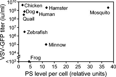
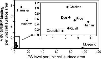
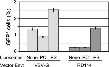
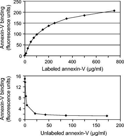
The envelope protein from vesicular stomatitis virus (VSV) has become an important tool for gene transfer and gene therapy. It is widely used mainly because of its ability to mediate virus entry into all cell types tested to date. Consistent with the broad tropism of the virus, the receptor for VSV is thought to be a ubiquitous membrane lipid, phosphatidylserine (PS). However, the evidence for this hypothesis is indirect and incomplete. Here, we have examined the potential interaction of VSV and PS at the plasma membrane in more detail. Measurements of cell surface levels of PS show a wide range across cell types from different organisms. We demonstrate that there is no correlation between the cell surface PS levels and VSV infection or binding. We also demonstrate that an excess of annexin V, which binds specifically and tightly to PS, does not inhibit infection or binding by VSV. While the addition of PS to cells does allow increased virus entry, we show that this effect is not specific to the VSV envelope. We conclude that PS is not the cell surface receptor for VSV, although it may be involved in a postbinding step of virus entry.


Figures

FIG. 1.
FIG. 1.
VSV-GFP titer versus surface PS levels for multiple cell types. Virus titer and PS levels were measured as described in Materials and Methods. X-axis data are the geometric means of the fluorescence of 10,000 live cells measured by flow cytometry, and the units are arbitrary fluorescence units (linear scale) generated by the cytometer. The PS levels shown are representative of at least two experiments for each cell line with different batches of annexin V. Vector titer data are for one complete experiment that was repeated with very similar results.
FIG. 2.
FIG. 2.
VSV-G/GFP binding versus surface PS levels for multiple cell types. The PS level determinations are the same as in Fig. 1 before scaling for cell size. VSV-G/GFP binding was measured at 4°C as described in Materials and Methods. To account for significant differences in cell size between cell types, both PS levels and binding levels are expressed as fluorescence per unit cell surface. Data shown are the geometric means of 10,000 cells for both PS and binding. The experiment was repeated with a different stock of VSV-G/GFP with very similar results. PS levels versus VSV-G/GFP binding for all cell types are shown. Inset, enlargement of the part of the graph containing the six cell types with the lowest levels of PS among those tested.
FIG. 3.
FIG. 3.
Effects of liposome addition on virus infection. PC and PS liposomes were generated as described and added to cells. Twenty-four hours later, the retrovirus vector LNCG, pseudotyped with either RD114 or VSV-G envelope proteins, was added to cells in the presence of 4 μg of Polybrene per ml. After 3 days, the cells were removed from the dishes and GFP-positive cells were counted by flow cytometry. Cells not incubated with the virus showed fewer than five GFP-positive cells in the 105 cells analyzed. Data shown are the means ± standard deviations of results from three replicates from one experiment. The experiment was repeated twice with very similar results.
FIG. 4.
FIG. 4.
Saturable annexin V binding to ZF4 cells. (Top) Alexa Fluor 488-labeled annexin V binding to ZF4 cells. Annexin V staining was performed as described in Materials and Methods, with variable amounts of annexin V. Data shown are the geometric means for 10,000 live cells minus the mean fluorescence of cells not exposed to annexin V. (Bottom) Competition of unlabeled annexin V with labeled annexin V on ZF4 cells. Staining with labeled annexin V was performed as described in Materials and Methods (5 μl of labeled annexin V per 100 μl of incubation mixture; ∼50 μl/ml) except that cells were exposed to various amounts of unlabeled annexin V for 15 min prior to the addition of labeled annexin. Each data point is the geometric mean fluorescence of 10,000 live cells minus the mean fluorescence of cells not exposed to annexin V.
FIG. 5.
FIG. 5.
Annexin V binding to ZF4 cells and effect on VSV-G/GFP binding. Cells were harvested for virus binding, treated with unlabeled annexin V (86 μg/ml) or buffer, and then measured for virus binding or labeled annexin V binding as described in Materials and Methods. The unlabeled cells are the same in both panels, and the grey curves in both panels represent the cells pretreated with 86 μg of unlabeled annexin V/ml. (Top) VSV-G/GFP binding to ZF4 cells with or without treatment of unlabeled annexin V (86 μg/ml). (Bottom) Labeled annexin V binding to cells with or without pretreatment of unlabeled annexin V (86 μg/ml). This experiment was repeated with virtually identical results.

Similar articles

Cited by

References

    1. Andree, H. A., C. P. Reutelingsperger, R. Hauptmann, H. C. Hemker, W. T. Hermens, and G. M. Willems. 1990. Binding of vascular anticoagulant alpha (VAC alpha) to planar phospholipid bilayers. J. Biol. Chem. 265:4923-4928. - PubMed
    1. Bartz, S. R., and M. A. Vodicka. 1997. Production of high-titer human immunodeficiency virus type 1 pseudotyped with vesicular stomatitis virus glycoprotein. Methods 12:337-342. - PubMed
    1. Boon, J. M., and B. D. Smith. 2002. Chemical control of phospholipid distribution across bilayer membranes. Med. Res. Rev. 22:251-281. - PubMed
    1. Boritz, E., J. Gerlach, J. E. Johnson, and J. K. Rose. 1999. Replication-competent rhabdoviruses with human immunodeficiency virus type 1 coats and green fluorescent protein: entry by a pH-independent pathway. J. Virol. 73:6937-6945. - PMC - PubMed
    1. Callahan, M. K., P. Williamson, and R. A. Schlegel. 2000. Surface expression of phosphatidylserine on macrophages is required for phagocytosis of apoptotic thymocytes. Cell Death Differ. 7:645-653. - PubMed

Publication types

LinkOut - more resources