. 2004 Oct;78(20):10995-1006.
doi: 10.1128/JVI.78.20.10995-11006.2004.

Major human cytomegalovirus structural protein pp65 (ppUL83) prevents interferon response factor 3 activation in the interferon response

Affiliations

Major human cytomegalovirus structural protein pp65 (ppUL83) prevents interferon response factor 3 activation in the interferon response

Davide A Abate et al. J Virol. 2004 Oct.

Abstract

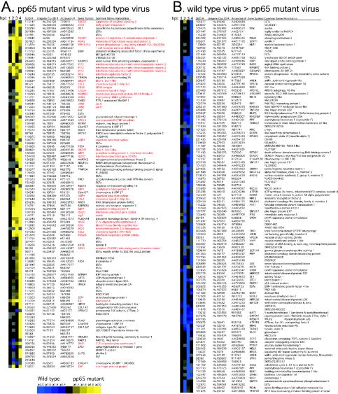
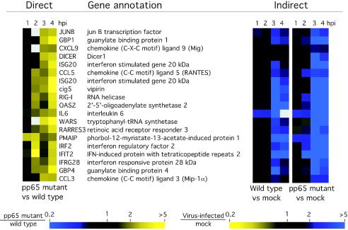
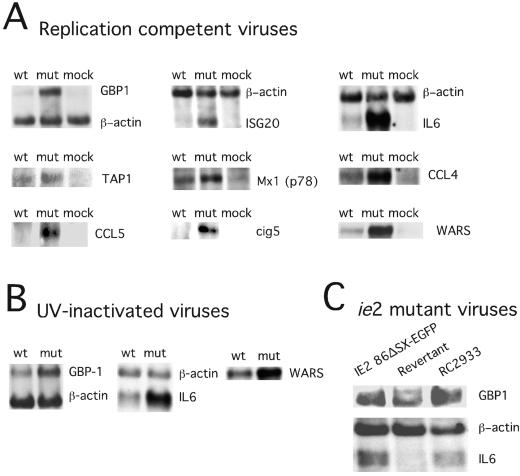
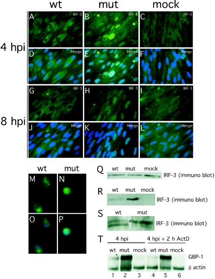
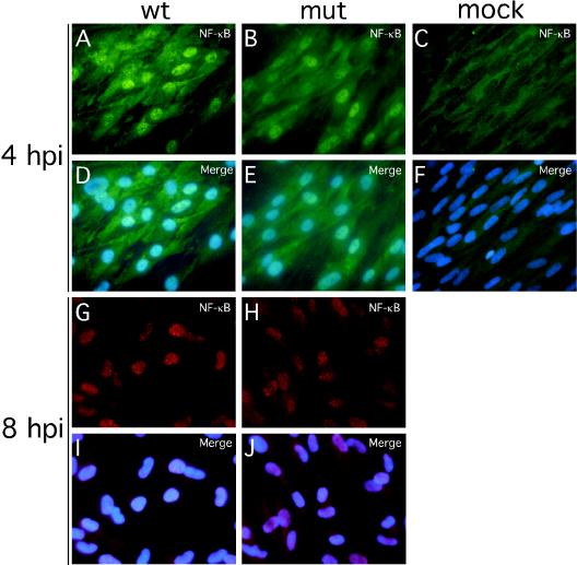
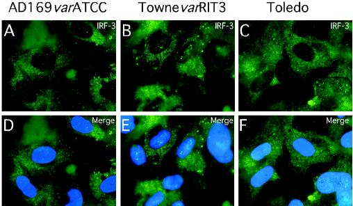
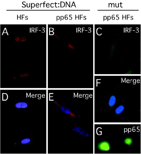
We have identified a cytomegalovirus virion protein capable of modulating the rapid induction of an interferon-like response in cells that follows virus binding and penetration. Functional genomics revealed a role for the major cytomegalovirus structural protein, pp65 (ppUL83), in counteracting this response. The underlying mechanism involves a differential impact of this structural protein on the regulation of interferon response factor 3 (IRF-3). In contrast, NF-kappaB is activated independent of pp65, and neither STAT1 nor STAT3 becomes activated by either virus. pp65 is sufficient to prevent the activation of IRF-3 when introduced alone into cells. pp65 acts by inhibiting nuclear accumulation of IRF-3 and is associated with a reduced IRF-3 phosphorylation state. Thus, this investigation shows that the major structural protein of cytomegalovirus is committed to the modulation of the IRF-3 response, a primary mediator of the type I interferon response. By subverting IRF-3, the virus escapes throwing a central alarm devoted to both immediate antiviral control and regulation of the immune response.


Figures

FIG. 1.
FIG. 1.
Cluster analysis (20) of genes regulated twofold or more in at least one time point (1, 2, 3, or 4 hpi) in a direct (type I) human cDNA microarray analysis comparing wt and pp65 mutant CMV. (A) Genes whose transcripts were more abundant upon infection with pp65 mutant RVAd65 than wt (pp65 mutant virus > wild-type virus). A total of 101 genes are represented in this panel. (B) Genes whose transcripts were more abundant upon infection with wt than pp65 mutant RVAd65 (wild-type virus > pp65 mutant virus). A total of 119 genes are represented in this panel. Each column of the microarray image represents a different time point, indicated at the top. Gray is used to indicate microarray spots that failed to pass the filtering criteria (see Materials and Methods). Red lettering denotes genes that are known to be IFN induced. Each row represents a different cDNA clone identified by a Stanford University identification number (SUID #). The Unigene cluster identification number (Clus ID #), GenBank accession number, gene symbol, and common gene name (or annotation) are also indicated for each clone. A color scale proportional to the factor of change is shown below panel A. Several cDNAs spotted in replicate on the microarrays (ISG20, 2 spots; dicer, 2 spots; MAX interacting protein 1, 2 spots; FZD-4, 2 spots; asparate β hydroxylase, 2 spots; GBP-1, 2 spots; JunB, 2 spots; C6orf37, 2 spots; and IFIT2, 3 spots) showed similar hybridization ratios. Complete data sets are available at http://genome-www5.stanford.edu.
FIG. 2.
FIG. 2.
Confirmation of pp65-induced genes by additional microarray experimental approaches. Cluster analysis of 19 genes for which transcripts were compared directly on a single microarray are shown at left. The transcripts shown here had a ratio of ≥2 for pp65 mutant virus-infected cell RNA versus wt virus-infected cell RNA in at least two time points (1, 2, 3, or 4 hpi) by using a direct analysis. A cluster analysis of the same genes following indirect analysis in which either wt or pp65 mutant virus infection was compared to mock infection at the same time point is shown at right. The color scales below the images indicate a factor of change ratio, shown with marks at 0.2-, 1-, 2-, and >5-fold spot intensity ratios. Ratios of mutant virus-infected to wt virus-infected cells are from a direct comparison (left), and ratios of either wt or pp65 mutant virus-infected cells to mock-infected cells are shown in the indirect comparisons (right). vs, versus.
FIG. 3.
FIG. 3.
RNA blot analysis of selected IFN response genes. RNA blots (5 μg of total cell RNA per lane) from infected cells collected at 4 hpi at an MOI of 4 and hybridized with the indicated IFN response gene probes prepared as described in Materials and Methods. (A) Comparisons of wt-, pp65 mutant (mut)-, and mock-infected (mock) HFs for expression of GBP1, ISG20, IL-6, Tap-1, Mx-1 (p78), CCL4, CCL5, cig5, and WARS with a β-actin control are shown for some samples. (B) Comparisons of UV-inactivated wt and pp65 mutant (mut) virus-induced expression of GBP1, IL-6, and WARS with a β-actin control are shown for some samples. (C) Comparisons are shown of RNA collected 4 hpi from IE2 86ΔSX-EGFP-, revertant-, and RC2933-infected cells probed for GBP1, IL-6, and β actin.
FIG. 4.
FIG. 4.
Impact of wt and pp65 mutant CMV infection on IRF-3 localization and phosphorylation state at early times after infection. (A to L) Immunofluorescence analysis of IRF-3 localization in HFs infected (MOI of 4) with wt virus at 4 (A and D) or 8 (G and J) hpi or with pp65 mutant virus (mut) at 4 (B and E) or 8 (H and K) hpi compared to mock-infected cells at 4 (C and F) or 8 (I and L) hpi. Overlays of Hoechst 44432-positive nuclei are shown in panels M to P (merge). Immunofluorescence analysis of IRF-3 localization in PBMC infected by wt (M and O) or mutant (N and P) virus (4 hpi with an MOI of 4). (Q to S) Immunoblot analyses of total (Q) and nuclear (R) IRF-3 levels in wt virus-, mutant virus- and mock-infected HFs, and immunoblot analysis of IRF-3 electrophoretic mobility forms revealing phosphorylation state in wt and mutant virus-infected cells (S). RNA stability assay (T) was performed with RNA extracted by wt CMV-, pp65 mutant CMV (mut)-, and mock-infected HFs (mock) in the absence (lanes 1 to 3) or presence of actinomycin D (2 h ActD; lanes 4 to 6). Infected-cell RNA was probed for GBP-1 and β-actin. Note that the monoclonal antibody we have employed in the experiments shown in Fig. 4 to 6 has been shown to give specific in situ immunofluorescence localization and immunoblot detection of IRF-3 (; M. G. Wathelet, personal communication) and has been used for this purpose in several incisive studies (5, 23, 49, 67, 72).
FIG. 5.
FIG. 5.
Impact of wt and pp65 mutant CMV infection on NF-κB localization. Shown are immunofluorescence assays of NF-κB localization in HFs infected (MOI of 4) with wt virus at 4 (A and D) or 8 (G and I) hpi or with pp65 mutant virus (mut) at 4 (B and E) or 8 (H and J) hpi compared to mock-infected cells at 4 (C and F) hpi. Texas Red-conjugated secondary antibody was used to generate the results shown in panels G through J. Overlays of Hoechst 44432-positive nuclei are shown (merge).
FIG. 6.
FIG. 6.
Impact of infection with additional CMV strains and strain variants on IRF-3 localization at early times after infection. Shown are immunofluorescence analyses of IRF-3 localization in HFs infected (MOI of 4) with AD169varATCC (A and D), TownevarRIT3 (B and E), and Toledo (passage level 10; C and F). Overlays of Hoechst 44432-positive nuclei are shown (merge).
FIG. 7.
FIG. 7.
Immunofluorescence analysis of IRF-3 localization in pp65-transduced cells. Immunofluorescence assays show IRF-3 localization 4 h after exposure to induction with pcDNA3-EYFP-loaded Superfect in control HFs (A) and pp65-transduced HFs (B) or at 4 hpi with pp65 mutant (mut) virus in pp65-transduced HFs (C). Overlays of IRF-3 stain with Hoechst 44432 show nuclei (D to F). Immunofluorescence assay shows pp65 localization in pp65-transduced HFs (G).

References

    1. Alcami, A., and U. H. Koszinowski. 2000. Viral mechanisms of immune evasion. Trends Microbiol. 8:410-418. - PMC - PubMed
    1. Arrode, G., and C. Davrinche. 2003. Dendritic cells and HCMV cross-presentation. Curr. Top. Microbiol. Immunol. 276:277-294. - PubMed
    1. Au, W. C., P. A. Moore, W. Lowther, Y. T. Juang, and P. M. Pitha. 1995. Identification of a member of the interferon regulatory factor family that binds to the interferon-stimulated response element and activates expression of interferon-induced genes. Proc. Natl. Acad. Sci. USA 92:11657-11661. - PMC - PubMed
    1. Barnes, B., B. Lubyova, and P. M. Pitha. 2002. On the role of IRF in host defense. J. Interferon Cytokine Res. 22:59-71. - PubMed
    1. Basler, C. F., A. Mikulasova, L. Martinez-Sobrido, J. Paragas, E. Muhlberger, M. Bray, H. D. Klenk, P. Palese, and A. Garcia-Sastre. 2003. The Ebola virus VP35 protein inhibits activation of interferon regulatory factor 3. J. Virol. 77:7945-7956. - PMC - PubMed

Publication types

MeSH terms