. 2004 Oct;78(20):11097-107.
doi: 10.1128/JVI.78.20.11097-11107.2004.

Molecular mechanisms of attenuation of the Sabin strain of poliovirus type 3

Affiliations

Molecular mechanisms of attenuation of the Sabin strain of poliovirus type 3

Stephen Guest et al. J Virol. 2004 Oct.

Abstract

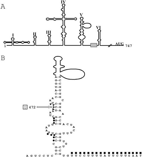
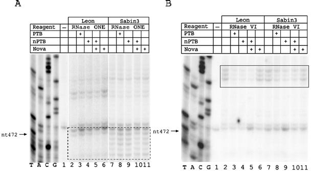
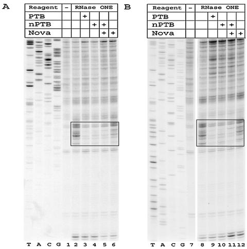
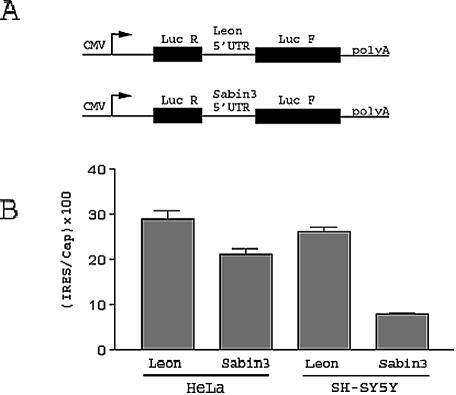
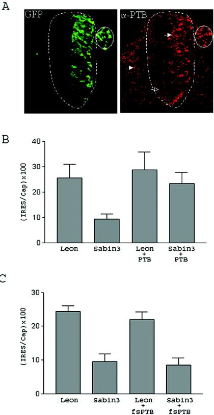
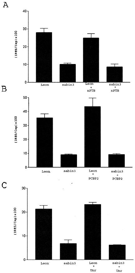
Mutations critical for the central nervous system (CNS) attenuation of the Sabin vaccine strains of poliovirus (PV) are located within the viral internal ribosome entry site (IRES). We examined the interaction of the IRESs of PV type 3 (PV3) and Sabin type 3 (Sabin3) with polypyrimidine tract-binding protein (PTB) and a neural cell-specific homologue, nPTB. PTB and nPTB were found to bind to a site directly adjacent to the attenuating mutation, and binding at this site was less efficient on the Sabin3 IRES than on the PV3 IRES. Translation mediated by the PV3 and Sabin3 IRESs in neurons of the chicken embryo spinal cord demonstrated a translation deficit for the Sabin3 IRES that could be rescued by increasing PTB expression in the CNS. These data suggest that the low levels of PTB available in the CNS, coupled to a reduced binding of PTB on the Sabin3 IRES, leads to its CNS-specific attenuation. This study also demonstrates the use of the chicken embryo to easily investigate translation of RNA within a neuron in the CNS of an intact living organism.


Figures

FIG. 1.
FIG. 1.
The PV 5′ UTR. (A) Schematic representation of the predicted secondary structure of the PV 5′ UTR. The shaded box represents the oligopyrimidine tract. (B) Detailed view of domain V and the oligopyrimidine tract for the Leon strain of PV. The mutation involved in attenuation of Sabin3 is boxed. Nucleotides susceptible to RNase ONE cleavage in the Sabin3, but not the Leon, IRES are indicated by triangles (see Fig. 2A). Nucleotides protected by PTB or nPTB from V1 cleavage are indicated by circles (see Fig. 2B), and nucleotides protected by PTB or nPTB from RNase ONE cleavage are indicated by squares (see Fig. 3).
FIG. 2.
FIG. 2.
PTB and nPTB bind the Leon and Sabin3 IRESs at a site adjacent to nt 472. Enzymatic footprint of the Leon and Sabin3 IRESs in the region surrounding nt 472 with RNase ONE (A) and RNase V1 (B). In this figure and the subsequent two figures, the table above the gel indicates whether RNA was incubated alone or with proteins and whether RNA-protein complexes were digested with an enzyme. RNA probes were extended with a primer complementary to nt 542 to 559. The region displaying enhanced cleavage by RNase ONE on the Sabin3 strain is indicated by the dashed box (see also triangles in Fig. 1B). The location of the PTB- or nPTB-binding site is indicated by the solid box (see also circles in Fig. 1B).
FIG. 3.
FIG. 3.
A second PTB- or nPTB-binding site in the PV IRES is located downstream of nt 472 in an oligopyrimidine tract. Enzymatic footprint of the Leon (A) and Sabin3 (B) IRESs in the region surrounding the oligopyrimidine tract. RNA probes were extended with a primer complementary to nt 665 to 682. The location of the PTB- or nPTB-binding site is indicated by the solid box (see also squares in Fig. 1B).
FIG. 4.
FIG. 4.
A binding site for Nova-1 is located immediately downstream of the polyprotein's translation start site. Enzymatic footprint of the Leon (A) and Sabin3 (B) IRESs in the region surrounding the initiation codon for the viral polyprotein. RNA probes were extended with a primer complementary to nt 815 to 834. The location of the Nova-1-binding site downstream of the initiation codon is indicated by the solid boxes. The locations of the initiating AUG and the consensus Nova-1-binding site are shown on the left.
FIG. 5.
FIG. 5.
The Sabin3 IRES has a translation defect in cultured SH-SY5Y neuroblastoma cells demonstrated by transfection with bicistronic reporter constructs. (A) Schematic representation of the bicistronic constructs containing either the Leon or the Sabin3 5′ UTR. The CMV promoter is represented by an arrow. The Renilla and firefly luciferase genes are represented by black boxes. The Leon or Sabin3 5′ UTR is located in the intercistronic region between the Renilla and firefly luciferase genes. (B) HeLa and SH-SY5Y cells were transiently transfected with bicistronic constructs. Cells were harvested 24 h posttransfection, and luciferase activities in the extracts were measured. IRES activity is shown as the ratio of firefly luciferase (IRES-driven translation) to Renilla luciferase (cap-driven translation) activity times 100.
FIG. 6.
FIG. 6.
The Sabin3 IRES has a translation defect in the CNS that can be rescued by coelectroporation of PTB. (A) Two-day-old chicken embryos were electroporated with a vector that expresses PTB and a vector that expresses GFP. Embryos were harvested at 24 h postelectroporation, sectioned, and overlaid with an anti-PTB mouse monoclonal antibody, followed by a Cy5-conjugated anti-mouse antibody. The section of the chicken embryo was then visualized under UV light with a filter for GFP or Cy5. The left panel shows GFP fluorescence in green, while the right panel shows the same field with anti-PTB staining in red. The spinal cord is outlined with a dotted line. As shown, neurons that received the electroporated GFP (and PTB expression construct) have migrated throughout one side of the spinal cord and the dorsal root ganglion (which is surrounded by a solid line). Cells that were electroporated have an increased PTB signal (closed arrow) compared to neurons on the other side of the spinal cord (open arrow) or nonneural cells (arrowhead). (B and C) Two-day-old chicken embryos were electroporated with the Leon or Sabin3 bicistronic construct with or without coelectroporation of the PTB expression construct (B) or with or without coelectroporation of a vector that expresses a truncated, nonfunctional fragment of PTB (pfs-PTB) (C).
FIG. 7.
FIG. 7.
Coelectroporation of nPTB, PCBP2, or unr does not rescue the translation defect of the Sabin3 IRES in the CNS. Two-day-old chicken embryos were electroporated with the Leon or Sabin3 bicistronic construct with or without coelectroporation of an nPTB expression vector (A), a PCBP2 expression vector (B), or a unr expression vector (C).
FIG. 8.
FIG. 8.
Knockdown of PTB expression in cultured cells by RNA interference demonstrates the importance of PTB in PV3 IRES translation. (A) Western immunoblot analysis of lysates from BHK-21 cells transfected with the plasmids indicated in the table above the gel. Lysates were subjected to electrophoresis, followed by immunostaining with anti-PTB antibody. The same membrane was then stripped and reprobed with anti-β-actin antibody. (B) BHK-21 cells were transfected with the Leon bicistronic reporter construct along with one of the following: control (control vector plus pfs-PTB), siRNA (siRNA construct plus pfs-PTB), or siRNA plus mmPTB (siRNA construct plus mmPTB).

References

    1. Belsham, G. J., and N. Sonenberg. 1996. RNA-protein interactions in regulation of picornavirus RNA translation. Microbiol. Rev. 60:499-511. - PMC - PubMed
    1. Blyn, L. B., K. M. Swiderek, O. Richards, D. C. Stahl, B. L. Semler, and E. Ehrenfeld. 1996. Poly(rC) binding protein 2 binds to stem-loop IV of the poliovirus RNA 5′ noncoding region: identification by automated liquid chromatography-tandem mass spectrometry. Proc. Natl. Acad. Sci. USA 93:11115-11120. - PMC - PubMed
    1. Blyn, L. B., J. S. Towner, B. L. Semler, and E. Ehrenfeld. 1997. Requirement of poly(rC) binding protein 2 for translation of poliovirus RNA. J. Virol. 71:6243-6246. - PMC - PubMed
    1. Boussadia, O., M. Niepmann, L. Creancier, A. C. Prats, F. Dautry, and H. Jacquemin-Sablon. 2003. Unr is required in vivo for efficient initiation of translation from the internal ribosome entry sites of both rhinovirus and poliovirus. J. Virol. 77:3353-3359. - PMC - PubMed
    1. Buckanovich, R. J., and R. B. Darnell. 1997. The neuronal RNA binding protein Nova-1 recognizes specific RNA targets in vitro and in vivo. Mol. Cell. Biol. 17:3194-3201. - PMC - PubMed

Publication types

Substances

LinkOut - more resources