. 2004 Oct;78(20):11303-12.
doi: 10.1128/JVI.78.20.11303-11312.2004.

Human immunodeficiency virus type 2 Gag interacts specifically with PRP4, a serine-threonine kinase, and inhibits phosphorylation of splicing factor SF2

Affiliations

Human immunodeficiency virus type 2 Gag interacts specifically with PRP4, a serine-threonine kinase, and inhibits phosphorylation of splicing factor SF2

Erin M Bennett et al. J Virol. 2004 Oct.

Abstract

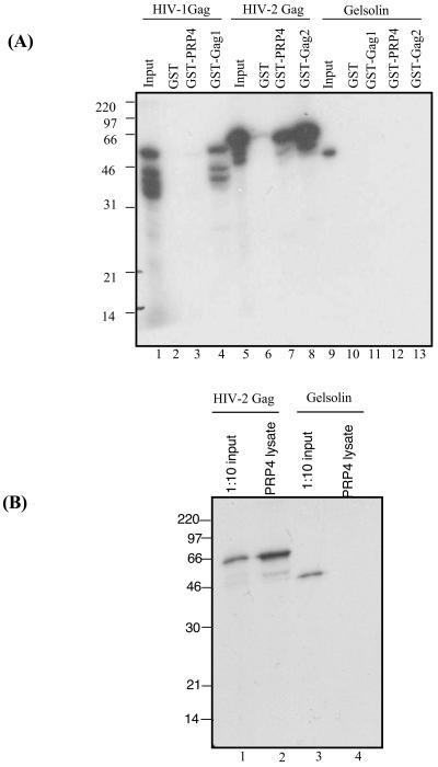
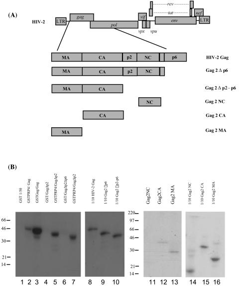
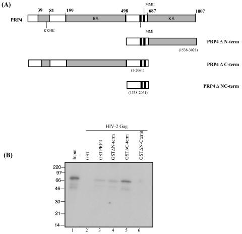
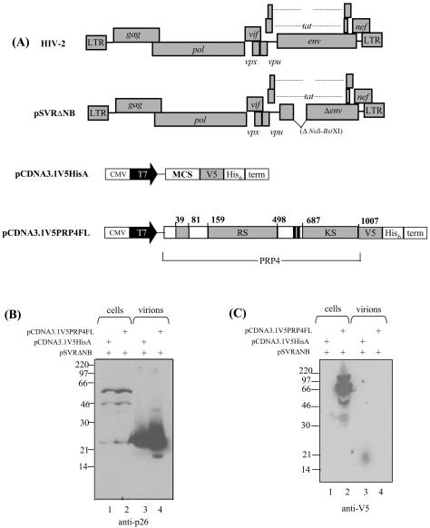
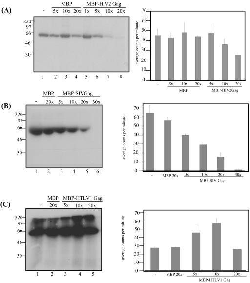
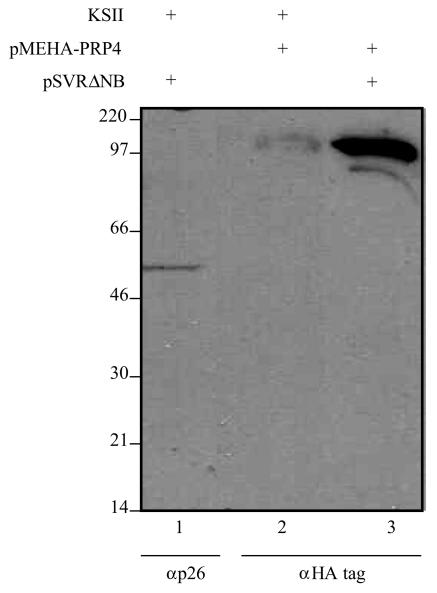
Using a yeast two-hybrid screen of a T-cell cDNA library to identify cellular proteins that bind to the human immunodeficiency virus type 2 (HIV-2) Gag polyprotein, we identified PRP4, a serine-threonine protein kinase. Specific interaction of PRP4 and HIV-2 Gag was confirmed in in vitro and in vivo assays. The interacting region of HIV-2 Gag is located in the conserved matrix and capsid domains, while both the RS (arginine-serine-rich) domain and the KS (kinase) domain of PRP4 are able to bind to HIV-2 Gag. PRP4 is not incorporated into virus particles. HIV-2 Gag is able to inhibit PRP4-mediated phosphorylation of the splicing factor SF2. This is also observed with Gag from simian immunodeficiency virus, a closely related virus, but not with Gag from human T-cell lymphotropic virus type 1. Our results provide evidence for a novel interaction between Gag and a cellular protein kinase involved in the control of constitutive splicing in two closely related retroviruses. We hypothesize that as Gag accumulates in the cell, down regulation of splicing occurs through reduced phosphorylation of SF2. At late stages of infection, this interaction may replace the function of the early viral regulatory protein Rev.


Figures

FIG. 1.
FIG. 1.
PRP4 binds specifically with HIV-2 Gag. (A) In vitro binding of PRP4 to HIV-1 and HIV-2 Gag proteins. In vitro-transcribed and -translated HIV-1 Gag (lanes 2 to 4), HIV-2 Gag (lanes 6 to 8), or gelsolin (lanes 10 to 13) was analyzed by GST fusion pull-down assays for the ability to bind GST, GST-PRP4, GST-Gag1, or GST-Gag2 immobilized on glutathione-Sepharose beads. Inputs were 10% of the amount of HIV-1 Gag (lane 1), HIV-2 Gag (lane 5), or gelsolin (lane 9) used in each assay. (B) PRP4 cellular lysate binding to HIV-2 Gag protein. In vitro-transcribed and -translated HIV-2 Gag (lane 2) or gelsolin (lane 4) was analyzed by pull-down assay for the ability to bind PRP4 protein extract from transfected 293T cells immobilized on protein A-protein G beads. Inputs were 10% of the amount of HIV-2 Gag (lane 1) or gelsolin (lane 3) used in each assay. The positions of marker proteins are indicated in kilodaltons.
FIG. 2.
FIG. 2.
Determination of the region of HIV-2 Gag involved in binding PRP4. (A) Schematic diagram of HIV-2 Gag domain proteins prepared for in vitro binding assays. 35S-labeled HIV-2 Gag truncations were prepared by TNT in vitro translation. (B) In vitro binding of HIV-2 Gag truncations to PRP4 protein. C-terminal protein truncations were analyzed by GST fusion pull-down assay for the ability to bind GST or GST-PRP4 (lanes 1 to 7) immobilized on glutathione-Sepharose beads. Individual Gag subdomains (MA, CA, and NC) were also assayed (lanes 11 to 13) Inputs (lanes 8 to 10 and 14 to 16) were 10% of the amount of the relevant 35S-labeled HIV-2 Gag protein used in each assay. The positions of marker proteins are in kilodaltons.
FIG. 3.
FIG. 3.
Determination of the region of PRP4 involved in binding to HIV-2 Gag. (A) Schematic diagram of PRP4 domains expressed as GST fusion proteins. Numbers in parentheses indicate nucleotide positions. Amino acid residues are shown in bold. Conserved domains are shaded. KKHK, KKHK box; RS, arginine-serine-rich domain; KS, kinase domain; MMI and MMII, conserved domains. (B) In vitro binding of PRP4 truncations to full-length HIV-2 Gag protein. PRP4 protein truncations (lanes 3 to 6) or GST (lane 2) were immobilized on glutathione-Sepharose beads and analyzed by GST fusion pull-down assay for the ability to bind in vitro-translated and radiolabeled HIV-2 Gag. Input (lane 1) is 10% of the amount of the 35S-labeled HIV-2 Gag protein used in the assay. Marker lane proteins are indicated in kilodaltons.
FIG. 4.
FIG. 4.
PRP4 is not incorporated into HIV-2 virus particles. (A) Schematic diagram of HIV-2 Gag and PRP4 expression constructs. CMV, cytomegalovirus immediate-early inhancer; T7, promoter/priming site; MCS, multiple cloning site; V5, V5 epitope; His6, polyhistidine tag; RS, arginine-serine-rich domain; KS, kinase domain. (B and C) 293T cells were transfected with PRP4 plasmid (pCDNA3.1V5PRP4FL) or control plasmid (pCDNA3.1V5HisA) and an env-deleted HIV-2 provirus as indicated. Cell and virus particle samples were prepared as described in Materials and Methods and subjected to analysis by Western blotting using monoclonal antibodies to either p26 (HIV-2 capsid) (B) or V5-tagged PRP4 (C). The positions of marker proteins are indicated in kilodaltons.
FIG. 5.
FIG. 5.
PRP4 phosphorylation observed in kinase assays is unique to SF2 protein. 293T cells were transfected with either empty vector (pCDNA3.1V5HisA) or vector containing full-length PRP4 fused to a V5 epitope tag (pCDNA3.1V5PRP4FL). Cells were lysed and protein was immunoprecipitated as described in Materials and Methods. Empty vector protein (lanes 1, 3, and 5) and PRP4 protein (lanes 2, 4, and 6) were used in a kinase assay as phosphorylating agents. GST fusion proteins were used as targets of phosphorylation: GST alone (lanes 1 and 2), GST fused to HIV-2 Gag (lanes 3 and 4), and GST fused to SF2 protein (lanes 5 and 6).
FIG. 6.
FIG. 6.
Competitive effect of lentiviral Gag protein on the kinase activity of PRP4. (A) PRP4 protein incorporating a V5 epitope tag immunoprecipitated from transfected 293T cells phosphorylates SF2 protein fused to glutathione-Sepharose beads (lane 1). Increasing molar excess of MBP protein (lanes 2, 3, and 4) or MBP-HIV-2 Gag protein (lanes 5, 6, and 7) are titrated against a constant molar amount of PRP4. (B) PRP4 protein phosphorylates SF2 alone (lane 1) and when a 20× molar excess of MBP is added (lane 2). MBP-SIV Gag protein is titrated against PRP4 protein at 5, 10, 20, and 30× the amount of PRP4 (lanes 3, 4, 5, and 6). (C) PRP4 phosphorylates SF2 (lane 1) and when 20× molar excess of MBP protein is added (lane 2). MBP-HTLV-1 Gag is titrated against the phosphorylation at 5, 10, and 20× the molar excess of PRP4 (lanes 3, 4, and 5). The data in each graph to the right represent the mean phosphorylation activity of three independent kinase assays for each lentivirus. Data were analyzed with NIH Image (A) and the Packard Instant Imager (B and C).
FIG. 7.
FIG. 7.
PRP4 and HIV-2 Gag protein interact in vivo. Cos-1 cells were transfected with an HIV-2 proviral construct (lane 1), a full-length PRP4 expression vector incorporating an HA tag (lane 2), or both (lane 3). Cell pellet extracts were blotted either with anti-p26 (lane 1) or anti-HA (lane 2) antibody to determine the amount of protein present. Cells were cotransfected with these plasmids, and HIV-2 Gag protein was immunoprecipitated with anti-p26 and then blotted with anti-HA monoclonal antibody to detect PRP4 (lane 3). Marker lane proteins are indicated in kilodaltons.

References

    1. Alahari, S. K., H. Schmidt, and N. F. Kaufer. 1993. The fission yeast prp4+ gene involved in pre-mRNA splicing codes for a predicted serine/threonine kinase and is essential for growth. Nucleic Acids Res. 21:4079-4083. - PMC - PubMed
    1. Arrigo, S., and K. Beemon. 1988. Regulation of Rous sarcoma virus RNA splicing and stability. Mol. Cell. Biol. 8:4858-4867. - PMC - PubMed
    1. Bevec, D., H. Jaksche, M. Oft, T. Wohl, M. Himmelspach, A. Pacher, M. Schebesta, K. Koettnitz, M. Dobrovnik, R. Csonga, F. Lottspeich, and J. Hauber. 1996. Inhibition of HIV-1 replication in lymphocytes by mutants of the Rev cofactor eIF-5A. Science 271:1858-1860. - PubMed
    1. Bouyac, M., M. Courcoul, G. Bertoia, Y. Baudat, D. Gabuzda, D. Blanc, N. Chazal, P. Boulanger, J. Sire, R. Vigne, and B. Spire. 1997. Human immunodeficiency virus type 1 Vif protein binds to the Pr55Gag precursor. J. Virol. 71:9358-9365. - PMC - PubMed
    1. Bukrinsky, M. I., N. Sharova, T. L. McDonald, T. Pushkarskaya, W. G. Tarpley, and M. Stevenson. 1993. Association of integrase, matrix, and reverse transcriptase antigens of human immunodeficiency virus type 1 with viral nucleic acids following acute infection. Proc. Natl. Acad. Sci. USA 90:6125-6129. - PMC - PubMed

Publication types

Substances