. 2004 Sep 27;32(17):5045-58.
doi: 10.1093/nar/gkh835. Print 2004.

Selection and evolution of NTP-specific aptamers

Affiliations

Selection and evolution of NTP-specific aptamers

Laure Weill et al. Nucleic Acids Res. .

Abstract

ATP occupies a central position in biology, for it is both an elementary building block of RNA and the most widely used cofactor in all living organisms. For this reason, it has been a recurrent target for in vitro molecular evolution techniques. The exploration of ATP-binding motifs constitutes both an important step in investigating the plausibility of the 'RNA world' hypothesis and a central starting point for the development of new enzymes. To date, only two RNA motifs that bind ATP have been characterized. The first one is targeted to the adenosine moiety, while the second one recognizes the 'Hoogsteen' face of the base. To isolate aptamers that bind ATP in different orientations, we selected RNAs on an affinity resin that presents ATP in three different orientations. We obtained five new motifs that were characterized and subsequently submitted to a secondary selection protocol designed to isolate aptamers specific for cordycepin. Interestingly, all the ATP-binding motifs selected specifically recognize the sugar-phosphate backbone region of the nucleotides. Three of the aptamers show some selectivity for adenine derivatives, while the remainder recognize any of the four nucleotides with similar efficiency. The characteristics of these aptamers are discussed along with implications for in vitro molecular evolution.


Figures

Figure 1
Figure 1
Sequences of the starting pool and of the five classes of ATP aptamers selected. The primer sequences are in lower case letters, and were omitted from the aptamer sequences, if not involved in pairings. Putative paired sequences are underlined, and conserved sequences are shaded. The number of occurrences of each sequence is indicated at the right.
Figure 2
Figure 2
Single sequences specifically bind ATP in solution. Cloned aptamer sequences were individually transcribed in the presence of [32P]UTP and loaded at 0.1 μM on a Tris-blocked or ATP–agarose column. Aptamers were pre-incubated or not with 5 mM ATP. The column was then washed with 10 column vol of binding buffer and subsequently eluted with 4 vol of ATP 5 mM. Each fraction was then counted in a scintillation counter. Equivalent results were obtained with one member from each class of ATP aptamer.
Figure 3
Figure 3
High RNA concentration inhibits class I and class III aptamer-binding. A constant amount of 32P-labelled RNA aptamer (50 pmol) was loaded onto an ATP–agarose column at increasing concentrations (10 nM to 1 μM). The column was then washed with 10 column vol of binding buffer and subsequently eluted with 4 vol of ATP 5 mM. Each fraction was then counted in a scintillation counter. The percentage of aptamer eluted by ATP is plotted against RNA concentration.
Figure 4
Figure 4
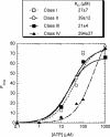
Affinity of the aptamers for ATP. The KD for each aptamer and FRNA, the fraction of RNA bound to ATP, were determined as described in the Materials and Methods section. Data points correspond to the mean of two (class I, II and IV) to four (class III) independent experiments.
Figure 5
Figure 5
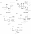
Secondary structure of the five ATP-binding motifs. Secondary structure models of the five aptamer motifs as defined by structure probing, sequence comparisons and energy minimization (42). The sequences shown are those of clone C14 (class I), C20 (class II), C27 (class III), C15 (class IV) and C25 (class V). Squares and circles indicate nucleotides hit by CMCT and DMS, respectively. A–U and G–U base pairs at the ends of helices have been implemented even if one of the residues is hit by a modifying agent. Pn's designate the paired segments. The sequences of conserved regions (see Figure 6) are shown in bold, the variability within theses sequences is indicated in Figure 7. Primer sequences were omitted from class V aptamers secondary structure model because they are not involved in any potential pairing. The number of nucleotides omitted is indicated in between brackets.
Figure 6
Figure 6
Alignments of the aptamer sequences yielded by the primary and secondary selections. The clone name is on the left, the number of occurrence of each sequence is mentioned when superior to one. The residues that do not vary in 95% of the sequences are shaded. Potentially paired nucleotides are underlined, and the paired regions are indicated by ‘Pn’ and an arrow at the top of each alignment. Alignments are based on sequence homology, potential pairings are sometimes slightly shifted from one clone to another. Arrows cover the whole region potentially involved in the pairing for all sequences. Primer sequences are in lower case letters and were not shaded even though they are invariant. They were omitted when not involved in potential pairings with a variable region, the number of nucleotides omitted is indicated in between brackets. The clones named ‘n+’ were selected under stringent conditions.
Figure 6
Figure 6
Alignments of the aptamer sequences yielded by the primary and secondary selections. The clone name is on the left, the number of occurrence of each sequence is mentioned when superior to one. The residues that do not vary in 95% of the sequences are shaded. Potentially paired nucleotides are underlined, and the paired regions are indicated by ‘Pn’ and an arrow at the top of each alignment. Alignments are based on sequence homology, potential pairings are sometimes slightly shifted from one clone to another. Arrows cover the whole region potentially involved in the pairing for all sequences. Primer sequences are in lower case letters and were not shaded even though they are invariant. They were omitted when not involved in potential pairings with a variable region, the number of nucleotides omitted is indicated in between brackets. The clones named ‘n+’ were selected under stringent conditions.
Figure 7
Figure 7
Minimal motifs proposed for the class I, class III and class IV aptamers. For the secondary structure models of the minimal motifs of class I, class III and class IV aptamers, alterations of the conserved sequences are indicated, and the number of occurrences of each change is mentioned in parentheses. D stands for deletion. These motifs show similar binding properties than the aptamers originally selected.
Figure 8
Figure 8
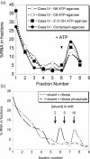
(a) Class IV binds to each of the three differently derivatized ATP–agarose affinity matrices and on cordycepin–agarose: 100 μl of a 0.1 μM solution of 32P-labelled class IV aptamer was loaded onto each of the four affinity matrices. The columns were then washed with 10 column vol of binding buffer, and subsequently eluted with 4 vol of 5 mM ATP. Each fraction was counted and the results were plotted as percentages of the total amount of RNA. Equivalent results were obtained with the four other classes of aptamer with the three different ATP–agarose matrices. (b) Class I aptamer minimal substrate is a ribose phosphate: 100 μl of a 0.1 μM solution of 32P-labelled class I aptamer was loaded onto a mixture of the three ATP–agarose matrices. The column was then washed with 10 column vol of binding buffer, and subsequently eluted with 4 vol of 2, 5 and 10 mM of ribose or ribose phosphate. A last elution with 10 mM ATP was performed to confirm that at least 75% of the bound aptamer was eluted. Each fraction was counted and the results were plotted as percentages of the total amount of RNA. This procedure was used for all the data presented in Figure 8.
Figure 9
Figure 9
Specificity of the five ATP-binding motifs. Selectivity of the different aptamers was determined as specified on Figure 8B, using a set of analogues. Eluant was used at three different concentrations: (+++), (++) and (+) indicate that aptamers were efficiently eluted at 2, 5 or 10 mM respectively. The (−) sign indicates that aptamers could not be eluted by the analogue considered.
Figure 10
Figure 10
Selection under more stringent condition results in consolidation of aptamer secondary structure. (A) Class III aptamer isolated from the primary selection. (B) Class III aptamer selected under more stringent conditions. The parts not shown are identical in the two secondary structure models. The ΔG values are those reported by the mfold software.

Similar articles

Cited by

References

    1. Famulok M. (1999) Oligonucleotide aptamers that recognize small molecules. Curr. Opin. Struct. Biol., 9, 324–329. - PubMed
    1. Gold L., Polisky,B., Uhlenbeck,O. and Yarus,M. (1995) Diversity of oligonucleotide functions. Annu. Rev. Biochem., 64, 763–797. - PubMed
    1. Jayasena S.D. (1999) Aptamers: an emerging class of molecules that rival antibodies in diagnostics. Clin. Chem., 45, 1628–1650. - PubMed
    1. Brody E.N. and Gold,L. (2000) Aptamers as therapeutic and diagnostic agents. J. Biotechnol., 74, 5–13. - PubMed
    1. Burgstaller P., Jenne,A. and Blind,M. (2002) Aptamers and aptazymes: accelerating small molecule drug discovery. Curr. Opin. Drug Discov. Devel., 5, 690–700. - PubMed

Publication types

MeSH terms