. 2004 Oct 5;101(40):14443-8.
doi: 10.1073/pnas.0401346101. Epub 2004 Sep 27.

Restoration of an absent G1 arrest and protection from apoptosis in embryonic stem cells after ionizing radiation

Affiliations

Restoration of an absent G1 arrest and protection from apoptosis in embryonic stem cells after ionizing radiation

Yiling Hong et al. Proc Natl Acad Sci U S A. .

Abstract

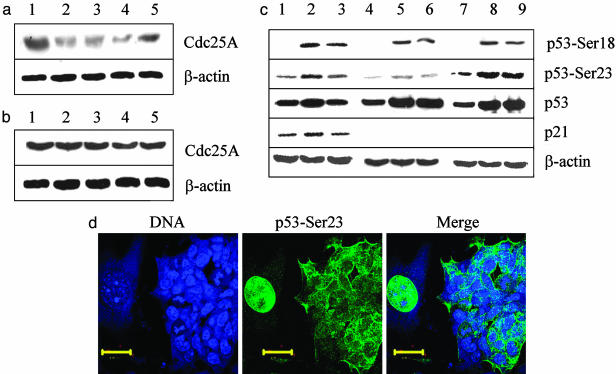
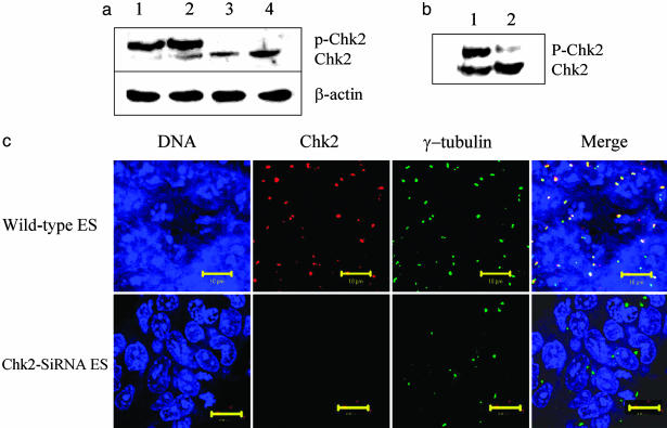
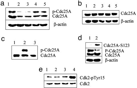
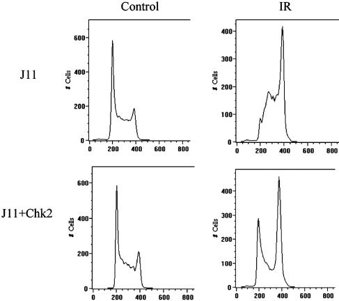
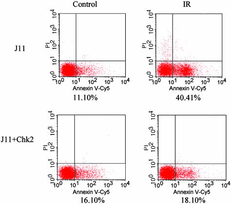
Response to DNA damage and cell-cycle regulation differ markedly between embryonic stem (ES) cells and somatic cells. ES cells require exquisitely sensitive mechanisms to maintain genomic integrity and do so, in part, by suppressing spontaneous mutation. Spontaneous mutation frequency in somatic cells is approximately 10(-4) compared with 10(-6) for ES cells. ES cells also lack a G(1) checkpoint and are hypersensitive to IR and other DNA-damaging agents. These characteristics facilitate apoptosis and the removal of cells with a mutational burden from the population, thereby keeping the population free of damaged cells. Here, we identify signaling pathways that are compromised and lead to a natural absence of aG(1) arrest in ES cells after DNA damage. The affected pathways are those mediated by p53 and p21 and by ATM, Chk2, Cdc25A, and Cdk2. In ES cells, Chk2 kinase is not intranuclear as in somatic cells but is sequestered at centrosomes and is unavailable to phosphorylate Cdc25A phosphatase and cause its degradation. Although ectopic expression of Chk2 does not rescue the p53/p21 pathway, its expression is sufficient to allow it to phosphorylate Cdc25A, activate downstream targets, restore a G(1) arrest, and protect the cell from apoptosis.


Figures

Fig. 1.
Fig. 1.
The pathways mediated by Cdc25A and p53 that lead to a G1 arrest after exposure to IR are compromised in ES cells. (a) MEFs were irradiated with 10 Gy of x-ray, lysed with RIPA buffer, and immunoblotted with Cdc25A mAb at increasing times after IR treatment. Lane 1, untreated sample; lane 2, 30 min after irradiation; lane 3, 60 min after irradiation; lane 4, 90 min after irradiation; lane 5, 120 min after irradiation. (b) J11 ES cells were irradiated with 10 Gy, and cell extracts were subjected to Western blots with the Cdc25A mAb. Lane 1, untreated; lane 2, 30 min after irradiation; lane 3, 60 min after irradiation; lane 4, 90 min after irradiation; lane 5, 120 min after irradiation. (c) MEFs (lanes 1-3), ES cells (lanes 4-6), and ES cells transfected with GFP-Chk2 (lanes 7-9) were subjected to 10 Gy of x-ray and analyzed by Western blot with a phosphospecific Ab to p53-Ser-18, a phosphospecific Ab to p53-Ser-23, a mAb to p53, a polyclonal Ab to p21, and a polyclonal Ab to β-actin as a control. Lanes 1, 4, and 7, blots from cells that were left untreated; lanes 2, 5, and 8, blots from cells collected 2 h after irradiation; lanes 3, 6, and 9, blots from cells collected 4 h after irradiation. (d) Colonies of ES cells and MEFs were cocultured on gelatinized coverslips and transfected with GFP-Chk2. The cells were irradiated with 10 Gy, fixed after 2 h, and stained with DRAQ5 to stain nuclei (Left) and phosphospecific Ab to p53-Ser-23 (Center). (Right) A merged image of Left and Center. (Scale bars, 10 μm.)
Fig. 2.
Fig. 2.
Stem cell Chk2 is hyperphosphorylated and colocalizes with γ-tubulin in centrosomes. (a) Lanes 1 and 3, untreated ES cells and MEFs were lysed with RIPA buffer containing 1% SDS and subjected to Western blot. Lanes 2 and 4, ES cells and MEFs were irradiated with 10 Gy and after 2 h were subjected to Western blot by using a mAb (A-12, Santa Cruz Biotechnology) directed at Chk2. Ab to β-actin was used as a control. (b) Aliquots of cell lysate from ES cells were subjected to Western blot analysis with (lane 2) or without (lane 1) treatment with calf intestinal alkaline phosphatase before electrophoretic fractionation. (c) Colonies of ES cells were grown overnight on gelatinized coverslips. Untreated cells (Upper) and cells transfected with Chk2-siRNA (Lower) were fixed with Formalde-Fresh and coimmunostained with Ab to detect endogenous Chk2 and γ-tubulin. In the leftmost panels, the ES cell nuclei were stained with DRAQ5. In the second panels from the left, cells were reacted with mAb to Chk2. In the third panels from the left, cells were reacted with Ab to γ-tubulin. In the rightmost panels, the images were merged to highlight colocalization. (Scale bars, 10 μm.)
Fig. 3.
Fig. 3.
Ectopic expression of Chk2 induces Cdc25A instability and accumulation of Cdk2 phosphorylated on tyrosine 15 after IR treatment. ES cells were trypsinized and transfected with a GFP-Chk2 fusion construct (a) or a control GFP-H2b construct (b) by using FuGENE 6 to effect the transfection. After 24 h, cells were irradiated with 10 Gy and harvested at half-hour intervals, and cell lysates were fractionated by SDS/PAGE and probed with a mAb to Cdc25A. In both a and b, the lane 1 sample was unirradiated, and samples in lanes 2, 3, 4, and 5 were obtained 30, 60, 90, and 120 min after irradiation, respectively. (c) Cell lysates from ES cells were fractionated by SDS/PAGE and probed by Western blot with mAb to Cdc25A. Lane 1, nontransfected J11 ES cells; lane 2, J11 ES cells transfected with a GFP-Chk2 cDNA construct; lane 3, J11 ES cells transfected with a GFP-Chk2 cDNA construct in which the cell lysate was treated with calf intestine alkaline phosphatase. (d) The phosphorylation status of Cdc25A serine 123 was assessed with a phosphospecific Ab after transfection with Chk2 minus irradiation (lane 1) and 1 h after irradiation (lane 2). The same blots were probed with mAb to Cdc25A (Cdc25A) and with Ab to β-actin as a loading control (β-actin). (e) The phosphorylation status of Cdk2 was determined by immunoprecipitation of Cyclin E with Ab from untreated (lanes 1 and 3) and IR-treated (lanes 2 and 4) ES cells. The extracts in lanes 3 and 4 are from ES cells transfected with Chk2. Cell lysates were fractionated by SDS/PAGE and probed with Ab to Cdk2 to show total Cdk2 present and with mAb directed at phosphorylated tyrosine 15 of Cdk2.
Fig. 4.
Fig. 4.
Ectopic expression of Chk2 restores the G1/S checkpoint in stem cell. Wild-type J11 ES cells or J11 ES cells transfected with Chk2 were subjected to 10 Gy of IR (Right) or left untreated (Left). Eight hours after irradiation, cells were trypsinized, resuspended in PBS, and stained with 1 mg/ml propidium iodide containing 0.1 mg/ml RNase A. Stained cells were subjected to flow cytometry (system ii software, Version 3.0) to cell-cycle distribution of cells.
Fig. 5.
Fig. 5.
Restoration of a G1 checkpoint by ectopic Chk2 expression protects ES cells from apoptosis. J11 ES cells transfected with Chk2 or mock-transfected ES cells were treated with 10 Gy of IR (Right) or left untreated (Left). Sixteen hours after treatment, both cell populations were harvested, stained with annexin V-Cy5 and 2.5 μg/ml propidium iodide, and analyzed by flow cytometry. Cells above the horizontal line are positive for propidium iodide staining and represent dead cells. Those displaced to the right of the vertical line are positive for annexin V-Cy5 and represent early apoptotic cells.

References

    1. Stambrook, P. J., Shao, C., Stockelman, M., Boivin, G., Engle, S. J. & Tischfield, J. A. (1996) Environ. Mol. Mutagen. 28, 471-482. - PubMed
    1. Shao, C., Deng, L., Hehegariu, O., Liang, L., Stambrook, P. J. & Tischfield, J. A. (2000) Proc. Natl. Acad. Sci. USA 97, 7405-7410. - PMC - PubMed
    1. Cervantes, R. B., Stringer, J. R., Shao, C., Tischfield J. A. & Stambrook, P. J. (2002) Proc. Natl. Acad. Sci. USA 99, 3586-3590. - PMC - PubMed
    1. Aladjem, M., Spike, B. T., Rodewald, L. W., Hope, T. J., Klemm, M., Jaenisch, R. & Wahl, G. M. M. (1998) Curr. Biol. 8, 145-155. - PubMed
    1. Hirao, A., Kong, Y.-Y., Matsuoka, S., Wakeham, A., Ruland, J., Yoshida, H., Liu, D., Elledge, S. J. & Mak, T. W. (2000) Science 289, 1824-1827. - PubMed

Publication types

MeSH terms