

Review
doi: 10.1016/j.cell.2004.09.020.
Sculpting the proteome with AAA(+) proteases and disassembly machines
Affiliations
- PMID: 15454077
- PMCID: PMC2717008
- DOI: 10.1016/j.cell.2004.09.020
Item in Clipboard
Review
Sculpting the proteome with AAA(+) proteases and disassembly machines
Cell.
.
Abstract
Machines of protein destruction-including energy-dependent proteases and disassembly chaperones of the AAA(+) ATPase family-function in all kingdoms of life to sculpt the cellular proteome, ensuring that unnecessary and dangerous proteins are eliminated and biological responses to environmental change are rapidly and properly regulated. Exciting progress has been made in understanding how AAA(+) machines recognize specific proteins as targets and then carry out ATP-dependent dismantling of the tertiary and/or quaternary structure of these molecules during the processes of protein degradation and the disassembly of macromolecular complexes.
Figures
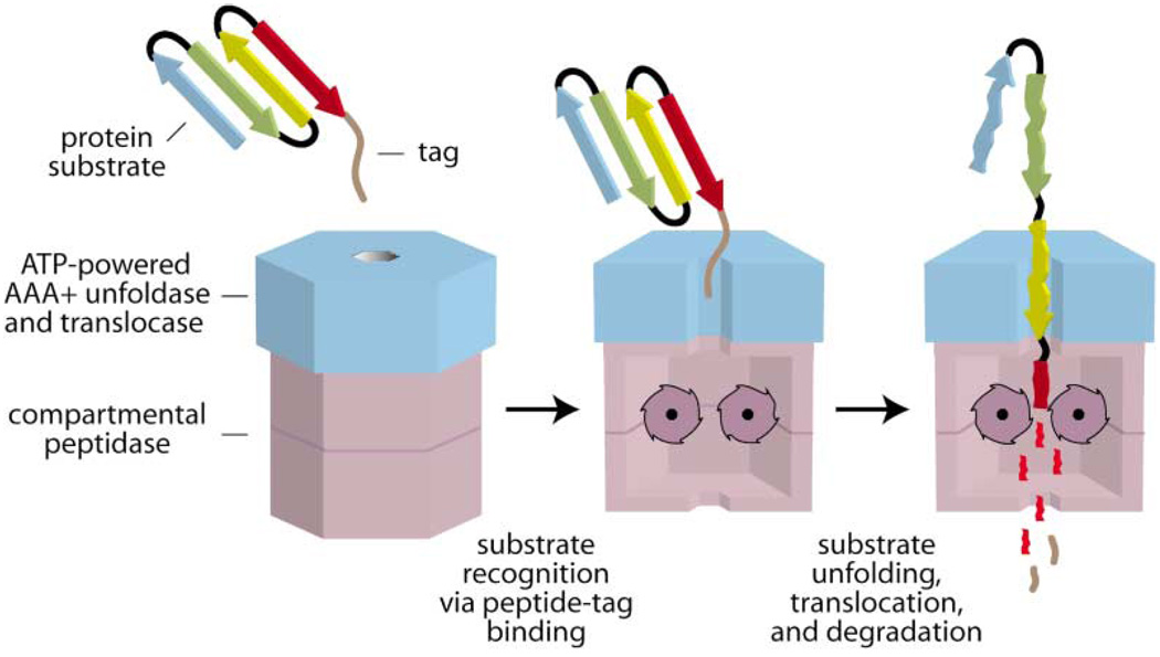
The recognition step is mediated by binding of a peptide tag (brown) on the protein substrate to a AAA+ ATPase (blue). In subsequent steps, the protein is unfolded, translocated into a compartmental peptidase (magenta), and degraded. Peptide fragments are shown diffusing out of the peptidase, but active participation of the ATPase may be required for exit of large fragments.
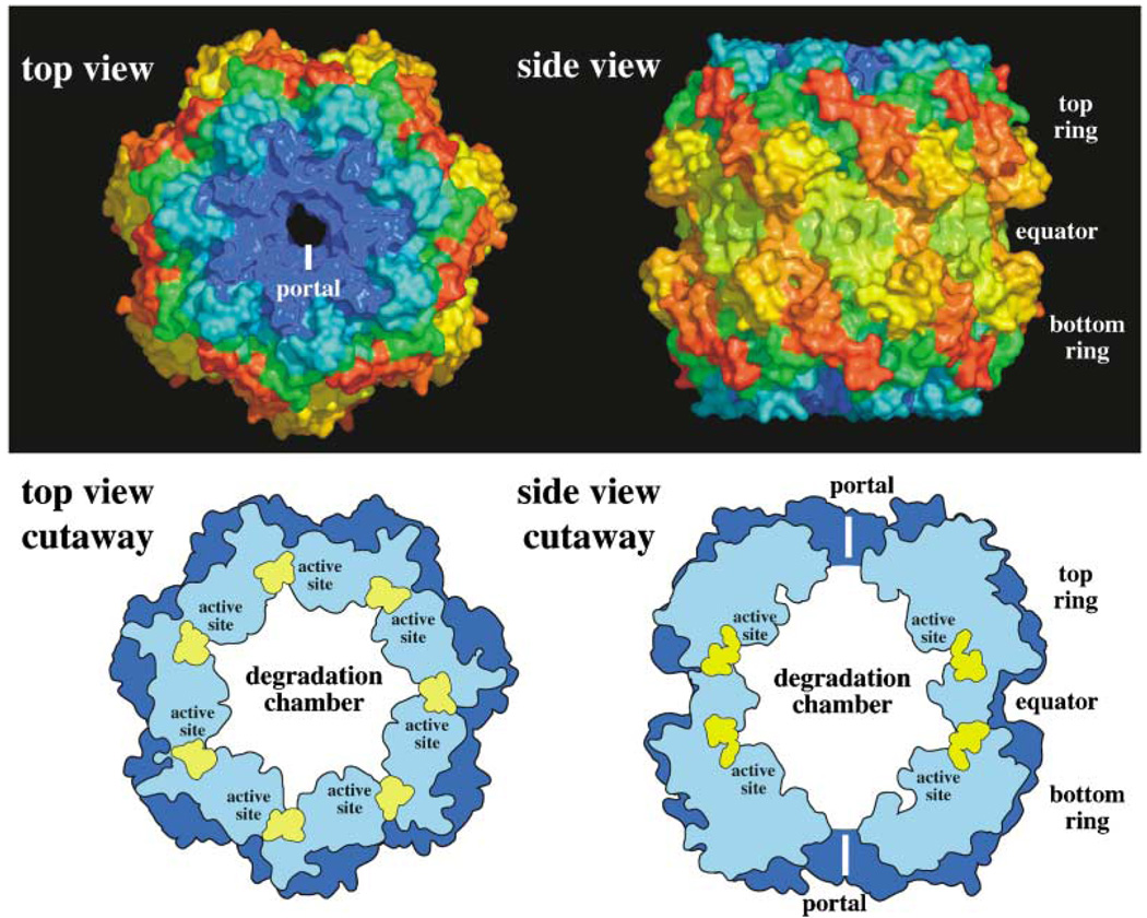
The figure is based on the structure reported in Wang et al., (1997). In the top panel, each of the 14 identical subunits of ClpP is shown colored from blue to red from the N terminus to the C terminus. The 7-fold symmetry of a single ClpP ring is evident in the top view, which also shows the entry/exit portal. The side view shows the face-to-face stacking of both ClpP rings. The bottom panel shows cutaway diagrams that illustrate the positions of the active site residues (yellow) within the degradation chamber.
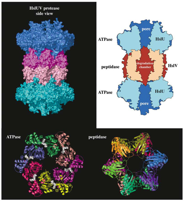
The figure is based on the structure reported in Sousa et al. (2000). The top left panel shows a surface representation with individual hexameric ringsofthe HslU ATPase colored blue/cyan and the HslV peptidase colored magenta/pink. The top right panel is a cutaway diagram showing the positions of the pore through HslU and the degradation chamber within HslV. The bottom panel shows axial views of the ATPase and peptidase in ribbon representation with individual subunits in different colors. ATP molecules (white) are shown in CPK representation.
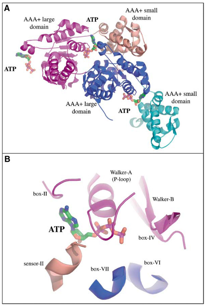
(A) Ribbon representation of two adjacent HslU subunits from a hexameric ring; stick representation of three ATP molecules. The large AAA+ domains are colored magenta and blue and the small AAA+ domains are colored pink and cyan. In this view, only the central ATP has a full complement of contacts, which are made from two large domains and one small domain. (B) Closeup view of the central ATP binding site. The box-II, Walker-A, box-IV, Walker-B, box-VI, box-VII, and sensor-II sequence motifs are found in all AAA+ ATPases (Schirmer et al., 1996; Neuwald et al., 1999).
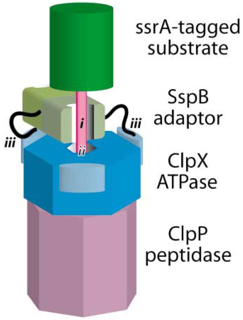
The protein substrate (green) has an ssrA-degradation tag (pink). One part of the tag (i) is bound by the SspB adaptor (light brown); another part (ii) is bound by the ClpX ATPase (blue). Flexible tails (black) of the SspB adaptor make tethering interactions (iii) with the N-terminal domains (light blue) of ClpX.
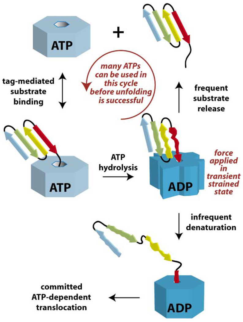
A tagged native protein binds to a AAA+ ATPase in its ATP bound state (light blue). A conformational change, which accompanies ATP hydrolysis, pulls the native protein into the central pore, creating an unfolding force and a highly strained enzyme (dark blue). At this point, the substrate can either dissociate or unfold. The probability of either event depends upon the stability of the structural elements in the substrate adjacent to the recognition tag. For very stable substrates, hundreds of cycles of protein binding and release may be required before denaturation is successful (Kenniston et al., 2003). Once denaturation occurs, additional rounds of ATP hydrolysis drive processive protein translocation. If a compartmental protease is bound to the ATPase, then the translocated protein is degraded; otherwise, it is released in a denatured state and has the opportunity to refold.
References
-
- Beuron F, Maurizi MR, Belnap DM, Kocsis E, Booy FP, Kessel M, Steven AC. At sixes and sevens: characterization of the symmetry mismatch of the ClpAP chaperone-assisted protease. J. Struct. Biol. 1998;123:248–259. - PubMed
-
- Bochtler M, Hartmann C, Song HK, Bourenkov GP, Bartunik HD, Huber R. The structures of HsIU and the ATP-dependent protease HsIU-HsIV. Nature. 2000;403:800–805. - PubMed
-
- Bolon DN, Wah DA, Hersch GL, Baker TA, Sauer RT. Bivalent tethering of the SspB adaptor to the AAA+ protease ClpXP is required for efficient substrate delivery: a protein-design study. Mol. Cell. 2004a;13:443–449. - PubMed
-
- Bolon DN, Grant RA, Baker TA, Sauer RT. Nucleotide-dependent substrate handoff from the SspB adaptor to the AAA+ ClpXP protease. Mol. Cell. 2004b in press. - PubMed
Publication types
MeSH terms
Substances
Grants and funding
LinkOut - more resources
Full Text Sources
Other Literature Sources
Molecular Biology Databases
