

Controlled ablation of microtubules using a picosecond laser
- PMID: 15454403
- PMCID: PMC1304929
- DOI: 10.1529/biophysj.104.049528
Controlled ablation of microtubules using a picosecond laser
Abstract
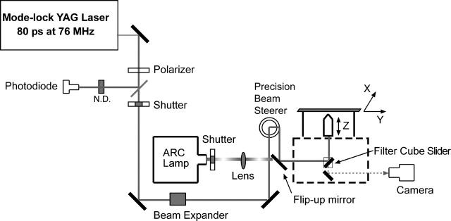
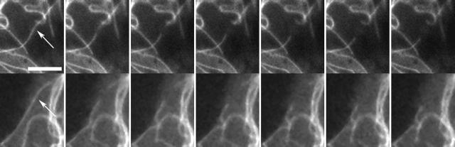
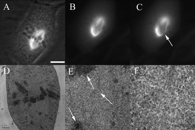
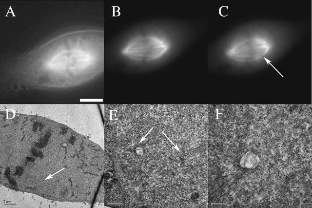
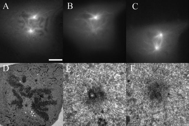
The use of focused high-intensity light sources for ablative perturbation has been an important technique for cell biological and developmental studies. In targeting subcellular structures many studies have to deal with the inability to target, with certainty, an organelle or large macromolecular complex. Here we demonstrate the ability to selectively target microtubule-based structures with a laser microbeam through the use of enhanced yellow fluorescent protein (EYFP) and enhanced cyan fluorescent protein (ECFP) variants of green fluorescent protein fusions of tubule. Potorous tridactylus (PTK2) cell lines were generated that stably express EYFP and ECFP tagged to the alpha-subunit of tubulin. Using microtubule fluorescence as a guide, cells were irradiated with picosecond laser pulses at discrete microtubule sites in the cytoplasm and the mitotic spindle. Correlative thin-section transmission electron micrographs of cells fixed one second after irradiation demonstrated that the nature of the ultrastructural damage appeared to be different between the EYFP and the ECFP constructs suggesting different photon interaction mechanisms. We conclude that focal disruption of single cytoplasmic and spindle microtubules can be precisely controlled by combining laser microbeam irradiation with different fluorescent fusion constructs. The possible photon interaction mechanisms are discussed in detail.
Figures
Similar articles
-
Laser nanosurgery of single microtubules reveals location-dependent depolymerization rates.J Biomed Opt. 2007 Mar-Apr;12(2):024022. doi: 10.1117/1.2718920. J Biomed Opt. 2007. PMID: 17477737
-
Isolation of FRET-positive cells using single 408-nm laser flow cytometry.Cytometry A. 2006 Apr;69(4):291-8. doi: 10.1002/cyto.a.20254. Cytometry A. 2006. PMID: 16498686
-
The interaction between human PEX3 and PEX19 characterized by fluorescence resonance energy transfer (FRET) analysis.Eur J Cell Biol. 2003 Jul;82(7):333-42. doi: 10.1078/0171-9335-00325. Eur J Cell Biol. 2003. PMID: 12924628
-
A synergy of technologies: combining laser microsurgery with green fluorescent protein tagging.Cell Motil Cytoskeleton. 1997;38(4):311-7. doi: 10.1002/(SICI)1097-0169(1997)38:4<311::AID-CM1>3.0.CO;2-6. Cell Motil Cytoskeleton. 1997. PMID: 9415373 Review.
-
Mitotic spindle: laser microsurgery in yeast cells.Curr Biol. 2004 Sep 21;14(18):R748-50. doi: 10.1016/j.cub.2004.09.010. Curr Biol. 2004. PMID: 15380083 Review.
Cited by
-
Chromosome tips damaged in anaphase inhibit cytokinesis.PLoS One. 2010 Aug 25;5(8):e12398. doi: 10.1371/journal.pone.0012398. PLoS One. 2010. PMID: 20811641 Free PMC article.
-
Laser cavitation rheology for measurement of elastic moduli and failure strain within hydrogels.Sci Rep. 2020 Aug 4;10(1):13144. doi: 10.1038/s41598-020-68621-y. Sci Rep. 2020. PMID: 32753667 Free PMC article.
-
Viscoelastic retraction of single living stress fibers and its impact on cell shape, cytoskeletal organization, and extracellular matrix mechanics.Biophys J. 2006 May 15;90(10):3762-73. doi: 10.1529/biophysj.105.071506. Epub 2006 Feb 24. Biophys J. 2006. PMID: 16500961 Free PMC article.
-
A subcellular cookie cutter for spatial genomics in human tissue.Anal Bioanal Chem. 2022 Jul;414(18):5483-5492. doi: 10.1007/s00216-022-03944-5. Epub 2022 Mar 2. Anal Bioanal Chem. 2022. PMID: 35233697 Free PMC article.
-
Laser microsurgery in the GFP era: a cell biologist's perspective.Methods Cell Biol. 2007;82:239-66. doi: 10.1016/S0091-679X(06)82007-8. Methods Cell Biol. 2007. PMID: 17586259 Free PMC article. Review.
References
-
- Aist, J. R., H. Liang, and M. W. Berns. 1993. Astral and spindle forces in PtK2 cells during anaphase B: a laser microbeam study. J. Cell Sci. 104:1207–1216. - PubMed
-
- Berns, M. W., J. Aist, J. Edwards, K. Strahs, J. Girton, P. McNeill, J. B. Rattner, M. Kitzes, M. Hammer-Wilson, L. H. Liaw, A. Siemens, M. Koonce, et al. 1981. Laser microsurgery in cell and developmental biology. Science. 213:505–513. - PubMed
-
- Berns, M. W., and A. D. Floyd. 1971. Chromosomal microdissection by laser. A cytochemical and functional analysis. Exp. Cell Res. 67:305–310. - PubMed
-
- Berns, M. W., R. S. Olson, and D. E. Rounds. 1969. In vitro production of chromosomal lesions with an argon laser microbeam. Nature. 221:74–75. - PubMed
Publication types
MeSH terms
Substances
Grants and funding
LinkOut - more resources
Full Text Sources
Other Literature Sources
Miscellaneous
