. 2004 Oct;87(4):2107-15.
doi: 10.1529/biophysj.104.040311.

Transmembrane peptide-induced lipid sorting and mechanism of Lalpha-to-inverted phase transition using coarse-grain molecular dynamics

Affiliations

Transmembrane peptide-induced lipid sorting and mechanism of Lalpha-to-inverted phase transition using coarse-grain molecular dynamics

Steve O Nielsen et al. Biophys J. 2004 Oct.

Abstract

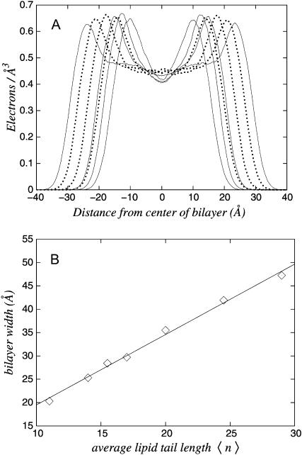
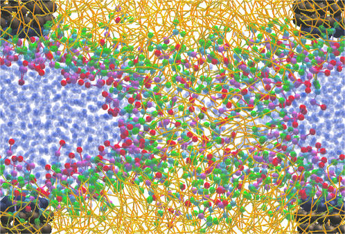
Molecular dynamics results are presented for a coarse-grain model of 1,2-di-n-alkanoyl-sn-glycero-3-phosphocholine, water, and a capped cylindrical model of a transmembrane peptide. We first demonstrate that different alkanoyl-length lipids are miscible in the liquid-disordered lamellar (Lalpha) phase. The transmembrane peptide is constructed of hydrophobic sites with hydrophilic caps. The hydrophobic length of the peptide is smaller than the hydrophobic thickness of a bilayer consisting of an equal mixture of long and short alkanoyl tail lipids. When incorporated into the membrane, a meniscus forms in the vicinity of the peptide and the surrounding area is enriched in the short lipid. The meniscus region draws water into it. In the regions that are depleted of water, the bilayers can fuse. The lipid headgroups then rearrange to solvate the newly formed water pores, resulting in an inverted phase. This mechanism appears to be a viable pathway for the experimentally observed Lalpha-to-inverse hexagonal (HII) peptide-induced phase transition.


Figures

FIGURE 1
FIGURE 1
Snapshot of the model transmembrane peptide assembly showing the van der Waals radius (top left) and the skeletal bonding (top right) structures. The assembly is ∼15 Å in radius and 20 Å long. The peptide consists of a hydrophobic cylinder capped with hydrophilic sites. The outer, middle, and inner capping rings are composed of identical sites, but have been colored differently (blue/purple/pink) for clarity. (Bottom left) The 11-site model of 1,2-di-undecanoyl-sn-glycero-3-phosphocholine (DC11PC) and (bottom right) the 23-site model of DC29PC. The choline and phosphate sites carry positive and negative electrostatic charges, respectively.
FIGURE 2
FIGURE 2
Panel A shows the lipid density distributions normal to the bilayer plane for the seven 160-lipid systems (S1–S7). Shown by solid line are the single-lipid systems and by dotted line the mixed-lipid systems. Panel B shows the bilayer width versus the average tail length (average n of the DCnPC constituent lipids) of these systems. The best linear fit is also shown. The error bars are smaller than the symbols.
FIGURE 3
FIGURE 3
The lipid density distributions for the mixed-lipid systems (shown here in solid line) in Fig. 2 A are broken down into their constituent lipid densities. Panel A is for 40 short lipids and 120 long lipids (S6). Panel B is for 80 short lipids and 80 long lipids (S5). Panel C is for 120 short lipids and 40 long lipids (S7). The short lipids are shown in heavy solid line and the long lipids in dotted line.
FIGURE 4
FIGURE 4
Schematic of the patchwork initial condition for the large mixed-lipid simulation (L1). Each patch, which includes both leaflets, contains only one of the two types of lipid.
FIGURE 5
FIGURE 5
Two-dimensional lipid-lipid radial distribution functions (RDFs) in the bilayer plane for the simulation whose initial condition is shown in Fig. 4 (L1). Panel A shows the initial distribution taken from the first 50 ps of the simulation. Panel B shows the equilibrated distribution taken from the last 1 ns of the MD simulation. The short lipid-short lipid RDF is shown in solid line, the long lipid-long lipid RDF in dotted line, and the short lipid-long lipid RDF in heavy solid line. The lipid location is taken to be the center of mass of the headgroup. Both leaflets are included, which is the reason the distributions do not go to zero at zero (projected) separation.
FIGURE 6
FIGURE 6
Two-dimensional radial distribution of lipids around the transbilayer peptide (system L2). Panel A shows the distribution immediately after the the peptide is inserted. The insertion location was chosen to correspond to a region of local enhancement of the long-lipid species. Panel B shows the distribution taken from the last 1 ns of the MD simulation. The short lipid is denoted by solid line, and the long lipid by dotted line. The lipid location is taken to be the center of mass of the headgroup. The peptide location is taken to be its center of mass. Both leaflets are included.
FIGURE 7
FIGURE 7
Extent of the lipid meniscus formed around the transbilayer peptide assembly (system L2). Separately for each of the two leaflets and each of the two lipid species, the average lipid headgroup center of mass height z (normal to the bilayer plane) relative to the peptide assembly center of mass is computed in the radial region in the membrane plane r to r + Δr, where r = 0 corresponds to the center of the peptide assembly. The short lipid is denoted by solid line and the long lipid by dotted line. The two leaflets appear as positive and negative height values.
FIGURE 8
FIGURE 8
Snapshot of the defective inverted phase formed from the 300:1 lipid/peptide system (L4). Compare to Fig. 9, which shows the defect-free 200:1 system. Coloring is as follows: water, blue; lipids, as in Fig. 1; peptide darkened for clarity.
FIGURE 9
FIGURE 9
The Lα-to-inverted phase transition for the 200:1 lipid/peptide system (L5) is shown in panels AC, with the simulation unit cell shown as a black rectangle. In panel A the meniscus induced by the peptide is evident. Accretion of interbilayer water into the meniscus allows for membrane contact (B). The lipid headgroups then rearrange to solvate the newly formed water pores (C). Coloring is as follows: water, blue; lipids as in Fig. 1; peptide darkened for clarity. Panel D shows a perspective view of the cylindrical water pores to further illustrate the structure of the inverted phase. All lipids have been removed for clarity.
FIGURE 10
FIGURE 10
Spontaneous symmetry breaking of the water network during the inverted phase transition (system L5). In the Lα phase the water is arranged in two-dimensional slabs in which the two directions are equivalent. The introduction of the peptide and the subsequent membrane contact does not break this symmetry, as seen immediately after fusion in the top panels. However, during the subsequent evolution this symmetry is spontaneously broken, as shown in the bottom panels.

References

    1. Betanzos, M., C. S. Chiang, H. R. Guy, and S. Sukharev. 2002. A large iris-like expansion of a mechanosensitive channel protein induced by membrane tension. Nat. Struct. Biol. 9:704–710. - PubMed
    1. Caffrey, M. 2000. A lipid's eye view of membrane protein crystallization in mesophases. Curr. Opin. Struct. Biol. 10:486–497. - PubMed
    1. Dan, N., P. Pincus, and S. A. Safran. 1993. Membrane-induced interactions between inclusions. Langmuir. 9:2768–2771.
    1. de Planque, M. R. R., E. Goormaghtigh, D. V. Greathouse, R. E. Koeppe II, J. A. W. Kruijtzer, R. M. J. Liskamp, B. de Kruijff, and J. A. Killian. 2001. Sensitivity of single membrane-spanning α-helical peptides to hydrophobic mismatch with a lipid bilayer: effects on backbone structure, orientation, and extent of membrane incorporation. Biochemistry. 40:5000–5010. - PubMed
    1. de Planque, M. R. R., D. V. Greathouse, R. E. Koeppe II, H. Schäfer, D. Marsh, and J. A. Killian. 1998. Influence of lipid/peptide hydrophobic mismatch on the thickness of diacylphosphatidylcholine bilayers. A 2H NMR and ESR study using designed transmembrane α-helical peptides and gramicidin A. Biochemistry. 37:9333–9345. - PubMed

Publication types

LinkOut - more resources