. 2005 Jan 15;105(2):735-41.
doi: 10.1182/blood-2003-11-3900. Epub 2004 Sep 28.

Functional defect of regulatory CD4(+)CD25+ T cells in the thymus of patients with autoimmune myasthenia gravis

Affiliations

Functional defect of regulatory CD4(+)CD25+ T cells in the thymus of patients with autoimmune myasthenia gravis

Anna Balandina et al. Blood. .

Abstract

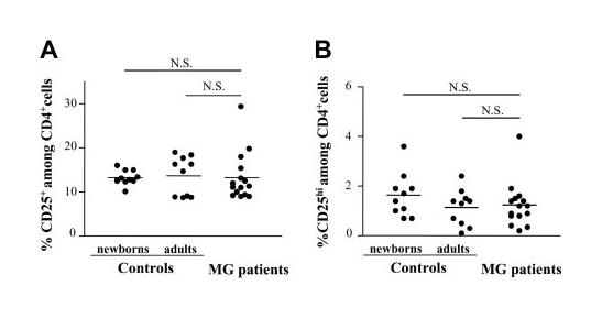
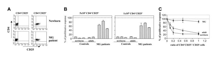
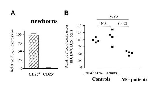
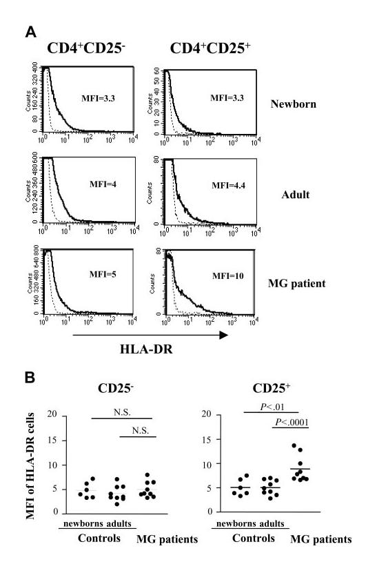
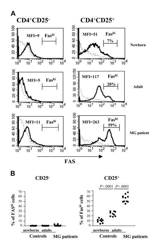
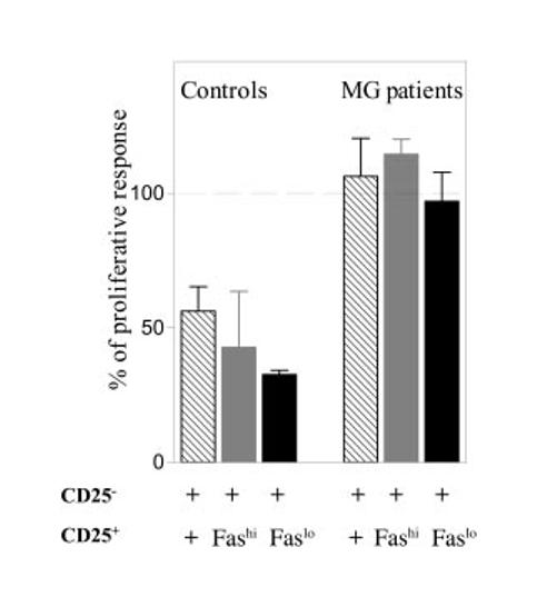
Thymus-derived CD4(+)CD25+ regulatory T (Treg) cells are essential for the maintenance of immunologic self-tolerance. Despite their critical role in the active suppression of experimental autoimmune disorders, little is known about their involvement in human autoimmune diseases. Myasthenia gravis (MG) is a CD4+ T cell-dependent autoimmune disease and the thymus is assumed to be the initiation site. To identify possible defects in the Treg cells in MG, we analyzed CD4(+)CD25+ cells in thymi from patients with MG compared to those from healthy subjects. We found a normal CD4(+)CD25+ number but a severe functional defect in their regulatory activity together with a decreased expression of the transcription factor, Foxp3, which is essential for T-cell regulatory function. The phenotypic analysis of CD4(+)CD25+ thymocytes revealed an increased number of activated effector cells with strong Fas expression in patients with MG. However, whatever their level of Fas, CD4(+)CD25+ thymocytes from patients with MG remained unable to suppress the proliferation of responding cells, indicating that the impaired Treg cell function is not due to contamination by activated effector T cells. These data are the first to demonstrate a severe functional impairment of thymic Treg cells in MG, which could contribute to the onset of this autoimmune disease.


Figures

Figure 1
Figure 1
The frequency of CD4+CD8CD25+ thymocytes in patients with MG is similar to healthy newborn and young adult thymi. Freshly isolated human thymocytes from hyperplastic thymi of 15 patients with MG as well as the thymocytes from 10 young adult and 10 newborn thymi were analyzed by 3-color flow cytometry for the expression of CD4, CD8, and CD25. Each point represents 1 patient or 1 control. The bars represent mean values. The proportion of CD4+CD8 cells with CD25+ (A) as well as CD25hi (B) expression was calculated. The differences between newborns, adults, and patients with MG are not significant (NS).
Figure 2
Figure 2
Myasthenic CD4+CD25+ thymocytes are responsive to mitogenic stimulation and their suppressive activity is impaired. (A) CD8-depleted thymocytes were purified by positive selection of CD4+ cells followed by positive or negative selection for CD25 expression using MACS magnetic microbeads. A representative experiment using newborn and myasthenic thymocytes is shown. (B) CD4+CD25+ thymocytes from 5 controls and 3 patients with MG were stimulated in the presence of irradiated allogenic T cell–depleted PBMCs and 5 μg/mL PHA. The results are expressed as the percent of proliferative response of CD4+CD25 thymocytes, with proliferation in the absence of CD4+CD25+ cells corresponding to 100%. Mean values (± SD) of triplicate wells are reported. (C) The CD4+CD25 thymocytes were stimulated in the presence of different doses of CD4+CD25+ thymocytes. Mean values (± SD) of 3 separate experiments are shown.
Figure 3
Figure 3
CD4+CD25+ thymocytes of patients with MG show decreased expression of Foxp3 mRNA. cDNA samples were obtained from CD4+CD25 and CD4+CD25+ thymocytes that had been purified using MACS magnetic microbeads and were subjected to real-time quantitative PCR using primers specific to FoxP3. The Foxp3 mRNA values were normalized to the 28S rRNA expression in each sample. (A) Quantification of relative Foxp3 mRNA levels for CD4+CD25+ and CD4+CD25+ thymocytes from newborns. Error bars indicate SD value. (B) Quantification of relative Foxp3 mRNA levels in CD4+CD25+ thymocytes from newborns, young adults, and patients with MG.9. ■ indicates 1 patient or 1 control. Bars represent mean values.
Figure 4
Figure 4
Distribution of CD25+ cells in normal and hyperplastic MG thymi. Frozen sections of normal (A-B) and hyperplastic MG (C-E) thymi were stained with anti-CD25 mAb (red color) and counterstained with Gill hematoxylin. The left photomicrographs (original magnification × 100) show that CD25 expression is observed in the medullary area of normal (A) and hyperplastic MG thymus (C-D) and, in addition, around the germinal centers of hyperplastic MG thymus (C,E). The right photomicrographs (original magnification × 400) are enlargements of the framed areas corresponding to medullary area of normal (B) and MG (D) thymus and germinal center of MG thymus (E).
Figure 5
Figure 5
Expression of HLA-DR is increased in CD4+CD25+ T cells from patients with MG. (A) Representative HLA-DR expression profile for newborn, young adult, and myasthenic CD8-depleted thymocytes stained with anti-HLA-DR, anti-CD25, anti-CD4 mAbs and analyzed by flow cytometry. HLA-DR expression is shown with a solid line and the staining with an isotype IgG control with a dotted line. (B) The percentage of HLA-DR was determined among CD4+CD25 and CD4+CD25+ cells in 9 MG patient, 9 young adult and 6 newborn thymi. ● represents 1 patient or 1 control. Bars indicate mean values.
Figure 6
Figure 6
CD4+CD25+ thymocytes from patients with MG are enriched in Fashi cells. (A) Representative Fas expression profile for newborn, young adult normal, and myasthenic CD8-depleted thymocytes stained with anti-CD25, anti-CD95, anti-CD4 mAbs and analyzed by flow cytometry. The IgG isotype control staining is shown with a dotted line. The percentage of Fashi cells is indicated above the brackets delimiting this cell subset. (B) The percentage of Fas expression was determined among CD4+CD25 and CD4+CD25+ cells in thymi from 11 patients with MG, 9 young adults, and 10 newborns.
Figure 7
Figure 7
The suppressive function of both CD4+CD25+Faslo and CD4+CD25+Fashi thymocytes of patients with MG is severely impaired. CD4+CD25 T cells from MG or normal thymi were stimulated in the presence of irradiated allogenic T cell–depleted PBMCs in coculture with sorted autologous unfractionated CD4+CD25+ (formula image), CD4+CD25+Fashi (formula image), or CD4+CD25+Faslo (formula image) thymocytes at a ratio of 1:1. The results are expressed as a percentage of the proliferative response versus that obtained when adding the 2 separate populations (defined as 100%). The experiments were done with thymic cells from 2 patients with MG and 2 healthy controls. The results are expressed as the mean ± SEM of these 2 experiments.

References

    1. Sakaguchi S, Sakaguchi N, Asano M, Itoh M, Toda M. Immunologic self-tolerance maintained by activated T cells expressing IL-2 receptor alpha-chains (CD25). Breakdown of a single mechanism of self-tolerance causes various autoimmune diseases. J Immunol. 1995;155:1151–1164. - PubMed
    1. Saoudi A, Seddon B, Fowell D, Mason D. The thymus contains a high frequency of cells that prevent autoimmune diabetes on transfer into prediabetic recipients. J Exp Med. 1996;184:2393–2398. - PMC - PubMed
    1. Shevach EM. CD4+ CD25+ suppressor T cells: more questions than answers. Nat Rev Immunol. 2002;2:389–400. - PubMed
    1. Stephens LA, Mottet C, Mason D, Powrie F. Human CD4(+)CD25(+) thymocytes and peripheral T cells have immune suppressive activity in vitro. Eur J Immunol. 2001;31:1247–1254. - PubMed
    1. Annunziato F, Cosmi L, Liotta F, et al. Phenotype, localization, and mechanism of suppression of CD4(+)CD25(+) human thymocytes. J Exp Med. 2002;196:379–387. - PMC - PubMed

Publication types

MeSH terms