. 2004 Nov;2(11):e332.
doi: 10.1371/journal.pbio.0020332. Epub 2004 Sep 28.

Developmental context determines latency of MYC-induced tumorigenesis

Affiliations

Developmental context determines latency of MYC-induced tumorigenesis

Shelly Beer et al. PLoS Biol. 2004 Nov.

Abstract

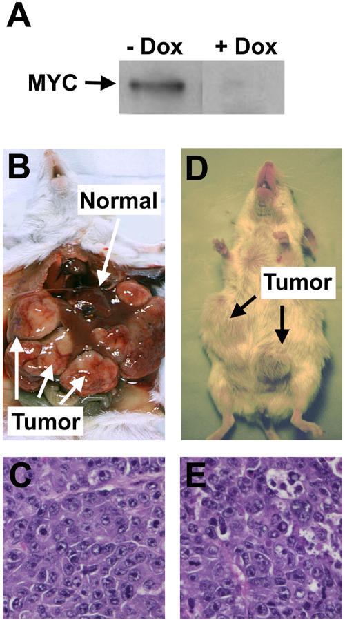
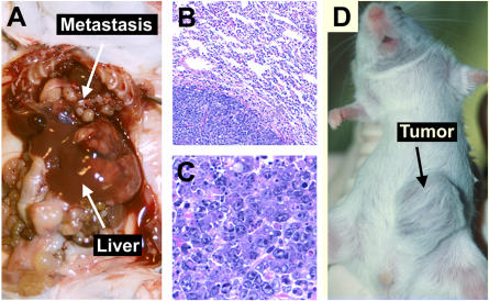
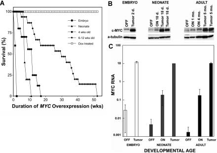
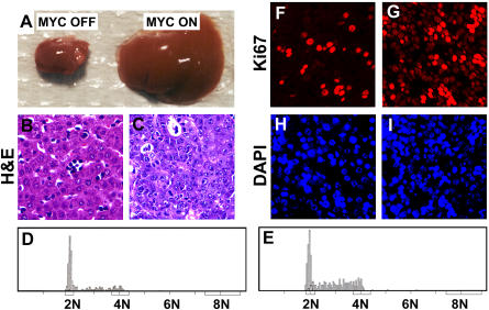
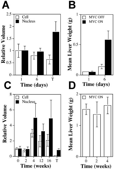
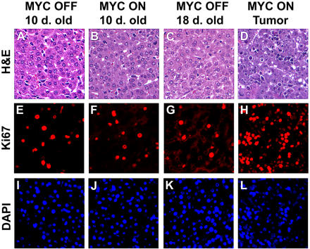
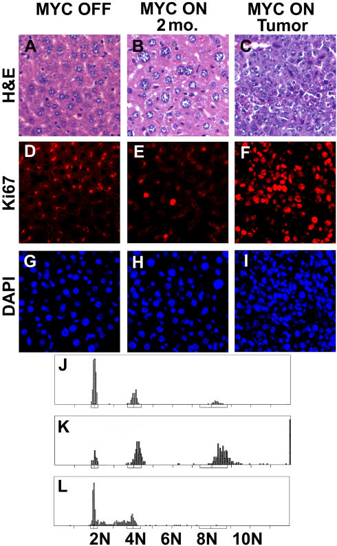
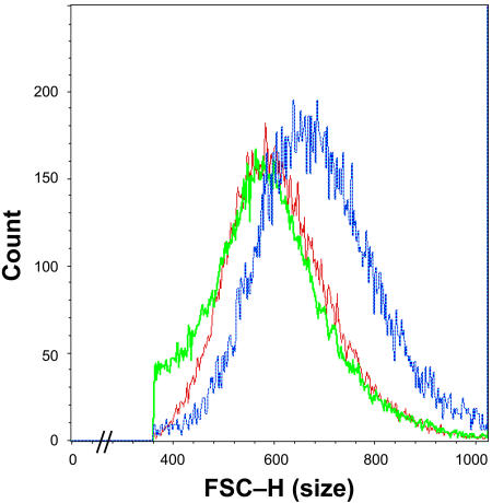
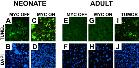
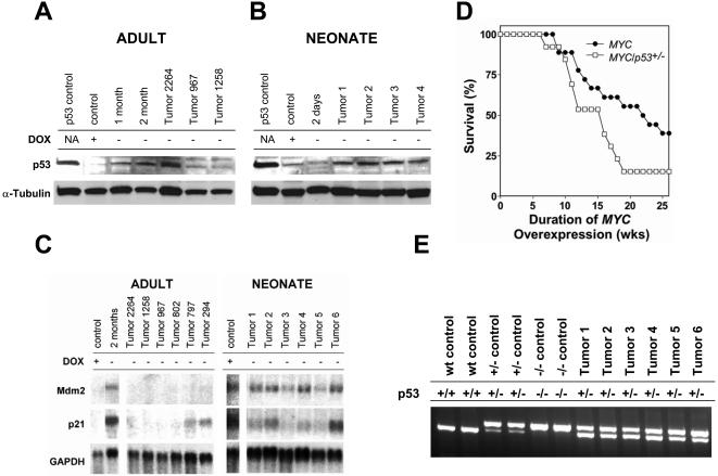
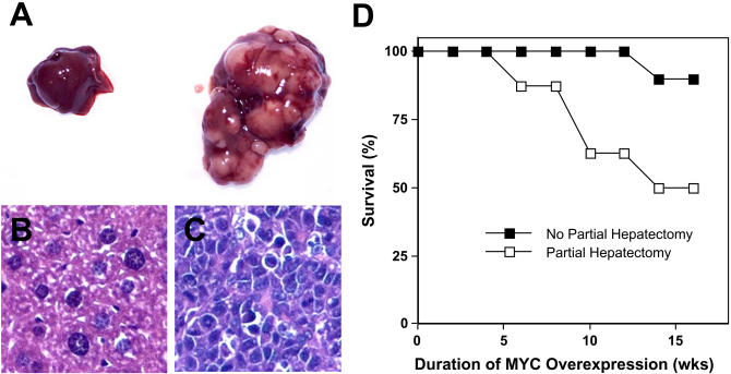
One of the enigmas in tumor biology is that different types of cancers are prevalent in different age groups. One possible explanation is that the ability of a specific oncogene to cause tumorigenesis in a particular cell type depends on epigenetic parameters such as the developmental context. To address this hypothesis, we have used the tetracycline regulatory system to generate transgenic mice in which the expression of a c-MYC human transgene can be conditionally regulated in murine hepatocytes. MYC's ability to induce tumorigenesis was dependent upon developmental context. In embryonic and neonatal mice, MYC overexpression in the liver induced marked cell proliferation and immediate onset of neoplasia. In contrast, in adult mice MYC overexpression induced cell growth and DNA replication without mitotic cell division, and mice succumbed to neoplasia only after a prolonged latency. In adult hepatocytes, MYC activation failed to induce cell division, which was at least in part mediated through the activation of p53. Surprisingly, apoptosis is not a barrier to MYC inducing tumorigenesis. The ability of oncogenes to induce tumorigenesis may be generally restrained by developmentally specific mechanisms. Adult somatic cells have evolved mechanisms to prevent individual oncogenes from initiating cellular growth, DNA replication, and mitotic cellular division alone, thereby preventing any single genetic event from inducing tumorigenesis.


Conflict of interest statement

The authors have declared that no conflicts of interest exist.

Figures

Figure 1
Figure 1. MYC Overexpression in Adult Hepatocytes Results in HCC
(A) Western blot analysis demonstrating that mice transgenic for both LAP-tTA and TRE-MYC conditionally express MYC protein in their hepatocytes in the absence (−) but not in the presence (+) of doxycycline. (B) Adult mouse with MYC-induced liver tumor. (C) Histology of an adult MYC-induced liver tumor. (D) Gross pathology of an adult liver tumor transplanted subcutaneously into a scid mouse. (E) Histology of an adult tumor transplanted subcutaneously into a scid mouse.
Figure 2
Figure 2. MYC-Induced Hepatic Tumors Are Invasive and Metastatic
(A) Adult mouse with MYC-induced liver tumor that has metastasized to the abdomen and the lungs. (B) Histology of an adult MYC-induced lung metastasis. (C) Histology of an adult MYC-induced liver tumor. (D) Gross pathology of a liver tumor from a neonatal host transplanted subcutaneously into a scid mouse.
Figure 3
Figure 3. MYC's Ability to Induce HCC Is Inversely Correlated with the Age of the Host at the Time of MYC Activation
(A) Survival of transgenic mice demonstrates that tumorigenesis in the liver is inversely correlated with the age of the host at the time of MYC induction. Shown are cases where MYC was constitutively expressed (▪), newborn mice in which MYC was activated at birth (▴), young mice in which MYC was activated at 4 weeks of age (•), adult mice in which MYC was activated at 6–12 weeks of age (♦), and transgenic mice treated with doxycycline (□). Cohorts consisted of 15–30 mice. Mice were scored when moribund. MYC transgene expression was induced to similar levels in the differently aged cohorts of mice. Survival time is measured as the time after MYC induction. (B) Western blot examining total MYC protein levels (human MYC and endogenous murine c-MYC) in mice when MYC is induced during embryonic development, activated at birth, and activated during adulthood (after 10 weeks of age). Adult mice exhibited a progressive increase in MYC protein levels during the course of MYC induction, with a significant increase in MYC protein in tumors. MYC protein levels in neonatal mice in which MYC was activated at birth were slightly increased at 10 d of age, and significantly increased at 18 d of age when these mice developed liver tumors. Liver tumors in 2- and 6-d-old neonatal mice that overexpressed MYC during embryonic development exhibited MYC protein levels similar to those observed in neonatal and adult tumors. (C) Real-time PCR analysis showing human MYC RNA levels in mice after different durations of MYC transgene induction. Adult livers exhibited a small increase in MYC RNA levels upon MYC activation, and a much greater increase in MYC RNA in MYC-induced tumors (black bars). In neonatal mice in which the MYC transgene was induced at birth, MYC RNA levels rose after 10 d of MYC activation. When these neonatal mice developed liver tumors, they exhibited MYC RNA levels similar to those seen in adult tumors (gray bars). Mice in which MYC was overexpressed during embryonic development developed liver tumors by 2 d of age and exhibited MYC RNA levels similar to those observed in neonatal and adult tumors (white bars).
Figure 4
Figure 4. MYC Activation during Embryonic Development Induces a Rapid Onset of Neoplasia
(A) A normal neonatal liver and a neonatal liver in which MYC was activated embryonically. (B) Histology of a normal neonatal liver in which MYC was not activated. (C) Histology of a neonatal liver in which MYC was activated embryonically. (D) DNA content of normal neonatal hepatocytes. (E) DNA content of neonatal hepatocytes in which MYC was activated embryonically. (F and H) Ki67 immunofluorescence and DAPI staining corresponding to a normal neonatal liver. (G and I) Ki67 immunofluorescence and DAPI staining corresponding to a neonatal liver in which MYC was activated embryonically.
Figure 8
Figure 8. MYC Activation in Adult Hepatocytes Causes Cellular Hypertrophy
(A) Relative volumes of neonatal hepatocytes and nuclei after MYC activation. Data are expressed as normalized volume plus or minus the standard error of the mean. The volume was normalized by dividing each measurement by the mean volume of normal 1-d-old neonatal mice. Three livers were measured per time point. T, tumor. (B) Neonatal liver weights of normal and MYC-activated livers. Three to five livers were weighed per time point. Data are expressed as the mean weight (grams) plus or minus the standard error of the mean. (C) Relative volumes of adult hepatocytes and nuclei after MYC activation. Volumes of cells are expressed as the mean volume divided by the mean volume of hepatocytes from normal mice plus or minus the standard error of the mean. Cells from two to three livers were measured per time point. (D) Adult liver weights after MYC activation. A total of nine livers were measured per time point after MYC activation. Data are expressed as the mean weight (grams) plus or minus the standard error of the mean.
Figure 5
Figure 5. MYC Activation at Birth Induces Proliferation of Neonatal Hepatocytes
(A) Histology of a normal 10-d-old neonatal liver. (B) Histology of a 10-d-old liver in which MYC was activated at birth. (C) Histology of a normal 18-d-old neonatal liver. (D) Histology of a MYC-induced neonatal liver tumor that developed after 18 d of MYC overexpression; MYC was activated at birth. (E–L) Ki67 immunofluorescence (E–H) and DAPI staining (I–L) of normal neonatal hepatocytes (E, G, I, and K), MYC-activated hepatocytes (F and J), and MYC-induced neonatal tumors (H and L). Upon initial MYC activation in neonatal mice, there was a small increase in Ki67-positive cells. MYC-induced neonatal tumors exhibited much higher levels of Ki67-positive cells.
Figure 6
Figure 6. MYC Activation in Adult Hepatocytes Induces Increased Cell Size and Endoreduplication, and Only Results in Cell Proliferation upon Neoplastic Conversion of Hepatocytes
(A) Histology of a normal liver. (B) Histology of a liver 2 months after MYC activation. (C) Histology of a MYC-induced liver tumor. (D and G) Ki67 immunofluorescence and DAPI staining of a normal adult liver. (E and H) Ki67 and DAPI staining of an adult liver after 8 weeks of MYC activation. (F and I) Ki67 and DAPI staining of a MYC-induced adult tumor. (J) DNA content measured in normal hepatocytes. (K) DNA content measured after MYC induction for 2 months. (L) DNA content of a representative MYC-induced liver tumor.
Figure 7
Figure 7. MYC Activation in Adult Hepatocytes Induces Increased Cell Size
A histogram obtained by FACS forward versus light scatter analysis of adult hepatocytes from normal FVB/N livers (green), doxycycline-treated transgenic livers (red), and livers in which the MYC transgene was overexpressed for 3 months (blue). The x-axis represents cell size and the y-axis represents cell count. Adult mice were matched for age.
Figure 9
Figure 9. MYC Activation Does Not Induce Apoptosis in Murine Hepatocytes
TUNEL assay (A, C, E, G, and I) and DAPI staining (B, D, F, H, and J) of normal (A, B, E, and F) and MYC-activated (C, D, and G–J) hepatocytes of neonatal (A–D) and adult (E–J) mice. After 4 weeks of MYC activation in adult hepatocytes, there was no evidence of apoptosis either by TUNEL assay (G) or by DAPI staining of nuclei (H). MYC activation is associated with increased apoptosis with the neoplastic conversion of neonatal (C) and adult (I) hepatocytes. Representative data from one of three experiments are shown. Identical results were seen when MYC was activated for 2 or 8 weeks.
Figure 10
Figure 10. MYC Activation Induces p53 Function, and Loss of p53 Function Is Necessary for MYC to Induce Tumorigenesis in Adult Hepatocytes
(A) Western blot analysis for p53 protein expression after MYC activation for 1 month, 2 months, and in MYC-induced tumors. As a positive control, we used a lymphoma cell line that overexpresses a mutant p53, kindly provided by Dr. Kevin Smith. (B) Western blot analysis for p53 protein expression in a normal neonatal liver, in a neonatal liver in which MYC was activated during embryonic development that was obtained from a 2-d-old mouse, and in MYC-induced neonatal tumors. As a positive control, we used a lymphoma cell line generated in our lab that overexpresses a mutant p53. (C) Northern blot analysis for p21 and MDM2 in neonatal and adult liver tumors. (D) Survival of adult mice after activation of MYC in the presence of the wild type or the loss of one p53 allele. (E) Loss of heterozygosity analysis of MYC/p53+/− tumors by PCR analysis.
Figure 11
Figure 11. Partial Hepatectomy Accelerates the Ability of MYC to Induce HCC in Adult Mice
(A) Liver from an adult mouse 7 weeks after MYC activation exhibited no gross phenotypic changes (left), whereas the liver from an adult mouse 7 weeks after MYC activation that had undergone partial hepatectomy exhibited a multifocal liver tumor (right). (B) The histology of the liver from an adult mouse 7 weeks after MYC activation exhibited no evidence of a tumor. (C) The histology of a liver from an adult mouse after MYC activation that had undergone partial hepatectomy exhibited a multifocal HCC. (D) Survival after MYC activation in adult mice that have either not undergone surgery (▪) or undergone a partial hepatectomy (□). Results are pooled from two independent experiments with a total of ten mice per group.

References

    1. Abou-Elella A, Gramlich T, Fritsch C, Gansler T. c-myc amplification in hepatocellular carcinoma predicts unfavorable prognosis. Mod Pathol. 1996;9:95–98. - PubMed
    1. Adams JM, Harris AW, Pinkert CA, Corcoran LM, Alexander WS, et al. The c-myc oncogene driven by immunoglobulin enhancers induces lymphoid malignancy in transgenic mice. Nature. 1985;318:533–538. - PubMed
    1. Artandi SE, Chang S, Lee SL, Alson S, Gottlieb GJ, et al. Telomere dysfunction promotes non-reciprocal translocations and epithelial cancers in mice. Nature. 2000;406:641–645. - PubMed
    1. Blyth K, Stewart M, Bell M, James C, Evan G, et al. Sensitivity to myc-induced apoptosis is retained in spontaneous and transplanted lymphomas of CD2-mycER mice. Oncogene. 2000;19:773–782. - PubMed
    1. Boige V, Laurent-Puig P, Fouchet P, Flejou JF, Monges G, et al. Concerted nonsyntenic allelic losses in hyperploid hepatocellular carcinoma as determined by a high-resolution allelotype. Cancer Res. 1997;57:1986–1990. - PubMed

Publication types

MeSH terms

Substances

Associated data