

Review
doi: 10.1523/JNEUROSCI.3604-04.2004.
Regulation of calcium/calmodulin-dependent protein kinase II activation by intramolecular and intermolecular interactions
Affiliations
- PMID: 15456810
- PMCID: PMC6729912
- DOI: 10.1523/JNEUROSCI.3604-04.2004
Item in Clipboard
Review
Regulation of calcium/calmodulin-dependent protein kinase II activation by intramolecular and intermolecular interactions
J Neurosci.
.
No abstract available
Figures
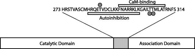
Schematic diagram of CaMKII domain structure. All CaMKII isozymes contain an N-terminal catalytic domain, an internal regulatory domain, and a C terminal that mediates holoenzyme formation. The regulatory domain, whose sequence is shown above the diagram, is bipartite. The proximal end (aa 282-300) contain residues that interact with the catalytic domain to inhibit phosphotransferase activity (indicated by gray bar below sequence). The distal portion of this domain (aa 293-310) binds to Ca2+/CaM (indicated by gray bar above sequence). Regulatory phosphorylation sites at Thr286, Thr305, and Thr306 are indicated by gray dots.
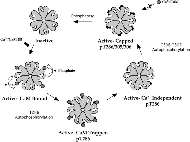
Regulation of CaMKII by autophosphorylation. Autophosphorylation within the regulatory domain of CaMKII defines several activity states for the kinase. In the absence of Ca2+/CaM and autophosphorylation, CaMKII is inactive (Inactive). Binding of Ca2+/CaM activates the kinase for substrate phosphorylation, bringing it to 100% of its maximal activity (CaM Bound). Binding of two Ca2+/CaMs to adjacent subunits stimulates inter-subunit phosphorylation of Thr286. The off-rate of Ca2+/CaM from pThr286 CaMKII is decreased by > 1000-fold, resulting in an enzyme that remains at 100% of its maximal Ca2+/CaM-stimulated activity even as calcium falls in the cell (CaM Trapped). Once Ca2+/CaM dissociates, the enzyme remains active but at a lower level than with saturating Ca2+/CaM, having between 20 and 80% of its maximal Ca2+/CaM-stimulated activity (Ca2+ Independent). The dissociation of Ca2+/CaM also uncovers additional sites in the regulatory domain (Thr305 and Thr306), which rapidly become autophosphorylated. The pThr286/pThr305/pThr306 CaMKII remains active at 20-80% of maximal activity because of pThr286 but is incapable of binding Ca2+/CaM (Capped). Phosphatase activity is required to reset the kinase to its basal state, and theoretically these sites could be individually regulated by dephosphorylation to produce pThr286 or pThr305/pThr306 states of the kinase from the triple-phosphorylated form. The pThr305/pThr306 state of the kinase would be completely inactive and unresponsive to Ca2+/CaM.
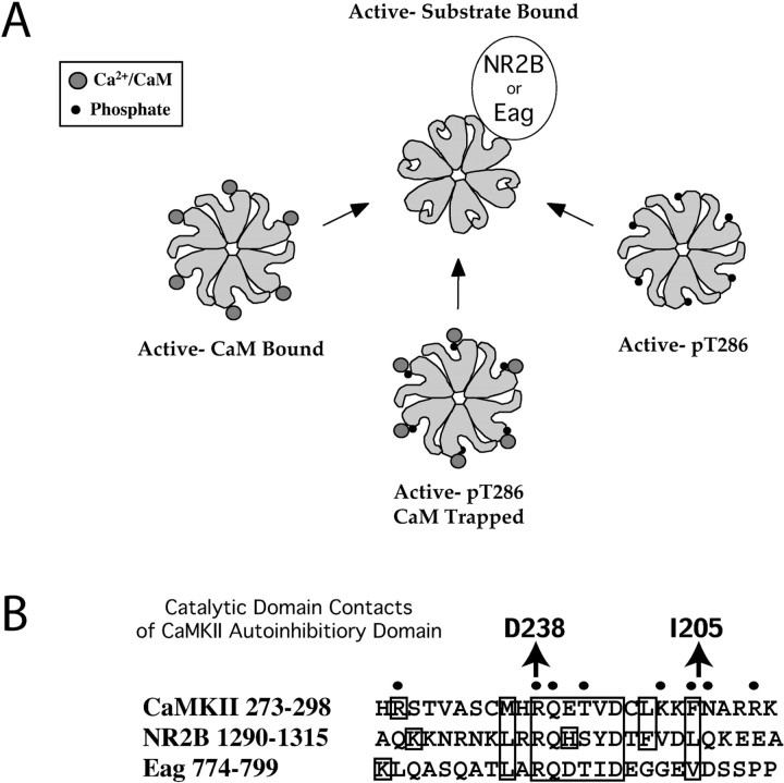
Regulation of CaMKII by binding interactions. Binding of CaMKII to exogenous proteins can regulate its activity. A, Proteins with domains that resemble the autoinhibitory domain of the kinase can bind to CaMKII in an activity-dependent manner. Activation of the kinase, by Ca2+/CaM binding or Thr286 autophosphorylation, causes a conformational change that reveals an interaction face on the catalytic domain. This interaction domain is blocked by the CaMKII autoinhibitory domain in the inactive state of the kinase. Interaction persists even in the absence of pThr286 or Ca2+/CaM and serves to block reassociation of the autoinhibitory domain with the catalytic site, thus rendering the kinase calcium independent. B, An alignment of the CaMKII autoinhibitory domain, NR2B, and Eag. Dots above the CaMKII sequence indicate residues of the autoinhibitory domain that were shown to contact the catalytic domain (Yang and Schulman, 1999). Catalytic domain contacts for selected residues are shown above the alignment. R283 is believed to contact D238, whereas F293 and N294 are believed to contact I205 in the catalytic domain. For a complete list of contacts, see Yang and Schulman (1999).
References
-
- Baines AJ (1996) Caenorhabditis elegans LIN-2A and mammalian neuronal CASK are prototypical members of a subfamily of MAGUKs (membrane-associated guanylate kinases) characterized by a common kinase-like domain and a guanylate kinase domain predicted to bind ATP. Biochem J 320: 694-696. - PMC - PubMed
-
- Bayer KU, De Koninck P, Leonard AS, Hell JW, Schulman H (2001) Interaction with the NMDA receptor locks CaMKII in an active conformation. Nature 411: 801-805. - PubMed
-
- Colbran RJ (1993) Inactivation of Ca2+/calmodulin-dependent protein kinase II by basal autophosphorylation. J Biol Chem 268: 7163-7170. - PubMed
Publication types
MeSH terms
Substances
Grants and funding
LinkOut - more resources
Full Text Sources