. 2004 Sep 29;24(39):8510-21.
doi: 10.1523/JNEUROSCI.1264-04.2004.

Abl tyrosine kinase promotes dendrogenesis by inducing actin cytoskeletal rearrangements in cooperation with Rho family small GTPases in hippocampal neurons

Affiliations

Abl tyrosine kinase promotes dendrogenesis by inducing actin cytoskeletal rearrangements in cooperation with Rho family small GTPases in hippocampal neurons

Shiloh B Jones et al. J Neurosci. .

Abstract

Nonreceptor tyrosine kinase Abl is an actin-binding protein and a key regulator of neuronal axonal development. Although Abl family kinases also are localized in dendrites and are implicated in postsynaptic functions, it is not clear how Abl kinases regulate dendritic morphogenesis. Using a developing hippocampal culture as a model, we found that the inhibition of Abl kinases by STI571 leads to a remarkable simplification of dendritic branching similar to the phenotype caused by an increased activity of small GTPase RhoA. Time-lapse microscopic imaging reveals a prominent reduction of dendritic branching. In contrast, neurons expressing a constitutively active v-abl construct (CA-Abl) show an exuberant microtubule-associated protein 2-positive (MAP2-positive) dendrite outgrowth, suggesting that Abl modulates dendritic growth. Biochemical assays using a glutathione S-transferase pull-down method to determine GTP-bound active Rho GTPases demonstrate that Abl inhibition increases RhoA activity but has no effect on the activity of Rac1 or Cdc42. At the cellular level the alteration of Abl also changes actin organization consistent with RhoA inhibition. Suppression of the RhoA downstream effector Rho kinase reverses STI571-induced dendritic simplification, demonstrating that activity of the Rho pathway is responsible for the Abl-induced changes in dendrogenesis. Furthermore, CA-Abl-induced neurite outgrowth is blocked by the expression of a constitutively active RhoA construct. The CA-Abl phenotype is not affected by destabilization of microtubules but is reversed partially when actin filaments are stabilized with jasplakinolide. Together, these studies support a critical role for Abl kinases in regulating dendrogenesis by inducing actin cytoskeletal rearrangements in cooperation with Rho GTPases.


Figures

Figure 1.
Figure 1.
Inhibition of Abl family tyrosine kinase activity disrupts dendrogenesis independently of axonogenesis. A, Developmental progression of cultured hippocampal neurons on etched-grid coverslips from 0.5 through 7 DIV. B, Treatment with the small molecule inhibitor STI571 from 4 DIV impairs dendrogenesis while axons continue to grow and branch. Arrows point to dendrites; arrowheads indicate axons. The asterisks indicate an approaching axon on 6 versus 7 DIV. Scale bar, 25 μm.
Figure 2.
Figure 2.
Inhibition of Abl family tyrosine kinase activity alters the developmental dynamics of dendrogenesis. A, Hippocampal neurons grown in culture for 4.5 d were incubated in the absence (DMSO) or presence (STI571) of Abl inhibitor for 48 hr. Time-lapse images were captured every 30 sec for 135 min. Only eight representative frames are shown here. Asterisks point to an elongating dendrite in neurons treated with DMSO. The number signs point to a retracting dendrite in neurons treated with 3 μm STI571. The arrowhead points to growth cones in neurons treated with DMSO or STI571. Scale bar, 15 μm. B, Comparison of histories of the percentage of change in dendrite length of neurons treated with DMSO and STI571. Shown here are the measurements of average length changes in 25 dendrites during a 135 min time-lapse recording. C, Comparison of the percentage of change in primary and secondary dendrite length of neurons treated with DMSO and STI571. Shown here are the measurements of average length changes in 25 dendrites before and after time-lapse recording. Asterisks indicate the significant differences between t = 0 and t = 135 min (*p < 0.05). D, Comparison of the histories of dendritic sprout formation between DMSO- and STI571-treated neurons during 30 min time-lapse recordings. In this case, sprouts were measured along 80 μm length of a dendrite, and a total of seven dendrites was selected for analysis from time-lapse light microscopy. Error bars indicate SEM. For details, see supplemental material, available at www.jneurosci.org.
Figure 3.
Figure 3.
Inhibition of Abl family tyrosine kinase activity disrupts axon-dendrite polarity. A, Immunofluorescent images of control neurons labeled with FITC phalloidin (a), anti-MAP2 (b), and anti-Tau (c). B, Hippocampal neurons at 4.5 DIV treated with 3 μm STI571 for 48 hr, stained with FITC phalloidin (a), and immunostained for anti-MAP2 (b) and anti-Tau (c). Note disruption of MAP2 and Tau segregation after Abl kinase inhibition. Arrows point to dendrites; arrowheads indicate axons. Asterisk points to the growth cone. Scale bar, 25 μm.
Figure 4.
Figure 4.
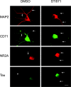
Immunofluorescent images of hippocampal neurons at 5 DIV treated with 0.03% DMSO (left) or 3 μm STI571 (right) for 48 hr and double-immunolabeled with anti-MAP2 and anti-CD71 (transferrin receptor) or anti-NR2A and anti-Tau. Arrows point to the dendrites; arrowhead points to the axon. Note the colocalization of MAP2 with CD71 in DMSO- and STI571-treated cells. Also note that, whereas Tau was localized to the axon, NR2A was localized more prominently in dendrites (arrow), with weaker staining in the axon (DMSO; compare Tau and NR2A staining). The segregation of Tau and NR2A was not clear in neurons treated with STI571. Scale bar, 15 μm.
Figure 5.
Figure 5.
Abl family tyrosine kinase activity influences primary dendrite formation and dendritic branching. A, Phase-contrast and double-immunofluorescent light microscopy of single neurons treated with DMSO or STI571 or transfected with CA-Abl. Neurons were treated for 48 hr from 5 to 7 DIV and immunostained with rabbit polyclonal anti-Abl and mouse monoclonal anti-MAP2. Note the simplification of dendritic profile in STI571-treated neurons in comparison to an exuberant outgrowth of MAP2-positive, highly branched processes in neurons transfected with CA-Abl. Scale bar, 15 μm. B, Quantification of primary and secondary dendrite outgrowth in neurons incubated with DMSO or STI571 or transfected with CA-Abl. C, Quantification of primary and secondary dendrite length in neurons incubated with DMSO or STI571 or transfected with CA-Abl. Error bars indicate SEM; *p < 0.05.
Figure 6.
Figure 6.
Inhibition of Abl tyrosine kinases by STI571 increases GTP-bound RhoA activity without affecting Rac1 or Cdc42 activity. A, Western blot showing RhoA, Rac1, or Cdc42 in GTP-bound forms. The 5 DIV neurons were treated with DMSO or STI571 for 48 hr or with STI571 for 48 hr, followed by ST1571 plus H2O2 for 10 min. The 6 DIV neurons were treated with Toxin B for 24 hr. The 7 DIV neurons were treated with 100 μm glutamate for 10 min. The 7 DIV cell lysates with different treatments were probed with anti-RhoA, Rac1, or Cdc42 antibodies to show their expression levels (left panels). The 7 DIV cell lysates from different treatments also were incubated with GST-RBD (RhoA) or GST-PBD (Rac1 and Cdc42) glutathione beads to pull down GTP-bound RhoA, Rac1, or Cdc42 as an indication of their activities. Bound proteins were analyzed by Western blot with anti-RhoA, Rac1, or Cdc42 antibodies (right panels). B, Relative RhoA, Rac1, and Cdc42 activity was determined semiquantitatively by using densitometric analysis as described in Materials and Methods. Data represent the means ± SEM of at least three independent experiments; *p < 0.05.
Figure 7.
Figure 7.
ROCK inhibition suppresses STI571 effect on dendrogenesis. A, Immunofluorescent light microscopy of anti-MAP2 and rhodamine phalloidin staining of 5 DIV hippocampal neurons treated with DMSO (STI571/- and Y-27632/-) or 3 μm STI571 for 48 hr or treated with 3 μm STI571 for 24 hr, followed by the addition of Y-27632 for an additional 18 hr. Cells were fixed on 7 DIV. Arrowheads point to focal actin filaments. Scale bar, 15 μm. B, Comparison of the percentage of change of dendrite numbers among neurons treated with DMSO, STI571, Y-27632, and STI571/Y-27632. Note that the reduction of dendrite numbers by STI571 treatment is reversed by coincubation of neurons with Y-27632. C, Comparison of the percentage of change of dendrite length among neurons treated with DMSO, STI571, Y-27632, and STI571/Y-27632. Note that the decrease in dendrite length by STI571 treatment is reversed by coincubation of neurons with Y-27632. The effect of Y-27632 is more dramatic on the primary dendrites than on the secondary dendrites in neurons 7 DIV. Error bars indicate SEM; *p < 0.05.
Figure 8.
Figure 8.
RhoA activation suppresses CA-Abl effect on dendrogenesis. Immunofluorescent light microscopy of hippocampal neurons transfected with pEGFP-C2, pEGFP-C2 plus CA-Abl, pGFP-CA-RhoA, and CA-Abl plus pGFP-CA-RhoA. Top, GFP fluorescence; bottom, anti-Abl. Note that the exuberant dendrite formation in pEGFP-C2 plus CA-Abl-transfected neurons was inhibited by expression of pGFP-CA-RhoA. Note that the Abl immunoreactivity in neurons transfected with pGFP-CA-RhoA is weak because of lower endogenous Abl expression when compared with neurons doubly transfected with pEGFP-C2 plus CA-Abl or CA-Abl plus pGFP-CA-RhoA. Scale bar, 15 μm.
Figure 9.
Figure 9.
The CA-Abl effect on dendrogenesis is dependent on the reorganization of actin cytoskeleton but is independent of the stability of microtubules. A, Immunofluorescent light microscopy of single neurons by anti-Abl antibody (top) and rhodamine phalloidin (bottom). The 5 DIV hippocampal neurons transfected with CA-Abl were treated either with 5 μm latrunculin A the next day for 24 hr to depolymerize actin filament or with 100 nm jasplakinolide on 7 DIV for 5 hr to stabilize actin filaments. Neurons were fixed on 7 DIV. Note that CA-Abl-induced process outgrowth is suppressed by jasplakinolide. In jasplakinolide-treated neurons, F-actin staining is weak because of competitive binding of jasplakinolide and phalloidin to F-actin. Note that F-actin staining in latrunculin A-treated neurons was enhanced to quantify the processes. Scale bar, 15 μm. B, Quantification of primary and secondary dendrite outgrowth in neurons incubated with DMSO, latrunculin A, and jasplakinolide as well as transfected with CA-Abl in the presence or absence of latrunculin A and jasplakinolide. C, Quantification of the relative length of primary and secondary dendrites in neurons incubated with DMSO, latrunculin A, and jasplakinolide as well as transfected with CA-Abl in the presence and absence of latrunculin A and jasplakinolide. Dendrite number and length were determined within 120 μm2 of the single neuronal cell bodies. Data represent the means ± SEM of three independent experiments; *p < 0.05.
Figure 10.
Figure 10.
The CA-Abl effect on dendrogenesis is independent of the stability of microtubules. Shown is double-immunofluorescent light microscopy of single neurons by rabbit polyclonal anti-Abl antibody (top) and mouse monoclonal anti-MAP2 (bottom). Control hippocampal neurons or CA-Abl-expressing cells were incubated with or without the presence of 5 μm vincristine for 5 hr. Note that the MAP2-positive microtubules were disorganized in the neurons treated with vincristine, which shows no significant effects on the dendrite morphology. Scale bar, 15 μm.

References

    1. Benard V, Bohl BP, Bokoch GM (1999) Characterization of Rac and Cdc42 activation in chemoattractant-stimulated human neutrophils using a novel assay for active GTPases. J Biol Chem 274: 13198-13204. - PubMed
    1. Bito H, Furuyashiki T, Ishihara H, Shibasaki Y, Ohashi K, Mizuno K, Maekawa M, Ishizaki T, Narumiya S (2000) A critical role for a Rho-associated kinase, p160ROCK, in determining axon outgrowth in mammalian CNS neurons. Neuron 26: 431-441. - PubMed
    1. Bradke F, Dotti CG (1999) The role of local actin instability in axon formation. Science 283: 1931-1934. - PubMed
    1. Buchdunger E, Zimmermann J, Mett H, Meyer T, Muller M, Druker BJ, Lydon NB (1996) Inhibition of the Abl protein-tyrosine kinase in vitro and in vivo by a 2-phenylaminopyrimidine derivative. Cancer Res 56: 100-104. - PubMed
    1. Buchdunger E, Cioffi CL, Law N, Stover D, Ohno-Jones S, Druker BJ, Lydon NB (2000) Abl protein-tyrosine kinase inhibitor STI571 inhibits in vitro signal transduction mediated by c-Kit and platelet-derived growth factor receptors. J Pharmacol Exp Ther 295: 139-145. - PubMed

Publication types

MeSH terms

LinkOut - more resources