. 2004 Sep 29;24(39):8629-39.
doi: 10.1523/JNEUROSCI.2877-04.2004.

Altered expression of the delta subunit of the GABAA receptor in a mouse model of temporal lobe epilepsy

Affiliations

Altered expression of the delta subunit of the GABAA receptor in a mouse model of temporal lobe epilepsy

Zechun Peng et al. J Neurosci. .

Abstract

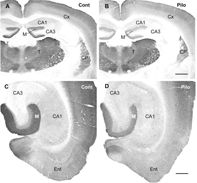
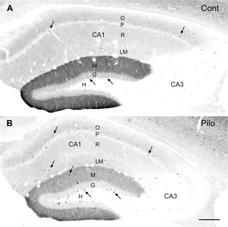
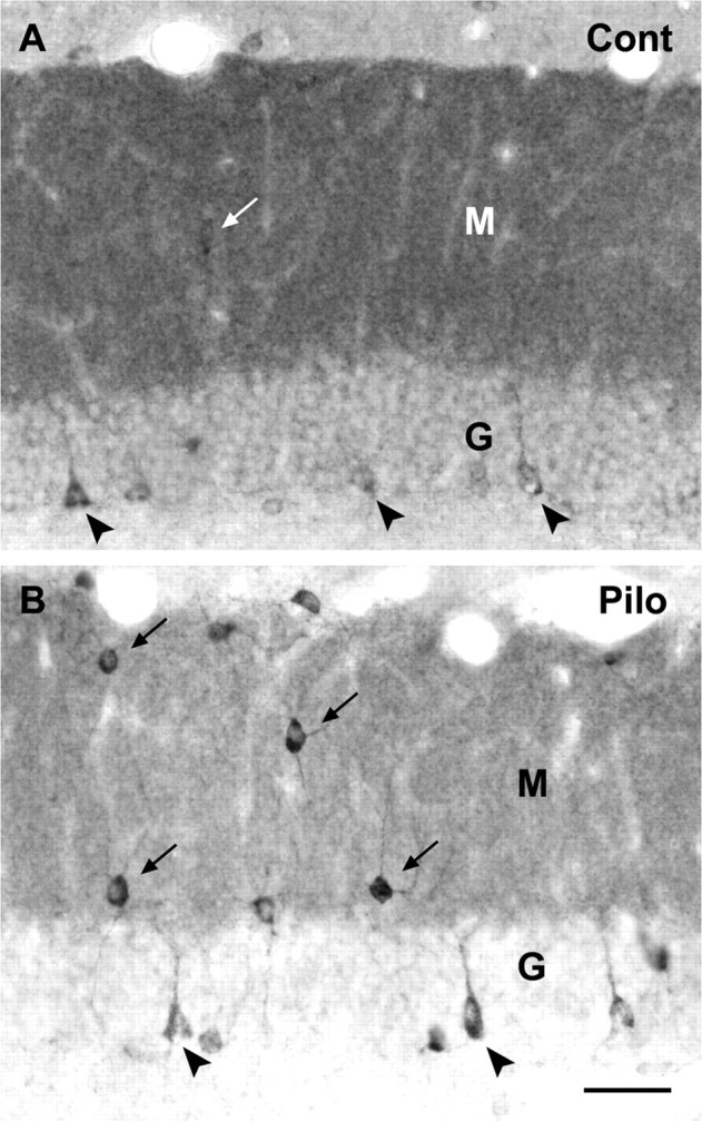
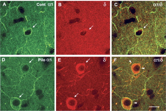
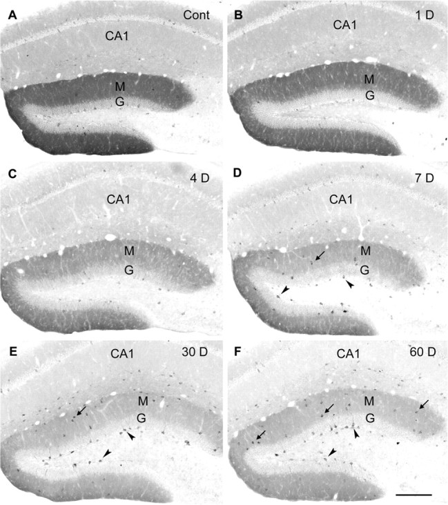
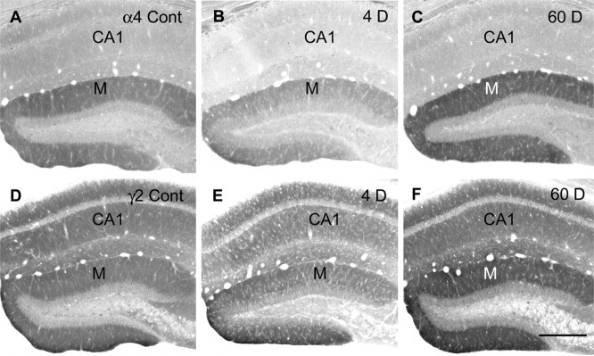
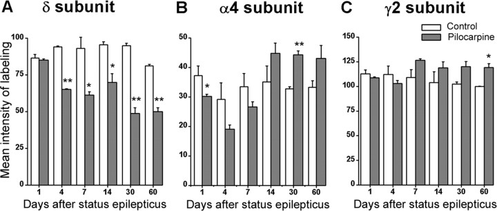
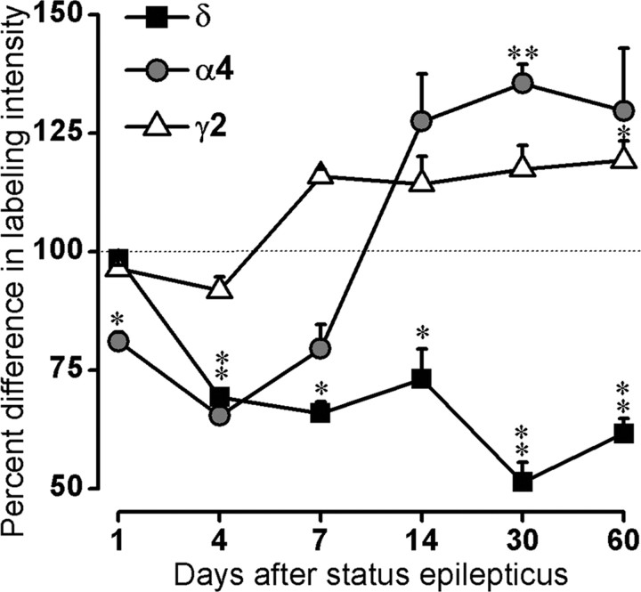
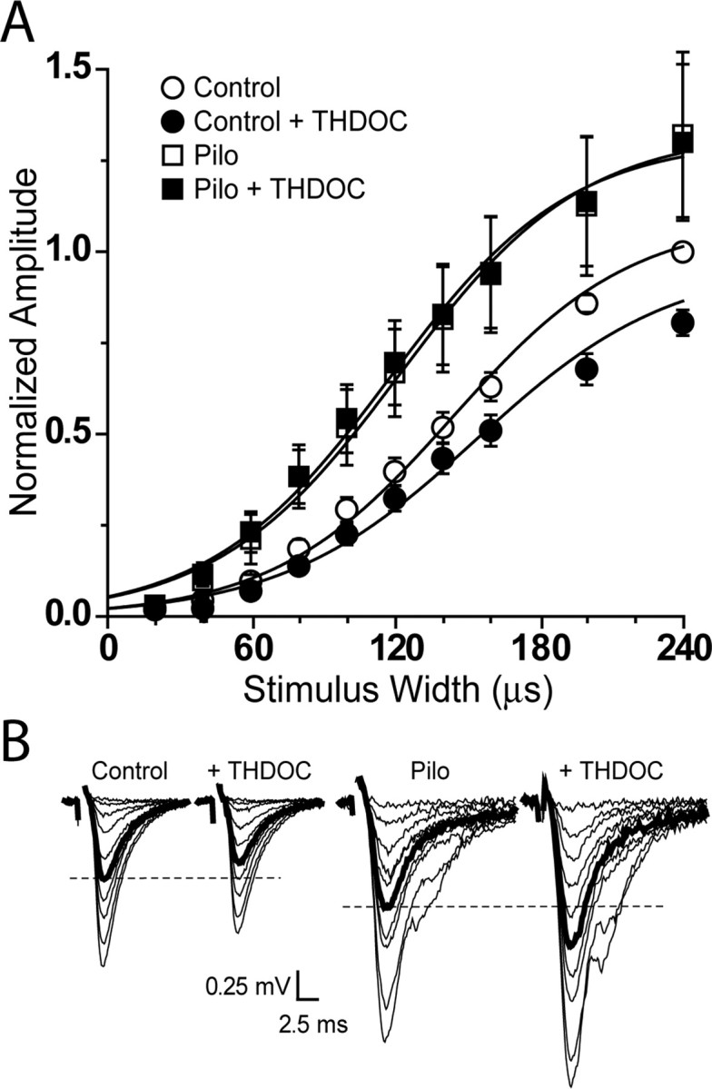
delta Subunit-containing GABA(A) receptors are located predominantly at nonsynaptic sites in the dentate gyrus where they may play important roles in controlling neuronal excitability through tonic inhibition and responses to GABA spillover. Immunohistochemical methods were used to determine whether delta subunit expression was altered after pilocarpine-induced status epilepticus in C57BL/6 mice in ways that could increase excitability of the dentate gyrus. In pilocarpine-treated animals, the normal diffuse labeling of the delta subunit in the dentate molecular layer was decreased by 4 d after status epilepticus (latent period) and remained low throughout the period of chronic seizures. In contrast, diffuse labeling of alpha4 and gamma2 subunits, potentially interrelated GABA(A) receptor subunits, was increased during the chronic period. Interestingly, delta subunit labeling of many interneurons progressively increased after pilocarpine treatment. Consistent with the observed changes in delta subunit labeling, physiological studies revealed increased excitability in the dentate gyrus of slices obtained from the pilocarpine-treated mice and demonstrated that physiological concentrations of the neurosteroid tetrahydrodeoxycorticosterone were less effective in reducing excitability in the pilocarpine-treated animals than in controls. The findings support the idea that alterations in nonsynaptic delta subunit-containing GABA(A) receptors in both principal cells and interneurons could contribute to increased seizure susceptibility in the hippocampal formation in a temporal lobe epilepsy model.


Figures

Figure 1.
Figure 1.
Comparison of immunohistochemical labeling for the δ subunit in control (Cont) (A, C) and pilocarpine-treated (Pilo) (B, D) mice at 2 months after status epilepticus. A, B, In coronal sections of the forebrain, decreased δ subunit labeling is most striking in the molecular layer (M) of the dentate gyrus in the pilocarpine-treated animal (B). Labeling is also moderately decreased in the neocortex (Cx) and slightly decreased in the caudate-putamen (CP) in this animal. No changes in δ subunit immunoreactivity are evident in the thalamus (T). C, D, In horizontal sections from the same animals, δ subunit immunoreactivity is also decreased in the molecular layer of the dentate gyrus at caudal levels (D), and the normally light labeling in CA1 (C) is further decreased in the pilocarpine-treated mouse (D). Decreased labeling of the δ subunit is also evident in the entorhinal cortex (Ent). Scale bars: A, B, 600 μm; C, D, 300 μm.
Figure 2.
Figure 2.
Alterations in δ subunit labeling in the hippocampal formation after pilocarpine-induced status epilepticus. A, In a control mouse, diffuse δ subunit labeling is high in the molecular layer (M) of the dentate gyrus. Moderate levels of diffuse labeling are evident in strata oriens (O), radiatum (R), and lacunosum-moleculare (LM) of CA1. Immunolabeling is low in CA3. Some moderately labeled interneurons (arrows) are evident along the base of the granule cell layer (G) and within CA1 where they are most numerous near the pyramidal cell layer (P). Very few δ subunit-labeled interneurons are present in the hilus (H). B, In a pilocarpine-treated mouse at 2 weeks after status epilepticus, diffuse δ subunit labeling in the molecular layer is substantially decreased. Diffuse labeling is also slightly decreased in CA1. In contrast, labeling of many interneurons (arrows) is increased. Strongly labeled interneurons are prominent along the base of the granule cell layer, within the molecular layer, and in strata pyramidale (P) and lacunosum-moleculare of CA1. Both sections were processed with a nickel-intensified labeling method. Scale bar: A, B, 200 μm.
Figure 3.
Figure 3.
Comparison of δ subunit-labeled interneurons in the dentate gyrus of control (Cont) (A) and pilocarpine-treated (Pilo) (B) mice. A, In control tissue, only lightly or moderately labeled interneurons can be detected within the molecular layer (M; arrow) and along the inner border of the granule cell layer (G; arrowheads). B, In a pilocarpine-treated animal at 60 d after status epilepticus, increased numbers of darkly labeled interneurons are evident in the molecular layer (arrows) and along the base of the granule cell layer (arrowheads) where many resemble basket cells. Immunolabeling is quite dense within the cytoplasm and extends into the proximal dendrites in many of the labeled interneurons. A decrease in diffuse labeling is evident within both the molecular and granule cell layers in the pilocarpine-treated animal. Scale bar: A, B, 40 μm.
Figure 4.
Figure 4.
Comparisons of δ subunit immunolabeling in α1 subunit-expressing interneurons in the molecular layer of control (Cont) (A-C) and pilocarpine-treated (Pilo) (D-F) mice with confocal microscopy. A-C, In a control animal, strong α1 immunolabeling is evident around the soma (arrow) and dendritic processes within the neuropil (A). Immunolabeling for the δ subunit is relatively low in the cytoplasm of the interneuron (B, arrow), and no δ subunit labeling can be detected on the surface of the interneuron (C). D-F, In a pilocarpine-treated animal, distinct α1 subunit labeling is evident on the surface of interneurons (arrows) in the molecular layer, and lighter labeling is present within the cytoplasm (D). Strong immunolabeling for the δ subunit is present in the cytoplasm of the same interneurons (E, arrows), and overlapping labeling for the δ and α1 subunits is present on the surface of the neurons (F, arrowheads). Scale bar: A-F, 10 μm.
Figure 5.
Figure 5.
Progressive changes in immunohistochemical labeling of the δ subunit in the dentate gyrus after pilocarpine-induced status epilepticus. In comparison with that in a control (Cont) mouse (A), diffuse immunolabeling is slightly decreased in the molecular (M) and granule cell (G) layers of the dentate gyrus at 1 d after status epilepticus (B) and decreases further by 4 d after pilocarpine treatment (C). At these early times, labeling of interneurons is also generally decreased (B, C). At 7 d (D), 30 d (E), and 60 d (F) after status epilepticus, the diffuse δ subunit immunoreactivity is substantially decreased throughout the molecular and granule cell layers of the dentate gyrus. In contrast, δ subunit labeling of many interneurons is increased, and labeled interneurons become particularly evident in the subgranular region (arrowheads) and molecular layer of the dentate gyrus (arrows). Scale bar: A-F, 200 μm.
Figure 6.
Figure 6.
Comparisons of immunolabeling for the α4 (A-C) and γ2 (D-F) subunits in the dentate gyrus in control (Cont) and pilocarpine-treated mice at 4 d (B, E) and 60 d (C, F) after status epilepticus. A, In a control mouse, labeling of the α4 subunit is prominent in the molecular layer (M) of the dentate gyrus. B, At 4 d after SE, α4 labeling is decreased in the molecular layer as well as in CA1. C, At 60 d after SE, α4 subunit labeling in the molecular layer is stronger than that in the control (compare C and A). D, In a control mouse, strong labeling for the γ2 subunit is evident in the molecular layer of the dentate gyrus and CA1. E, At 4 d after pilocarpine treatment, γ2 labeling is slightly decreased throughout the hippocampal formation. F, At 60 d after SE, γ2 labeling in the molecular layer is increased and is stronger than that in the control mouse (compare F and D). Illustrated sections were processed in parallel for each subunit, and sections for both subunits are from the same animal. Scale bar: A-F, 300 μm.
Figure 7.
Figure 7.
Comparisons of the mean intensity of immunolabeling for the α4, γ2, and δ subunits in control and pilocarpine-treated animals at selected times after status epilepticus. **p < 0.01; *p < 0.05.
Figure 8.
Figure 8.
Comparisons of percentage differences in intensity of labeling for δ, α4, and γ2 subunits in pilocarpine-treated animals compared with controls at 1-60 d after status epilepticus. Control values are represented as 100% (dotted line) for all subunits. Intensity of δ subunit labeling is below control values at 4-60 d after status epilepticus. In contrast, after initial small decreases, the intensity of both α4 and γ2 labeling increases above control values and remains elevated through the remainder of the study. **p < 0.01; *p < 0.05.
Figure 9.
Figure 9.
A physiological concentration of THDOC (10 nm) does not affect fEPSP slope in slices from pilocarpine-treated animals. A, Stimulus-response curves from control (○, •) and pilocarpine-treated animals (□, ▪) in control conditions (○, □) and after a 20 min perfusion of THDOC (•, ▪). Data (±SEM) are normalized to the slope of the EPSP240 (evoked by stimulus width, 240 μsec) under control conditions (○). Lines represent averages of Boltzman function generated by the averages of the parameters fitted to individual experiments. B, Representative traces from individual experiments. Bolded traces are approximate W50 responses (W50, stimulus width required to elicit a half-maximal response), and dashed lines are drawn to facilitate comparison of W50 responses.

Comment in

References

    1. Adkins CE, Pillai GV, Kerby J, Bonnert TP, Haldon C, McKernan RM, Gonzalez JE, Oades K, Whiting PJ, Simpson PB (2001) α4β3δ GABAA receptors characterized by FRET-derived measurements of membrane potential. J Biol Chem 276: 38934-38939. - PubMed
    1. Araujo F, Ruano D, Vitorica J (1998) Absence of association between δ and γ2 subunits in native GABAA receptors from rat brain. Eur J Pharmacol 347: 347-353. - PubMed
    1. Belelli D, Casula A, Ling A, Lambert JJ (2002) The influence of subunit composition on the interaction of neurosteroids with GABAA receptors. Neuropharmacology 43: 651-661. - PubMed
    1. Bencsits E, Ebert V, Tretter V, Sieghart W (1999) A significant part of native γ-aminobutyric acidA receptors containing α4 subunits do not contain γ or δ subunits. J Biol Chem 274: 19613-19616. - PubMed
    1. Brickley SG, Cull-Candy SG, Farrant M (1996) Development of a tonic form of synaptic inhibition in rat cerebellar granule cells resulting from persistent activation of GABAA receptors. J Physiol (Lond) 497: 753-759. - PMC - PubMed

Publication types

MeSH terms