. 2004 Oct;24(20):8847-61.
doi: 10.1128/MCB.24.20.8847-8861.2004.

Bone-specific transcription factor Runx2 interacts with the 1alpha,25-dihydroxyvitamin D3 receptor to up-regulate rat osteocalcin gene expression in osteoblastic cells

Affiliations

Bone-specific transcription factor Runx2 interacts with the 1alpha,25-dihydroxyvitamin D3 receptor to up-regulate rat osteocalcin gene expression in osteoblastic cells

Roberto Paredes et al. Mol Cell Biol. 2004 Oct.

Abstract

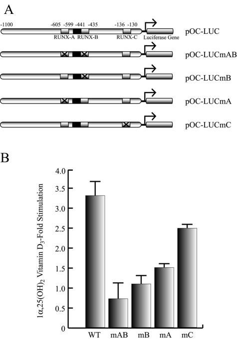
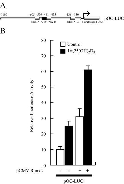
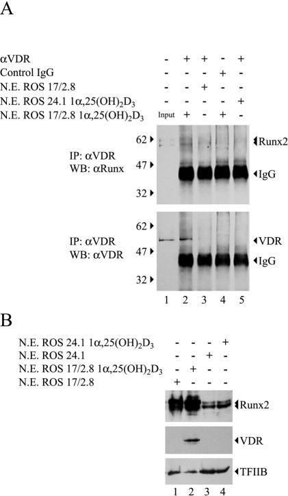
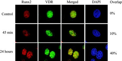
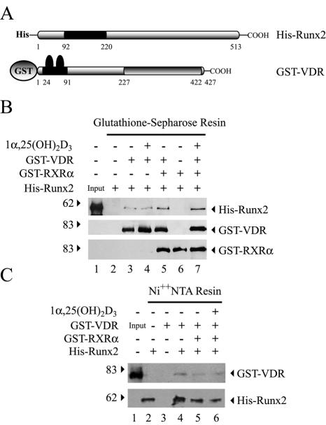
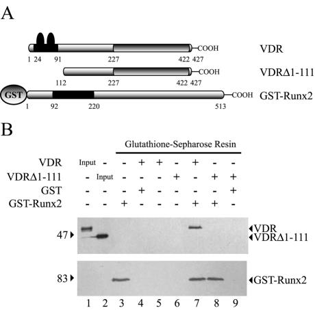
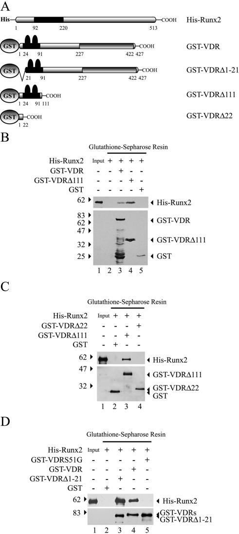
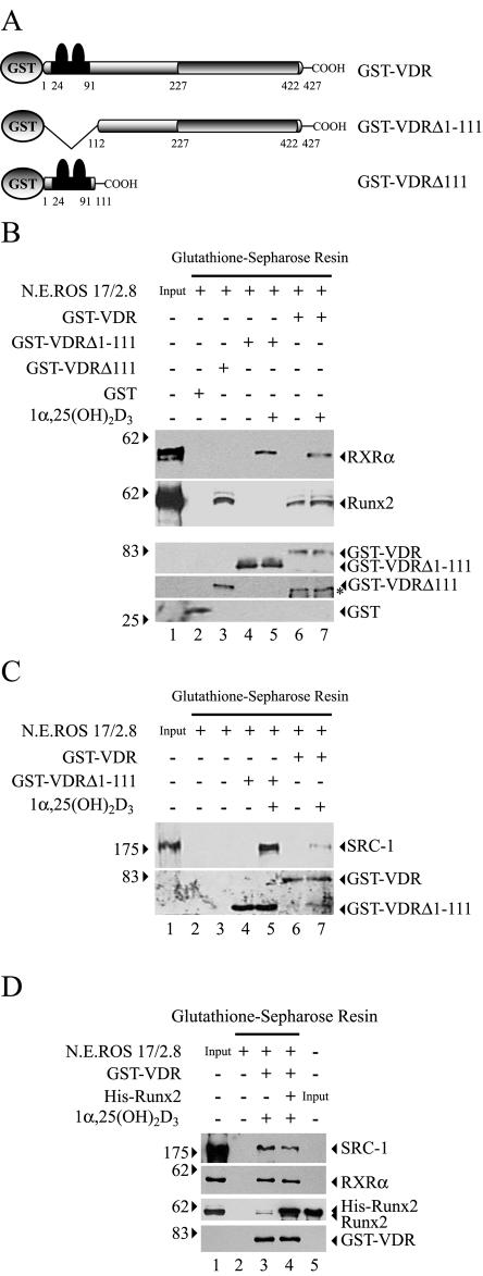
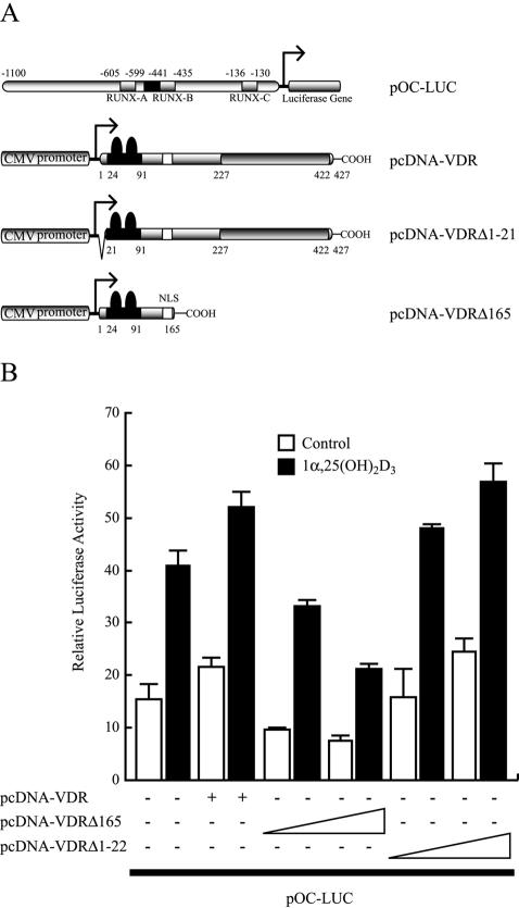
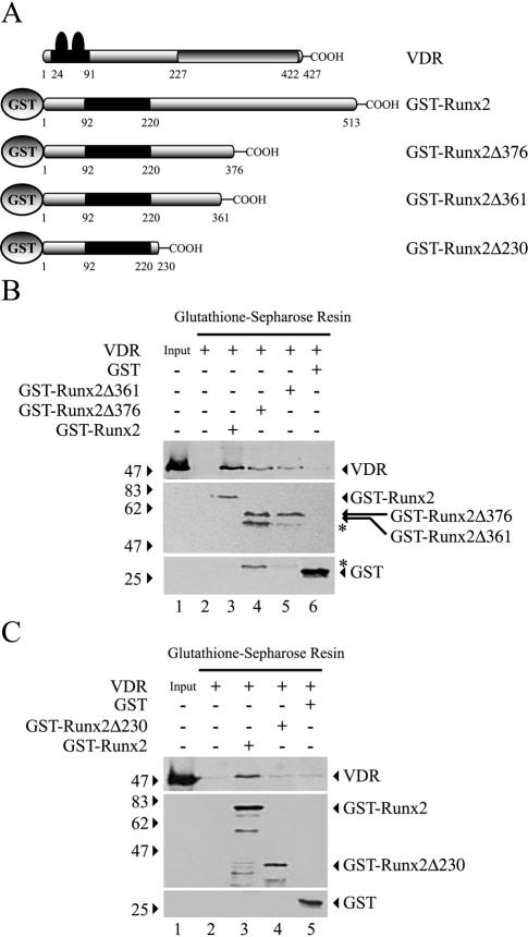
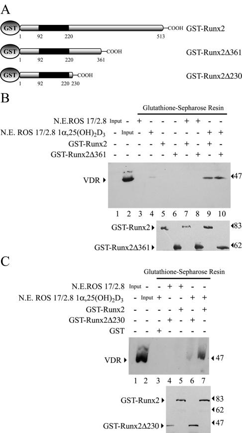
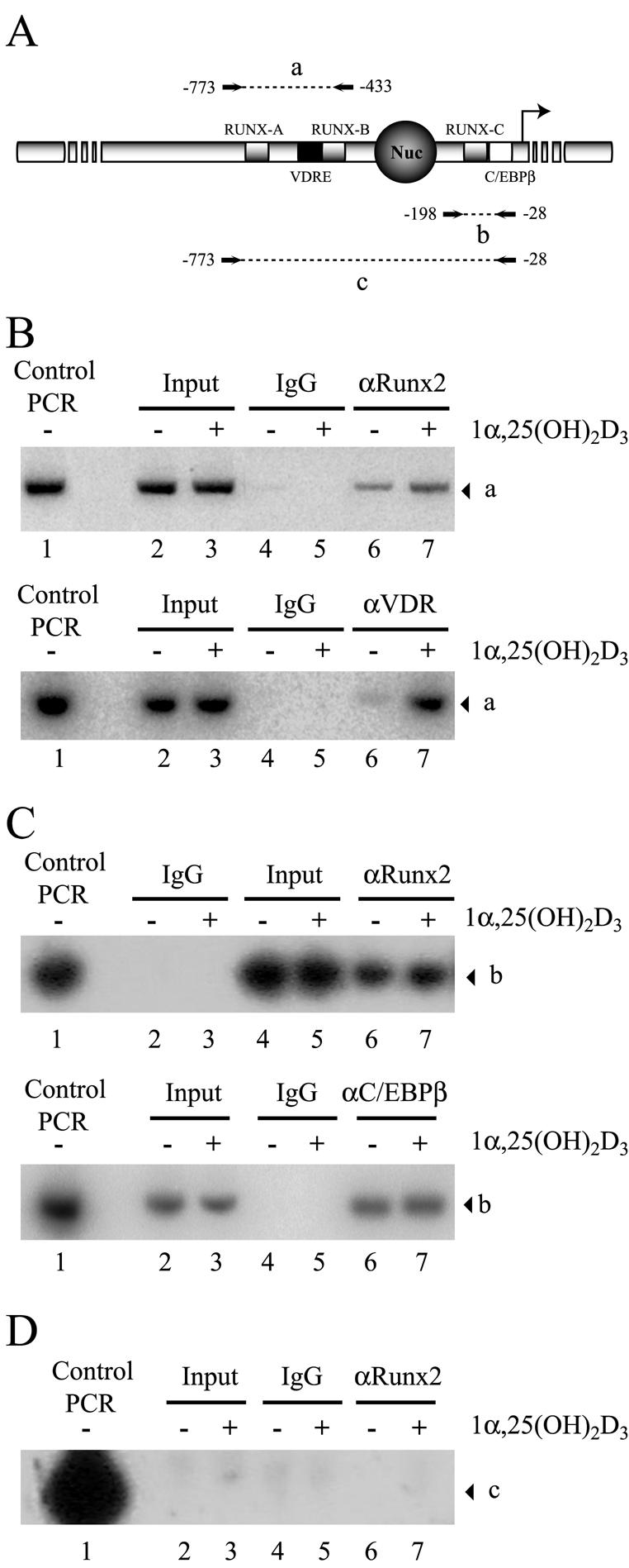
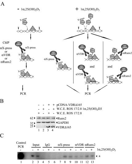
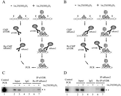
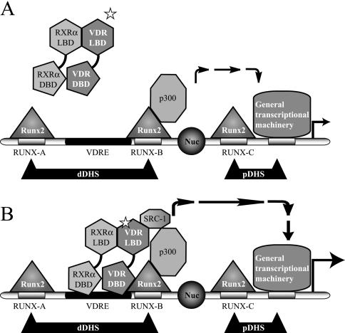
Bone-specific transcription of the osteocalcin (OC) gene is regulated principally by the Runx2 transcription factor and is further stimulated in response to 1alpha,25-dihydroxyvitamin D3 via its specific receptor (VDR). The rat OC gene promoter contains three recognition sites for Runx2 (sites A, B, and C). Mutation of sites A and B, which flank the 1alpha,25-dihydroxyvitamin D3-responsive element (VDRE), abolishes 1alpha,25-dihydroxyvitamin D3-dependent enhancement of OC transcription, indicating a tight functional relationship between the VDR and Runx2 factors. In contrast to most of the members of the nuclear receptor family, VDR possesses a very short N-terminal A/B domain, which has led to the suggestion that its N-terminal region does not contribute to transcriptional enhancement. Here, we have combined transient-overexpression, coimmunoprecipitation, in situ colocalization, chromatin immunoprecipitation, and glutathione S-transferase pull-down analyses to demonstrate that in osteoblastic cells expressing OC, VDR interacts directly with Runx2 bound to site B, which is located immediately adjacent to the VDRE. This interaction contributes significantly to 1alpha,25-dihydroxyvitamin D3-dependent enhancement of the OC promoter and requires a region located C terminal to the runt homology DNA binding domain of Runx2 and the N-terminal region of VDR. Together, our results indicate that Runx2 plays a key role in the 1alpha,25-dihydroxyvitamin D3-dependent stimulation of the OC promoter in osteoblastic cells by further stabilizing the interaction of the VDR with the VDRE. These studies demonstrate a novel mechanism for combinatorial control of bone tissue-specific gene expression. This mechanism involves the intersection of two major pathways: Runx2, a "master" transcriptional regulator of osteoblast differentiation, and 1alpha,25-dihydroxyvitamin D3, a hormone that promotes expression of genes associated with these terminally differentiated bone cells.


Figures

FIG. 1.
FIG. 1.
The Runx2 sites are required for 1α,25-dihydroxyvitamin D3-dependent enhancement of the OC promoter. (A) Schematic representation of various constructs containing wild-type or mutated versions of the rat OC gene promoter controlling the luciferase reporter gene. Each Runx2 site is indicated with an open box, and the mutated Runx2 sites are marked with an X. The VDRE is marked with a black box. pOC-LUC, wild-type rat OC gene promoter; pOC-LUCmAB, rat OC gene promoter with mutated Runx2 sites A and B; pOC-LUCmB, rat OC gene promoter with mutated Runx2 site B; pOC-LUCmA, rat OC gene promoter with mutated Runx2 site A; pOC-LUCmC, rat OC gene promoter with mutated Runx2 site C. (B) Each pOC-LUC construct (1 μg) was transiently transfected into ROS 17/2.8 cells, and luciferase activity was measured to evaluate responsiveness to 24 h of 1α,25-dihydroxyvitamin D3 [1α,25-(OH)2 vitamin D3] treatment. The effect of 1α,25-dihydroxyvitamin D3 is reported as fold stimulation of basal promoter activity levels. All transfections were normalized by cotransfection of the pCMV-β-galactosidase expression plasmid and represent several independent experiments, each assayed in triplicate. Each bar represents the mean ± standard error of the mean (n = 3; P < 0.03). WT, wild type.
FIG. 2.
FIG. 2.
Runx2 up-regulates basal and 1α,25-dihydroxyvitamin D3-enhanced OC gene promoter activity. (A) Schematic representation of the construct containing the rat OC gene promoter controlling the luciferase reporter gene (pOC-LUC) utilized in this analysis. See the legend to Fig. 1 for an explanation of the symbols. (B) ROS 17/2.8 cells were transiently cotransfected with a Runx2 expression plasmid (2 μg) and the pOC-LUC reporter construct (1 μg). The presence (+) or absence (−) of the Runx2 plasmid is shown below the bars. Luciferase activity was assayed after culturing the cells for 24 h in the presence (black bars) or absence (white bars) of 10−8 M 1α,25-dihydroxyvitamin D3 [1α,25-(OH)2D3]. The data were normalized to values for pCMV-β-galactosidase activity as an internal control. Each bar represents the mean ± standard error of the mean (n = 6; P < 0.03).
FIG. 3.
FIG. 3.
Runx2 and VDR are components of the same nuclear protein complexes in osteoblastic cells expressing OC. (A) Nuclear extracts (N.E.) (200 μg) from ROS 17/2.8 or ROS 24/1 cells cultured in the presence of 10−8 M 1α,25-dihydroxyvitamin D3 [1α,25-(OH)2D3] for 4 h were immunoprecipitated (IP) with an anti-VDR (αVDR) monoclonal antibody (Oncogene) or with nonspecific mouse immunoglobulin G fraction (Santa Cruz Biotechnology). Immunoprecipitated complexes were fractionated by SDS-PAGE (8% acrylamide) and analyzed by Western blotting (WB) with anti-Runx2 (upper panel) or anti-VDR (lower panel) antibodies (Santa Cruz Biotechnology). (B) Aliquots (15 μg) of N.E. samples from the ROS 17/2.8 or ROS 24.1 cells shown in panel A were analyzed by Western blotting with anti-Runx2 (upper panel) or anti-VDR (middle panel) antibodies. To control for equal loading, the membrane was reblotted against TFIIB (lower panel). The different combinations of antibodies and N.E. included in each lane are indicated at the top of the blots. The positions of molecular markers are shown to the left of the blots.
FIG. 4.
FIG. 4.
Runx2 and VDR colocalize in the nuclei of osteoblastic cells. Untreated ROS 17/2.8 cells (control) or cells treated with 1α,25-dihydroxyvitamin D3 were processed for in situ immunofluorescence as described in Materials and Methods. A weak nuclear-cytoplasmic signal for VDR is detected in the control cells, while Runx2 exhibits a characteristic punctate nuclear staining. The overlap between the two signals is undetectable as assessed by the measure colocalization function of Metamorph software (top panels). The treatment of cells with 1α,25-dihydroxyvitamin D3 for 45 min (middle panels) or 24 h (bottom panels) significantly increases nuclear staining for the VDR and the subsequent overlap with the Runx2 signal (10 and 40% overlap, respectively). The DAPI panel shows the stained nuclei in the field.
FIG. 5.
FIG. 5.
Runx2 and VDR interact in vitro. Full-length Runx2 and VDR were expressed as His- and GST-tagged fusion proteins, respectively, in bacteria, and their ability to interact in vitro was evaluated by pull-down assays. (A) Schematic representation of the His-Runx2 and GST-VDR recombinant proteins. The runt homology domain of Runx2, localized between amino acid residues 92 and 220, is indicated with a black box. The zinc finger motif of VDR (DBD), localized between amino acid residues 24 and 91, and the LBD of VDR, localized between residues 227 and 422, are also indicated. (B) GST-VDR (1.5 μg) bound to 20 μl of glutathione-Sepharose resin was incubated with His-Runx2 (1.5 μg) and/or GST-RXRα (1.5 μg) for 2 h at 4°C. Precipitated proteins were then analyzed by Western blotting with anti-Runx2 (upper panel) and anti-VDR (lower panel) antibodies. (C) His-Runx2 (1.5 μg) bound to 20 μl of Ni2+-nitrilotriacetic acid (NTA) resin was incubated with GST-VDR (1.5 μg) and/or GST-RXRα (1.5 μg) for 2 h at 4°C. Precipitated proteins were analyzed by Western blotting with specific antibodies against VDR (upper panel) and Runx2 (lower panel). The different combinations of recombinant proteins and the presence of 10−8 M 1α,25-dihydroxyvitamin D3 [1α,25-(OH)2D3] are indicated at the top of each panel. The positions of molecular mass markers are shown at the left of the blots.
FIG. 6.
FIG. 6.
VDR requires a specific domain to interact with Runx2. (A) Schematic representation of the GST-Runx2 and GST-free VDR recombinant proteins utilized in these GST pull-down experiments. See the legend to Fig. 5 for an explanation of the symbols. The VDRΔ1-111 protein corresponds to a mutated VDR form in which amino acid residues 1 to 111, which include the zinc finger domain, have been deleted. (B) GST-Runx2 (1.5 μg) bound to 20 μl of glutathione-Sepharose beads was incubated with 1.5 μg of either GST-free VDR or GST-free VDRΔ1-111 for 2 h at 4°C. Precipitated VDR (upper panel) and GST-Runx2 (lower panel) proteins were then determined by Western blotting. The combinations of proteins used in each binding reaction are indicated at the top of the blots. The positions of molecular mass markers are shown to the left of the blots.
FIG. 7.
FIG. 7.
The N terminus of VDR is sufficient for interaction with Runx2. (A) Schematic representation of the His-Runx2 and GST-VDR recombinant proteins utilized in these GST pull-down experiments. See the legend to Fig. 5 for an explanation of the symbols. The GST-VDRΔ111 protein corresponds to a mutated form of VDR that lacks amino acid residues 112 to 427. The GST-VDRΔ1-21 protein corresponds to a mutated form of VDR that lacks amino acid residues 1 to 21. The GST-VDRΔ22 protein corresponds to a mutated form of VDR that lacks amino acid residues 23 to 427. (B) GST-VDR (1.5 μg) or GST-VDRΔ111 (1.5 μg) bound to glutathione-Sepharose beads were incubated with 1.5 μg of His-Runx2 for 2 h at 4°C. Precipitated Runx2 (upper panel) and GST-VDR (lower panel) proteins were visualized by Western blotting with anti-Runx2 and anti-GST antibodies, respectively. (C) GST-VDRΔ111 (1.5 μg) and GST-VDRΔ22 (1.5μg) bound to glutathione-Sepharose beads were incubated with 1.5 μg of His-Runx2 for 2 h at 4°C. Precipitated Runx2 (upper panel) and GST-VDR (lower panel) proteins were visualized by Western blotting with anti-Runx2 and anti-GST antibodies, respectively. (D) GST-VDRS51G corresponds to a mutated VDR protein in which serine residue 51 has been changed to glycine. GST-VDRS51G (1.5 μg), GST-VDR (1.5 μg), and GST-VDRΔ1-21 (1.5 μg) were incubated with His-Runx2 protein and analyzed as described above. The combinations of proteins used in each binding reaction are indicated at the top of the blots. The positions of molecular mass markers are shown to the left of the blots.
FIG. 8.
FIG. 8.
Runx2 and VDR are components of multisubunit nuclear complexes in osteoblastic cells. (A) Schematic representation of the GST-VDR proteins used in the GST pull-down assays. See the legend to Fig. 5 for an explanation of the symbols. (B) One hundred fifty micrograms of nuclear extract (N.E.) from ROS 17/2.8 cells was incubated with either GST-VDR, GST-VDRΔ111, or GST-VDRΔ1-111 (1 μg of each) bound to glutathione-Sepharose beads. Binding reactions were carried out in the presence (+) or absence (−) of 10−8 M 1α,25-dihydroxyvitamin D3 [1α,25-(OH)2D3]. The precipitated proteins were resolved by SDS-PAGE (10% acrylamide) and revealed byWestern blotting with specific antibodies against RXRα (upper panel), Runx2 (second panel), VDR (third and fourth panels), or GST (lower panel). The different VDR protein forms were detected as follows: GST-VDR and GST-VDRΔ1-111 (third panel) with a specific antibody against the C terminus of VDR; GST-VDRΔ111 (fourth panel) with an antibody against the N terminus of VDR. The asterisk indicates a cleavage product of the GST-VDR protein. (C) Aliquots of the precipitated protein samples shown in panel B were resolved by SDS-PAGE (7% acrylamide) and analyzed by Western blotting detecting the presence of SRC-1 (upper panel) as well as GST-VDR and GST-VDRΔ1-111 (lower panel). (D) One hundred fifty micrograms of nuclear extract from ROS 17/2.8 cells was enriched in Runx2 by adding 2 μg of purified His-Runx2 (lanes 3 and 4). The samples were then incubated with GST-VDR and analyzed by Western blotting as described above. The combinations of proteins used in each binding reaction are indicated at the top of the blots. The positions of molecular mass markers are shown to the left of the blots.
FIG. 9.
FIG. 9.
Expression of the VDRΔ165 domain inhibits OC promoter activity in osteoblastic cells. (A) Schematic representation of the construct carrying the full-length OC promoter in front of the luciferase reporter gene (pOC-LUC) and the expression plasmids encoding wild-type VDR (pcDNA-VDR) and the N-terminal domain of VDR (pcDNAΔ165). (B) pOC-LUC and VDR expression plasmids were cotransfected in ROS 17/2.8 cells cultured in the presence (black bars) or absence (white bars) of 1α,25-dihydroxyvitamin D3 [1α,25-(OH)2D3] for 24 h. The presence or absence of each plasmid is shown below the bars. pOC-LUC was added at a concentration of 1 μg/well (35 mm), whereas pcDNAΔ165 and pcDNA-VDR were added at 0.25 and 0.5 μg/well. Luciferase activity was evaluated as described in the legend to Fig. 1. Each bar represents the mean ± standard error of the mean (n = 6; P < 0.03).
FIG. 10.
FIG. 10.
Runx2 requires a specific domain to interact with VDR. (A) Schematic representation of the GST-Runx2 and VDR proteins used in the GST pull-down assays. GST-Runx2Δ376, GST-Runx2Δ361, and GST-Runx2Δ230 correspond to mutated Runx2 proteins that lack amino acid residues 377 to 513, 362 to 513, and 231 to 513, respectively. (B) A 1.5-μg amount (each) of GST-Runx2, GST-Runx2Δ376, and GST-Runx2Δ361 proteins bound to glutathione-Sepharose beads was incubated with 1.5 μg of GST-free VDR. The precipitated VDR (upper panel) and Runx2 (middle panel) proteins were determined by Western blotting with specific antibodies. The asterisks mark cleavage products of GST-Runx2Δ376. (C) GST pull-down assays were repeated as for panel B but with GST-Runx2 and GST-Runx2Δ230, which lacks most of the C-terminal region of Runx2. The precipitated proteins were detected by Western blotting as indicated for panel B. The combinations of proteins used in each binding reaction are indicated at the top of the blots. The positions of molecular mass markers are shown to the left of the blots.
FIG. 11.
FIG. 11.
GST-Runx2 interacts with nuclear VDR. (A) GST-Runx2 proteins. (B and C) The ability of GST-Runx2 proteins to interact with the VDR present in nuclear extracts (N.E.) from ROS 17/2.8 cells treated with 10−8 M 1α,25-dihydroxyvitamin D3 [1α,25-(OH)2D3] for 4 h or with vehicle was evaluated by GST pull-down assays. (B) GST-Runx2 and GST-Runx2Δ361 (1.5 μg each) bound to Sepharose beads (lower panel) were incubated with 150 μg of nuclear extracts from ROS 17/2.8 cells for 2 h at 4°C. The precipitated VDR was then detected by Western blotting (upper panel). (C) Similarly, GST-Runx2 and GST-Runx2Δ230 (1.5 μg each) (lower panel) were incubated with 150 μg of nuclear extracts from ROS 17/2.8 cells, and the precipitated VDR was detected by Western blotting (upper panel). The combinations of proteins used in each binding reaction are marked at the top of the blots. The positions of molecular mass markers are shown to the left of the blots.
FIG. 12.
FIG. 12.
Enhanced binding of Runx2 and VDR to the distal OC promoter in 1α,25-dihydroxyvitamin D3-treated osteoblastic cells. (A) Schematic representation of the rat OC gene promoter, indicating the relative positions of binding sites for VDR, Runx2, and C/EBPβΒ transcription factors. The combination of primers used in the PCR amplification of immunoprecipitated DNA fragments is indicated, and the amplified fragments are marked by letters a (−773 to −433), b (−198 to −28), and c (−773 to −28). Nuc, positioned nucleosome. (B to D) ChIP of formaldehyde-cross-linked chromatin from ROS17/2.8 cells cultured in the presence (+) or absence (−) of 1α,25-dihydroxyvitamin D3 [1α,25-(OH)2D3], using anti-Runx2 antibody (B [upper panel], C [upper panel], and D), anti-VDR (B [lower panel]), and anti-C/EBPβ (C [lower panel]). Control PCR indicates the signal obtained when the pOC-3.4 plasmid, containing 1.1 kb of rat OC promoter, was used as a template. IgG, immunoglobulin G.
FIG. 13.
FIG. 13.
The VDRΔ165 domain binds to the OC promoter and competes with VDR for interaction with the OC VDRE. (A) Schematic representation of the ChIP analysis at the OC gene promoter in cells that overexpress the VDRΔ165 domain (VDR DBD). The transcription factors bound to the promoter are indicated, and the white star indicates the ligand 1α,25-dihydroxyvitamin D3 [1α,25-(OH)2D3]. The anti-X-press (αX-press) antibody used in the ChIP and the X-press tag present in the VDR DBD are also indicated. (B) Aliquots (15 μg) of whole cellular extracts (W.C.E.) from ROS 17/2.8 cells transiently transfected with the pcDNA-VDRΔ165 or pcDNA3.1His plasmid were analyzed by Western blotting with anti-Runx2 (upper panel) or anti-VDR (lower panel) antibodies. To control for equal loading, the membrane was reblotted against GAPDH (glyceraldehyde-3-phosphate dehydrogenase) (middle panel). The different combinations of antibodies and W.C.E. included in each lane are indicated at the top of the blots. The positions of molecular size markers are shown to the left of the blots. (C) ChIP of formaldehyde-cross-linked chromatin from ROS17/2.8 cells that overexpress the VDRΔ165 domain (lanes 4, 5, and 8 to 13) cultured in the presence (+) or absence (−) of 1α,25-dihydroxyvitamin D3, using anti X-press antibody (lanes 6 to 9), anti-VDR antibody (lanes 10 and 11), and anti-Runx2 antibody (lanes 12 and 13). ROS17/2.8 cells transiently transfected with the pcDNA3.1His empty plasmid were used as a negative control for ChIP with anti-X-press antibody (lanes 6 and 7). Control PCR indicates the signal obtained when the pOC-3.4 plasmid, containing 1.1 kb of rat OC promoter, was used as a template. The DNA fragment corresponding to the distal OC gene promoter amplified by PCR is marked by the letter a (see Fig. 12A for an illustration). IgG, immunoglobulin G.
FIG. 14.
FIG. 14.
Runx2 and VDR bind simultaneously to the distal OC promoter in osteoblastic cells treated with 1α,25-dihydroxyvitamin D3. (A and B) Schematic representation of the Re-ChIP analysis at the rat OC gene promoter. The different transcription factors bound to the promoter are indicated, and the white star indicates the ligand 1α,25-dihydroxyvitamin D3 [1α,25-(OH)2D3]. The antibodies used in each ChIP step are indicated at the left of each panel. (C and D) Re-ChIP of formaldehyde-cross-linked chromatin from ROS17/2.8 cells cultured in the presence (+) or absence (−) of 1α,25-dihydroxyvitamin D3, using anti-VDR (αVDR) antibody followed by anti-Runx2 antibody (C) or anti-Runx2 antibody followed by anti-VDR antibody (D). Control PCR indicates the signal obtained when the pOC-3.4 plasmid, containing 1.1 kb of rat OC promoter, was used as a template. The DNA fragment corresponding to the distal OC gene promoter amplified by PCR is marked by the letter a. IgG, immunoglobulin G.
FIG. 15.
FIG. 15.
Schematic view of OC gene promoter transcribing at basal levels (A) or enhanced by 1α,25-dihydroxyvitamin D3 (B). The OC gene promoter and its various regulatory elements are represented by different shadings. The circle indicates a positioned nucleosome (Nuc) flanked by a distal and proximal DNase I-hypersensitive sites (dDHS and pDHS). The different transcription factors bound to the promoter are indicated, and the white star indicates the ligand 1α,25-dihydroxyvitamin D3. The stimulatory effect of the transcription factors on the general transcription machinery is represented by arrows.

Similar articles

Cited by

References

    1. Banerjee, C., L. R. McCabe, J. Y. Choi, S. W. Hiebert, J. L. Stein, G. S. Stein, and J. B. Lian. 1997. Runt homology domain proteins in osteoblast differentiation: AML3/CBFA1 is a major component of a bone-specific complex. J. Cell Biochem. 66:1-8. - PubMed
    1. Breen, E. C., A. J. van Wijnen, J. B. Lian, G. S. Stein, and J. L. Stein. 1994. In vivo occupancy of the vitamin D responsive element in the osteocalcin gene supports vitamin D-dependent transcriptional upregulation in intact cells. Proc. Natl. Acad. Sci. USA 91:12902-12906. - PMC - PubMed
    1. Christakos, S., P. Dhawan, Y. Liu, X. Peng, and A. Porta. 2003. New insights into the mechanisms of vitamin D action. J. Cell Biochem. 88:695-705. - PubMed
    1. Ducy, P., R. Zhang, V. Geoffroy, A. L. Ridall, and G. Karsenty. 1997. Osf2/Cbfa1: a transcriptional activator of osteoblast differentiation. Cell 89:747-754. - PubMed
    1. Gutierrez, J., J. Sierra, R. Medina, M. Puchi, M. Imschenetzky, A. van Wijnen, J. Lian, G. Stein, J. Stein, and M. Montecino. 2000. Interaction of CBF alpha/AML/PEBP2 alpha transcription factors with nucleosomes containing promoter sequences requires flexibility in the translational positioning of the histone octamer and exposure of the CBF alpha site. Biochemistry 39:13565-13574. - PubMed

Publication types

MeSH terms

LinkOut - more resources