. 2004 Oct;24(20):8963-9.
doi: 10.1128/MCB.24.20.8963-8969.2004.

RNA polymerase II carboxy-terminal domain phosphorylation is required for cotranscriptional pre-mRNA splicing and 3'-end formation

Affiliations

RNA polymerase II carboxy-terminal domain phosphorylation is required for cotranscriptional pre-mRNA splicing and 3'-end formation

Gregory Bird et al. Mol Cell Biol. 2004 Oct.

Abstract

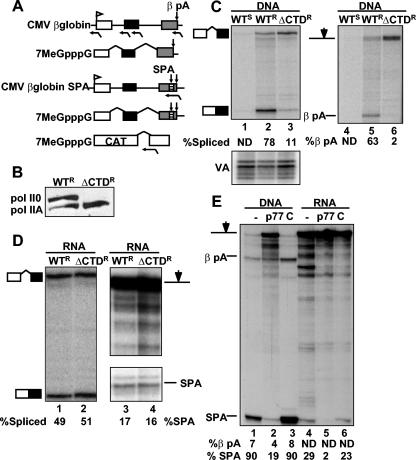
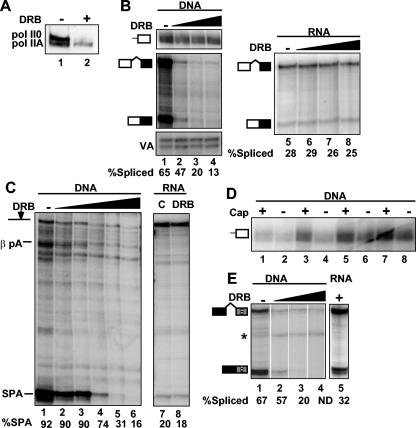
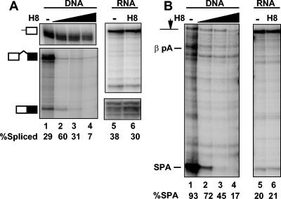
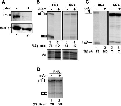
We investigated the role of RNA polymerase II (pol II) carboxy-terminal domain (CTD) phosphorylation in pre-mRNA processing coupled and uncoupled from transcription in Xenopus oocytes. Inhibition of CTD phosphorylation by the kinase inhibitors 5,6-dichloro-1beta-D-ribofuranosyl-benzimidazole and H8 blocked transcription-coupled splicing and poly(A) site cleavage. These experiments suggest that pol II CTD phosphorylation is required for efficient pre-mRNA splicing and 3'-end formation in vivo. In contrast, processing of injected pre-mRNA was unaffected by either kinase inhibitors or alpha-amanitin-induced depletion of pol II. pol II therefore does not appear to participate directly in posttranscriptional processing, at least in frog oocytes. Together these experiments show that the influence of the phosphorylated CTD on pre-mRNA splicing and 3'-end processing is mediated by transcriptional coupling.


Figures

FIG. 1.
FIG. 1.
pol II ΔCTD does not support cotranscriptional pre-mRNA processing and does not inhibit processing uncoupled from transcription in Xenopus oocytes. (A) Maps of the CMV β-globin gene and capped (7MeGpppG) pre-mRNAs with and without synthetic poly(A) site (SPA, striped box) in exon 3. Poly(A) cleavage sites βpA and SPA are marked by downward arrows. Capped CAT pre-mRNA containing the SV40 t-intron is shown at the bottom. Antisense RNase protection probes are marked below the maps. (B) Western blot of Rpb1WTR (WTR) and Rpb1ΔCTDR (ΔCTDR) expressed in oocytes detected with antibody against the B10 epitope tag. Hyper- and hypophosphorylated wild-type pol II0 and pol IIA are indicated. (C) RPA of CMV β-globin transcripts in α-amanitin-injected oocytes expressing α-amanitin-sensitive (WTS) or -resistant Rpb1 with full-length (WTR) or deleted CTD (ΔCTDR) with intron 1 and poly(A) site probes. VA RNA transcribed by pol III is a control for injection efficiency. The CMV β-globin (0.2 ng in 4.6 nl) and VA plasmids were coinjected with α-amanitin 15 h after injection of the Rpb1 expression plasmids (0.2 ng in 4.6 nl). Percent spliced or cleaved relative to total RNA (see Materials and Methods) is noted below lane numbers. Values too small to be accurately determined are labeled ND. (D) RPA of β-globin SPA pre-mRNA coinjected with α-amanitin into oocytes expressing Rpb1WTR or Rpb1ΔCTDR with intron 1 and SPA probes. Note that expression of Rpb1ΔCTDR has no effect on processing uncoupled from transcription. (E) RPA of CMV β-globin SPA (lanes 1 to 3) or injected β-globin SPA transcripts with SPA probe (lanes 4 to 6). Oocytes were coinjected with either no antibody (−), anti-CstF p77 (p77) (5.3 ng/oocyte), or irrelevant control (C) rabbit immunoglobulin G (5.3 ng/oocyte).
FIG. 2.
FIG. 2.
Inhibition of CTD phosphorylation by DRB inhibits cotranscriptional processing but not processing uncoupled from transcription. (A) Western blot of Rpb1 from oocytes preincubated without (−) or with (+) DRB (75 μM) using anti-pol II CTD Ser 5-PO4. (B) RPA of β-globin RNAs from oocytes incubated in 0, 15, 30, and 60 μM DRB and injected with pCMV β-globin SPA or capped β-globin SPA pre-mRNA. 5′ end (lanes 1 to 4, top panel) and intron 1 probes (lanes 1 to 4, middle panel, and lanes 5 to 8) were used for RPA. VA is a control for injection efficiency. (C) RPA of CMV β-globin SPA transcripts from oocytes treated with DRB at 0, 3.75, 7, 15, 30, and 60 μM with SPA probe (lanes 1 to 5). β-globin RNAs from oocytes incubated in 0 and 75 μM DRB and injected capped β-globin SPA pre-mRNA are also shown (lanes 6 and 7). (D) RPA of capped (+) and uncapped (−) transcripts from panel B selected by GST-eIF4E binding with 5′ CMV β-globin probe (amount of DRB in lanes 1 and 2, 0 μM; lanes 3 and 4, 15 μM; lanes 5 and 6, 30 μM; lanes 7 and 8, 60 μM). (E) Cap-selected transcripts from the RPA shown in panel B were assessed for splicing of β-globin intron 2. Note that capped transcripts made in 30 and 60 μM DRB are predominantly unspliced (lanes 3 and 4), whereas those made with in the absence of DRB (lane 1) are predominantly spliced. Capped β-globin pre-mRNA was injected into oocytes treated with 75 μM DRB (lane 6). The asterisk indicates an irrelevant undigested probe.
FIG. 3.
FIG. 3.
The protein kinase inhibitor H8 specifically inhibits pre-mRNA processing coupled to transcription. (A) RPA of β-globin intron 1 splicing from oocytes injected with CMV β-globin SPA and treated with 0, 25, 50, or 100 μg of H8/ml (lanes 1 to 4). RPA of capped β-globin pre-mRNA injected into oocyte treated with 0 or 200 μg of H8/ml (lanes 5 and 6). (B) RPA of SPA site cleavage with the same RNA samples as those used for the experiment depicted in panel A.
FIG. 4.
FIG. 4.
Degradation of RNA pol II by α-amanitin does not inhibit pre-mRNA processing uncoupled from transcription. (A) Western blot of total protein from oocytes with (+) and without (−) α-amanitin (α-Am) injection. The blot was probed with anti-pol II 8WG16 and anti-CstF p77 as a loading control. (B) RPA of β-globin intron 2 splicing from oocytes injected with either CMV β-globin DNA or β-globin pre-mRNA with (+) or without (−) preinjection of α-amanitin 15 h beforehand. VA is a control for injection efficiency. (C) RPA of β-globin poly(A) site cleavage with the same RNA samples as those used for the experiment shown in panel B. (D) RPA of SV40 t intron splicing in oocytes injected with capped CAT pre-mRNA (Fig. 1A) with (+) or without (−) preinjection of α-amanitin. ND, not determined.

References

    1. Ahn, S. H., M. Kim, and S. Buratowski. 2004. Phosphorylation of serine 2 within the rna polymerase II C-terminal domain couples transcription and 3′ end processing. Mol. Cell. 13:67-76. - PubMed
    1. Bauren, G., S. Belikov, and L. Wieslander. 1998. Transcriptional termination in the Balbiani ring 1 gene is closely coupled to 3′-end formation and excision of the 3′-terminal intron. Genes Dev. 12:2759-2769. - PMC - PubMed
    1. Bauren, G., and L. Wieslander. 1994. Splicing of Balbiani ring 1 gene pre-mRNA occurs simultaneously with transcription. Cell 76:183-192. - PubMed
    1. Bentley, D. 2002. The mRNA assembly line: transcription and processing machines in the same factory. Curr. Opin. Cell Biol. 14:336-342. - PubMed
    1. Beyer, A. L., and Y. N. Osheim. 1988. Splice site selection, rate of splicing, and alternative splicing on nascent transcripts. Genes Dev. 2:754-765. - PubMed

Publication types

LinkOut - more resources