. 2004 Oct;24(20):9006-18.
doi: 10.1128/MCB.24.20.9006-9018.2004.

c-Jun-deficient cells undergo premature senescence as a result of spontaneous DNA damage accumulation

Affiliations

c-Jun-deficient cells undergo premature senescence as a result of spontaneous DNA damage accumulation

Ann MacLaren et al. Mol Cell Biol. 2004 Oct.

Abstract

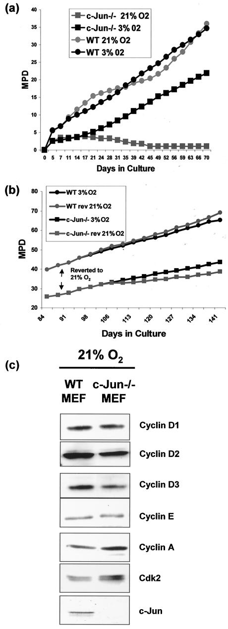
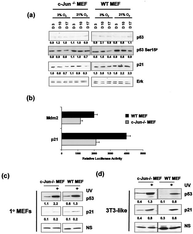
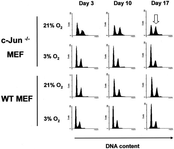
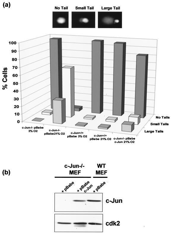
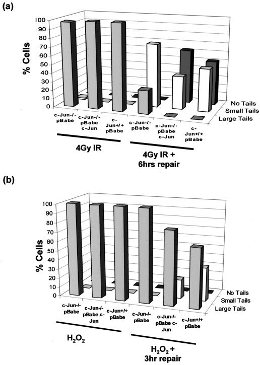
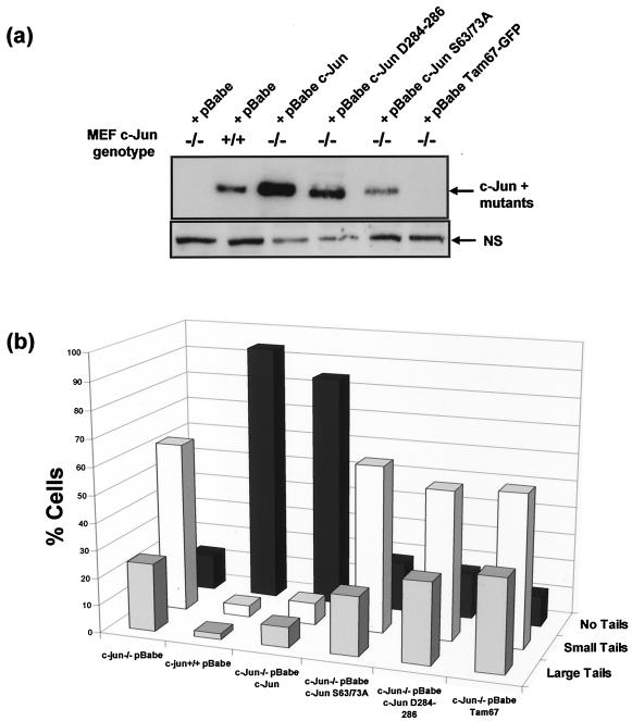
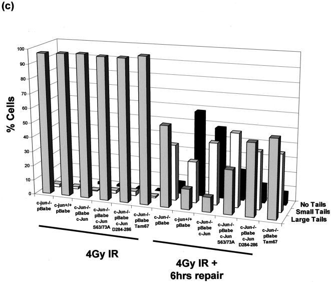
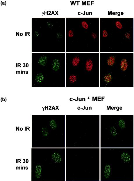
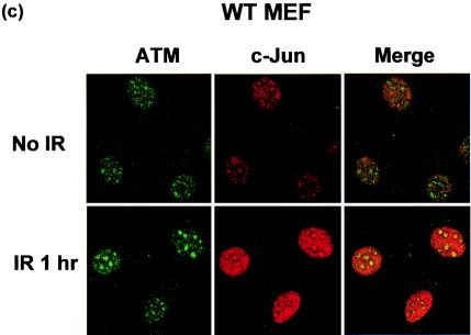
Mouse embryo fibroblasts deficient for the c-Jun proto-oncogene (c-Jun-/- MEF) undergo p53-dependent premature senescence in conventional culture. This phenotype becomes evident only after several cell divisions, suggesting that senescence may result from exposure to unknown environmental factors. Here, we show that c-Jun-/- MEF can proliferate successfully in low oxygen (3% O2), indicating that premature senescence under conventional culture conditions is a consequence of hyperoxic stress. c-Jun-/- MEF exhibit higher basal levels of DNA damage compared to normal fibroblasts in high but not low oxygen, implying that senescence results from chronic accumulation of spontaneous DNA damage. This accumulation may be attributable, at least in part, to inefficient repair, since DNA damage induced by gamma ionizing radiation and H2O2 persists for longer in c-Jun-/- MEF than in wild-type MEF. Unexpectedly, p53 expression, phosphorylation, and transcriptional activity are largely unaffected by oxygen exposure, indicating that the accumulation of spontaneous DNA damage does not result in chronic activation of p53 as judged by conventional criteria. Finally, we find that c-Jun associates with nuclear foci containing gammaH2AX and ATM following irradiation, suggesting a potential role for c-Jun in DNA repair processes per se.


Figures

FIG. 1.
FIG. 1.
Premature senescence associated with c-Jun deficiency can be rescued by culture at low oxygen. (a) WT (filled circles) and c-Jun−/− (filled squares) MEF were cultured at either 21% oxygen (grey symbols) or 3% oxygen (black symbols) continuously for 70 days (24 passages). Cells were split every 3 days, and the total numbers ofcells were counted and mean population doublings (MPD) were determined. Following isolation at 3% oxygen, cells were grown until 2 × 10⁵ cells of each genotype were available (approximately 3 to 4 days), and then 1 × 10⁵ cells of each genotype were plated at either high or low oxygen and assigned an MPD of 0. (b) WT (filled circles) and c-Jun−/− (filled squares) MEF cultured at low oxygen were split into two aliquots after 90 days of culture; one flask was subsequently passaged at 21% oxygen (grey symbols), and the other was passaged at 3% oxygen (black symbols). Cells were split every 3 days, and the total numbers of cells were counted and MPD were determined. (c) Western blotting analysis of cyclin D1, D2, D3, E, A, Cdk2, and c-Jun expression in cell extracts prepared from early-passage WT and c-Jun−/− MEF cultured in 21% O2.
FIG. 2.
FIG. 2.
c-Jun deficiency does not alter p53 modification or function. (a) Early-passage c-Jun−/− and WT MEF were cultured at either 3 or 21% oxygen, and samples were harvested for protein analysis at days 3, 10, and 17 (i.e., passage 2 [P2], P4, and P6). Whole-cell extracts were prepared and analyzed by SDS-PAGE and Western blotting for p53, Ser15p p53, p21, and Erk expression. Band intensities were quantified by laser densitometry, and relative expression levels were calculated in arbitrary units (shown below the corresponding bands), using ERK values to correct for variations in protein loading. (b) Mdm2 and p21 luciferase constructs were transfected into early-passage WT and c-Jun−/− MEF cultured at 21% O2, and luciferase activity was measured 48 h later. Results represent the means of three independent transfections ± standard errors of the means. (c) Early-passage 1° MEF (c) and late passage 3T3-like cell lines generated from these cultures (d) were cultured in 21% O2 and either not treated or treated with 80 J of UV/m2. Cells were harvested 8 h following UV treatment, and whole-cell extracts were prepared. Following SDS-PAGE analysis, membranes were probed for either p53 or p21 expression. Relative expression levels were quantified and calculated in arbitrary units (shown below the corresponding bands) as for panel a, using a nonspecific band (NS) to correct for variations in protein loading.
FIG. 3.
FIG. 3.
Lack of c-Jun results in an accumulation in G2/M. Early-passage c-Jun−/− and WT MEF were cultured at either 3 or 21% oxygen over a 17-day period as described in the legend to Fig. 2a. Subconfluent cultures of cells were harvested at days 3, 10, and 17 (i.e., P2, P4, and P6) and fixed prior to being stained with propidium iodide. DNA content was analyzed by flow cytometry. The open arrow indicates enrichment of G2/M cells in c-Jun−/− cultures after 17 days of culture.
FIG. 4.
FIG. 4.
c-Jun deficiency results in increased levels of spontaneous DNA damage. (a) c-Jun−/− MEF, c-Jun−/− MEF reconstituted with ectopic c-Jun, and WT MEF were cultured at either 3 or 21% oxygen for 4 days prior to analysis. For control purposes the WT and c-Jun−/− MEF were infected with the same empty retroviral vector (pBabe) used to express ectopic c-Jun in the reconstituted cells. A denaturing comet assay was performed, and DNA was stained and examined by fluorescence microscopy. (b) Western blotting analysis of c-Jun expression levels in the cell cultures used for panel a.
FIG. 5.
FIG. 5.
Prolonged persistence of DNA damage induced by IR and H2O2 in c-Jun-deficient MEF. (a) A neutral comet assay was performed on c-Jun−/− MEF, c-Jun−/− cells reconstituted with ectopic c-Jun, and WT MEF cultured at 3% O2. Cells were treated with 4Gy of IR to induce double-strand breaks and harvested immediately or left for 6 h to repair damage prior to analysis. Cells were scored as having no tail, small tails, or large tails, and results were plotted as the percentage of cells in each group. The results are representative of three independent experiments. (b) Denaturing comet analysis of c-Jun−/− MEF, c-Jun−/− MEF reconstituted with ectopic c-Jun, and WT MEF treated with 100 μM H2O2 for 15 min. Cells were either harvested immediately or washed and incubated in fresh medium for 3 h to allow repair prior to analysis.
FIG. 6.
FIG. 6.
Restoration of normal DNA damage levels requires functional c-Jun DNA binding and transactivation domains. (a) WT and c-Jun−/− MEF were transduced with an empty retroviral vector (pBabe) or a vector encoding either c-Jun, c-Jun mutant D284-286 or S63/73A, or Tam67-GFP. Protein extracts were prepared from selected cultures and analyzed by Western blotting for expression of endogenous and ectopic c-Jun proteins. (b) WT or c-Jun−/− MEF reconstituted with c-Jun or the indicated mutants were cultured in 21% O2 for 7 days, after which the cells were harvested and analyzed for basal levels of DNA damage using a denaturing comet assay and scored by fluorescence microscopy. (c) WT or c-Jun−/− MEF reconstituted with c-Jun or the indicated mutants cultured in 3% O2 were irradiated with 4 Gy of IR, after which the cells were either harvested immediately or left for 6 h to allow repair prior to analysis. Cells were analyzed for DNA damage using a neutral comet assay and scored by fluorescence microscopy.
FIG. 6.
FIG. 6.
Restoration of normal DNA damage levels requires functional c-Jun DNA binding and transactivation domains. (a) WT and c-Jun−/− MEF were transduced with an empty retroviral vector (pBabe) or a vector encoding either c-Jun, c-Jun mutant D284-286 or S63/73A, or Tam67-GFP. Protein extracts were prepared from selected cultures and analyzed by Western blotting for expression of endogenous and ectopic c-Jun proteins. (b) WT or c-Jun−/− MEF reconstituted with c-Jun or the indicated mutants were cultured in 21% O2 for 7 days, after which the cells were harvested and analyzed for basal levels of DNA damage using a denaturing comet assay and scored by fluorescence microscopy. (c) WT or c-Jun−/− MEF reconstituted with c-Jun or the indicated mutants cultured in 3% O2 were irradiated with 4 Gy of IR, after which the cells were either harvested immediately or left for 6 h to allow repair prior to analysis. Cells were analyzed for DNA damage using a neutral comet assay and scored by fluorescence microscopy.
FIG. 7.
FIG. 7.
c-Jun colocalizes to sites of DNA damage. WT and c-Jun−/− MEF cultured in 21% O2 for 4 days were treated with 4 Gy of IR and left for 30 min. Cells were then fixed and stained with anti-phosphorylated H2AX (γH2AX; green) and anti-c-Jun (red) antibodies (a and b) or WT MEF were stained with anti-ATM (green) or anti-Jun (red) antibodies (c). Images were analyzed and merged by confocal microscopy and are representative of the majority of cells in the cultures.
FIG. 7.
FIG. 7.
c-Jun colocalizes to sites of DNA damage. WT and c-Jun−/− MEF cultured in 21% O2 for 4 days were treated with 4 Gy of IR and left for 30 min. Cells were then fixed and stained with anti-phosphorylated H2AX (γH2AX; green) and anti-c-Jun (red) antibodies (a and b) or WT MEF were stained with anti-ATM (green) or anti-Jun (red) antibodies (c). Images were analyzed and merged by confocal microscopy and are representative of the majority of cells in the cultures.

Similar articles

Cited by

References

    1. Baus, F., V. Gire, D. Fisher, J. Piette, and V. Dulic. 2003. Permanent cell cycle exit in G2 phase after DNA damage in normal human fibroblasts. EMBO J. 22:3992-4002. - PMC - PubMed
    1. Black, E. J., W. Clark, and D. A. Gillespie. 2000. Transient deactivation of ERK signalling is sufficient for stable entry into G0 in primary avian fibroblasts. Curr. Biol. 10:1119-1122. - PubMed
    1. Burma, S., B. P. Chen, M. Murphy, A. Kurimasa, and D. J. Chen. 2001. ATM phosphorylates histone H2AX in response to DNA double-strand breaks. J. Biol. Chem. 276:42462-42467. - PubMed
    1. Chevray, P. M., and D. Nathans. 1992. Protein interaction cloning in yeast: identification of mammalian proteins that react with the leucine zipper of Jun. Proc. Natl. Acad. Sci. USA 89:5789-5793. - PMC - PubMed
    1. Chiang, Y. C., S. C. Teng, Y. N. Su, F. J. Hsieh, and K. J. Wu. 2003. c-Myc directly regulates the transcription of the NBS1 gene involved in DNA double-strand break repair. J. Biol. Chem. 278:19286-19291. - PubMed

Publication types

MeSH terms

LinkOut - more resources