. 2004 Oct;24(20):9026-37.
doi: 10.1128/MCB.24.20.9026-9037.2004.

Transgenic analysis reveals that thyroid hormone receptor is sufficient to mediate the thyroid hormone signal in frog metamorphosis

Affiliations

Transgenic analysis reveals that thyroid hormone receptor is sufficient to mediate the thyroid hormone signal in frog metamorphosis

Daniel R Buchholz et al. Mol Cell Biol. 2004 Oct.

Abstract

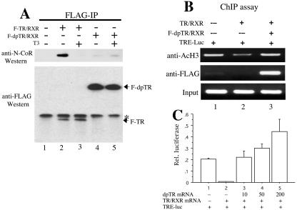
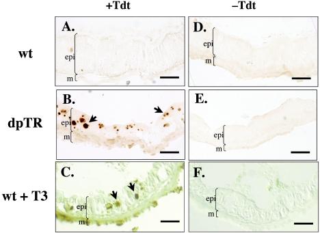
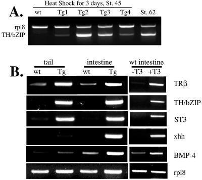
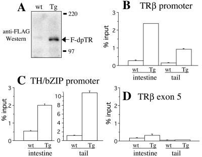
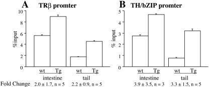
Thyroid hormone (T3) has long been known to be important for vertebrate development and adult organ function. Whereas thyroid hormone receptor (TR) knockout and transgenic studies of mice have implicated TR involvement in mammalian development, the underlying molecular bases for the resulting phenotypes remain to be determined in vivo, especially considering that T3 is known to have both genomic, i.e., through TRs, and nongenomic effects on cells. Amphibian metamorphosis is an excellent model for studying the role of TR in vertebrate development because of its total dependence on T3. Here we investigated the role of TR in metamorphosis by developing a dominant positive mutant thyroid hormone receptor (dpTR). In the frog oocyte transcription system, dpTR bound a T3-responsive promoter and activated the promoter independently of T3. Transgenic expression of dpTR under the control of a heat shock-inducible promoter in premetamorphic tadpoles led to precocious metamorphic transformations. Molecular analyses showed that dpTR induced metamorphosis by specifically binding to known T3 target genes, leading to increased local histone acetylation and gene activation, similar to T3-bound TR during natural metamorphosis. Our experiments indicated that the metamorphic role of T3 is through genomic action of the hormone, at least on the developmental parameters tested. They further provide the first example where TR is shown to mediate directly and sufficiently these developmental effects of T3 in individual organs by regulating target gene expression in these organs.


Figures

FIG. 1.
FIG. 1.
The fusion of the RID of N-CoR to a dominant negative TR leads to partial derepression. (A) Diagram of TR fusion proteins. The fusion constructs F-TR-l and F-TR-s include a long and short fragment of Xenopus N-CoR containing the RID. The black boxes labeled 3, 2, and 1 represent the corepressor nuclear receptor binding motifs (I/LXXI/VI) of N-CoR that directly bind TR (21). The dominant negative Xenopus TRα (dnTRα) part of the fusion protein corresponds to Xenopus TRα without the terminal helix 12 (3, 44). Both fusion proteins also contain an N-terminal FLAG tag. (B) The fusion proteins have reduced abilities to repress T3 target genes in the frog oocyte transcription system. The plasmids for the dual luciferase reporter system, where the firefly luciferase was driven by the T3-inducible Xenopus TRβA promoter (TRE-luc) and the Renilla luciferase was driven by the T3-independent TK promoter, were injected into the oocyte nucleus. The mRNAs for RXR and FLAG-tagged wild-type TRα (F-TR), F-TR-l, or F-TR-s were injected into the cytoplasm. After overnight incubation in the absence of T3, the oocytes were harvested for luciferase assay, and the relative expression of the reporter firefly luciferase over the control Renilla luciferase was determined. The experiment was repeated with similar results. (C) Western blot showing the expression of the receptor proteins in the frog oocyte. Protein extracts from luciferase assay from lanes 1 to 4 as shown in panel B were separated by SDS-PAGE, and FLAG-tagged wild-type and mutant TRs were detected by anti-FLAG Western blotting. Molecular weight markers are shown, and the asterisk indicates a cross-reacting band.
FIG. 2.
FIG. 2.
The transactivation domain of VP16 fused to wild-type TR, F-TR-l, and F-TR-s induces gene activation in the absence of T3. (A) Diagram of TR fusion proteins. The activation domain of VP16 was inserted between the FLAG tag and the N terminus of wild-type Xenopus TRα, F-TR-l, and F-TR-s (Fig. 1) to produce F-VP-TR, F-dpTR-l, and F-dpTR, respectively. (B) The VP16 fusion proteins activate transcription in Xenopus oocytes. The oocytes were injected as described in the legend to Fig. 1, except with the indicated receptor mRNAs. Luciferase activity was measured after overnight incubation in the presence or absence of 10 nM T3. Note that all TR fusion proteins not only failed to repress the promoter but also induced transcription above basal levels in the absence of T3. The experiment was repeated with similar results. (C) Western blot showing the expression of the receptor proteins in the frog oocyte. Protein extracts from the luciferase assay from lanes 1 to 6 in panel B were separated by SDS-PAGE and detected by anti-FLAG Western blotting. For unknown reasons, the VP16 fusion proteins were expressed at higher levels than the FLAG-tagged wild-type TR when the same amount of mRNA was injected, but this result does not affect the conclusion that they activate the promoter in the absence of T3. Molecular weight markers are shown.
FIG. 3.
FIG. 3.
F-dpTR has drastically reduced ability to associate with N-CoR in vivo, correlating with its failure to induce histone deacetylation in the absence of T3, and competes with wild-type TR for transcription regulation. (A) Coimmunoprecipitation revealed dramatically weakened interaction between N-CoR and F-dpTR. Xenopus oocytes were injected with RXR and F-TR or F-dpTR and treated with or without T3. After overnight incubation, anti-FLAG antibodies were added to oocyte lysates to immunoprecipitate the receptor complexes. Immunoprecipitates were run on SDS-PAGE and blotted with anti-N-CoR or anti-FLAG antibodies. Note that N-CoR associated with wild-type TR in the absence but not the presence of T3 (lanes 2 and 3), whereas little N-CoR bound to F-dpTR independently of T3 (lanes 4 and 5). The weak signals in both lane 4 and lane 5 were due to residual binding of endogenous N-CoR to the TR moiety or immunoprecipitation background. Lane 1 is a no-injection control, and the asterisk indicates a cross-reacting band. (B) Coexpression of F-dpTR prevents histone deacetylation caused by wild-type TR in oocytes. Oocytes were injected with the reporter DNA and the indicated receptor mRNAs as described in the legend to Fig. 1 and incubated overnight in the absence of T3. ChIP was carried out on oocyte homogenates with antibodies against acetylated H3 or FLAG, and the ChIP DNA was detected by using a primer set flanking the TRE region in the reporter DNA. Note that the F-dpTR was bound to the TRE and prevented histone deacetylation (lane 3) as seen with wild-type TR alone (lane 2). (C) F-dpTR induces transcription even in the presence of unliganded wild-type TR. Oocytes were injected with reporter plasmids and the indicated receptor mRNAs as described in the legend to Fig. 1. Wild-type TR and RXR were injected at a concentration of 50 ng/μl, and dpTR was injected at 10, 50, and 200 ng/μl. After an overnight incubation in the absence of T3, the relative luciferase activity was measured. Note that the mutant receptor can completely counter the repression by wild-type TR and at higher mutant TR concentrations induce transcription above basal levels.
FIG. 4.
FIG. 4.
Expression of F-dpTR in transgenic tadpoles initiates metamorphosis. Wild-type tadpoles (wt) and sibling tadpoles transgenic for F-dpTR (Tg) under the control of a heat shock-inducible promoter were reared together in methimazole to block endogenous T3 synthesis and were heat shocked daily for 8 days. For comparison, wild-type tadpoles were treated with 5 nM T3 for 3 days. At day 0, tadpoles were the same size and at the same stage (stage 52 to 54). Note that after 8 days of heat shock, transgenic and wild-type animals without T3 treatment were morphologically distinct, as highlighted by the gills (white brackets) and the hind limbs (white arrowheads), and the morphology of the transgenic animal looked similar to that of the T3-treated wild-type tadpoles. The color differences among animals are due to photography and do not affect the conclusion. This experiment was repeated more than 10 times with similar results.
FIG. 5.
FIG. 5.
Expression of F-dpTR causes larval intestinal epithelial cells to undergo apoptosis. Wild-type and F-dpTR transgenic tadpoles were reared in methimazole to block metamorphosis and then heat shocked. A TUNEL assay to detect apoptotic cells was carried out on cross sections of intestine after 4 days of heat shock. For comparison, wild-type tadpoles were treated with 5 nM T3 for 3 days before the TUNEL assay. Wild-type tadpoles without T3 treatment showed no TUNEL labeling (A), whereas the intestine of F-dpTR transgenic and T3-treated tadpoles had many stained cells, indicating cell death (B and C, black arrows). Lack of staining in the sections performed without terminal TdT revealed the specificity of the reaction (D to F). Brackets indicate boundaries of muscles (m) and larval intestinal epithelium (epi) facing the intestinal lumen (not marked is a thin layer of connective tissue present between the muscles and epithelium). The color differences among sections were due to photography but do not affect the conclusion. This experiment was repeated three times with similar results. Bars, 25 μm.
FIG. 6.
FIG. 6.
Expression of F-dpTR initiated the development of the adult intestine. Wild-type and sibling transgenic tadpoles were reared in methimazole to block metamorphosis and then heat shocked for 10 days. For comparison, wild-type tadpoles were treated with 5 nM T3 for 3 days. The intestines were isolated, and sections were stained with methyl green-pyronine Y. Note that wild-type intestine remained typical of tadpole intestine, as seen by the presence of a thin muscle layer, little connective tissue, and no adult intestinal precursor cells (A). Histology of the F-dpTR and T3-treated wild-type intestines revealed increases in muscle layer thickness, proliferation of connective tissue, and appearance of adult epithelial precursor cells (B and C). Staining intensity differed among the sections due to staining conditions but does not affect the conclusions. This experiment was repeated four times with similar results. Bars, 25 μm.
FIG. 7.
FIG. 7.
Transgenic expression of F-dpTR leads to the activation of known T3-regulated genes in the absence of T3. (A) The T3 response gene, TH/bZIP, is upregulated in whole bodies of early premetamorphic tadpoles (stage 45). Total RNA was extracted from whole bodies of individual stage (St.) 45 wild-type (wt) and transgenic (Tg) tadpoles after 3 days of heat shock and subjected to RT-PCR. No expression was detected in wild-type tadpoles, whereas variable levels of upregulation were observed in four different F-dpTR transgenic tadpoles, and this range of expression encompassed that seen in the tails of wild-type tadpoles at the climax of metamorphosis (stage 62). The gene rpl8 was used as an RNA-loading control. (B) F-dpTR regulates gene expression as in T3-treated wild-type tadpoles. Wild-type and F-dpTR transgenic tadpoles blocked at stage 52 by methimazole were heat shocked for 4 days, and RT-PCR analysis was carried out on RNA isolated from tails and intestines for the expression of T3 response genes, with the T3-independent gene rpl8 as an internal control. For comparison, the same analysis was carried out on intestines from wild-type tadpoles treated with 5 nM T3 for 3 days. The following genes were analyzed: TRβ and TH/bZIP, ubiquitously expressed, direct T3 response genes; stromelysin-3 (ST3), a fibroblast-specific, direct T3 response gene; sonic hedgehog (xhh), an intestinal, epithelium-specific, direct T3 response gene; and bone morphogenic protein 4 (BMP-4), a fibroblast-specific, late upregulated gene. The experiments were repeated with similar results for six transgenic tadpoles showing a phenotype similar to that seen in Fig. 4.
FIG. 8.
FIG. 8.
ChIP assay using quantitative PCR shows that F-dpTR binds specifically to TRE of T3 target promoters in transgenic animals. After 4 days of heat shock, proteins from the head region were isolated from wild-type and transgenic tadpoles for Western blotting, and nuclei from tails and intestines were isolated for the ChIP assay. (A) Western blotting revealed the expression of F-dpTR in transgenic but not wild-type tadpoles after heat shock. The proteins were separated by SDS-PAGE and detected with anti-FLAG antibody. (B to D) ChIP from isolated nuclei with anti-FLAG antibodies followed by quantitative PCR shows that F-dpTR binds specifically to TRE of T3 target promoters in transgenic animals. Quantitative PCR was carried out on purified ChIP DNA. The anti-FLAG ChIP showed background levels of 0.5% or less of input DNA in wild-type (wt) tadpoles, compared to 1 to 10% in transgenic (Tg) tadpoles for the T3 response genes TRβ and TH/bZIP (B and C). Less than 0.5% of input DNA was immunoprecipitated in both wild-type and transgenic tadpoles for TRβ exon 5 in the coding region (D), demonstrating the specificity of F-dpTR binding to chromatin targets in vivo. These experiments were repeated two to four times with similar results.
FIG. 9.
FIG. 9.
Histone acetylation levels at T3 target promoters in tails and intestines increase after heat shock in dpTR transgenic tadpoles. Wild-type and transgenic tadpoles were heat shocked for between 4 and 10 days before chromatin was isolated from intestines and tails for anti-acetyl-H4 ChIP. Acetylation levels were higher at the TRβ (A) and TH/bZIP (B) promoters in both the intestines and the tails. The error bars are from duplicate PCR samples of a representative ChIP experiment. The values below the bars represent the average ± standard error for the increase (n-fold) in acetylation levels between wild-type and dpTR transgenic tadpoles from three to five independent ChIP experiments.

Similar articles

Cited by

References

    1. Amano, T., K. Leu, K. Yoshizato, and Y.-B. Shi. 2002. Thyroid hormone regulation of a transcriptional coactivator in Xenopus laevis: implication for a role in postembryonic tissue remodeling. Dev. Dyn. 223:526-535. - PubMed
    1. Atkinson, B. G. 1994. Metamorphosis: model systems for studying gene expression in postembryonic development. Dev. Genet. 15:313-319.
    1. Buchholz, D. R., S.-C. V. Hsia, L. Fu, and Y.-B. Shi. 2003. A dominant negative thyroid hormone receptor blocks amphibian metamorphosis by retaining corepressors at target genes. Mol. Cell. Biol. 23:6750-6758. - PMC - PubMed
    1. Buckbinder, L., and D. D. Brown. 1993. Expression of the Xenopus laevis prolactin and thyrotropin genes during metamorphosis. Proc. Natl. Acad. Sci. USA 90:3820-3824. - PMC - PubMed
    1. Chen, J. D., and H. Li. 1998. Coactivation and corepression in transcriptional regulation by steroid/nuclear hormone receptors. Crit. Rev. Eukaryot. Gene Expr. 8:169-190. - PubMed

MeSH terms

LinkOut - more resources