. 2004 Oct;24(20):9137-51.
doi: 10.1128/MCB.24.20.9137-9151.2004.

PHR1, a PH domain-containing protein expressed in primary sensory neurons

Affiliations

PHR1, a PH domain-containing protein expressed in primary sensory neurons

Shunbin Xu et al. Mol Cell Biol. 2004 Oct.

Abstract

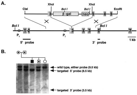
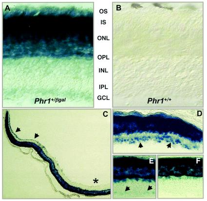
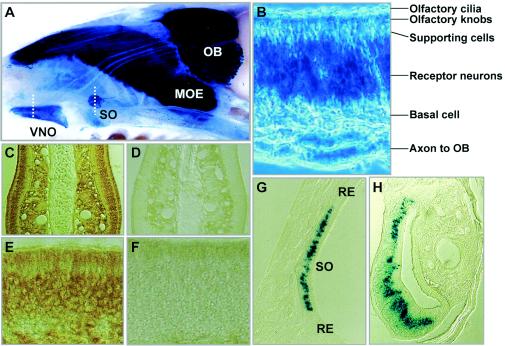
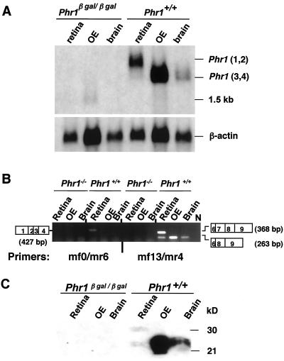
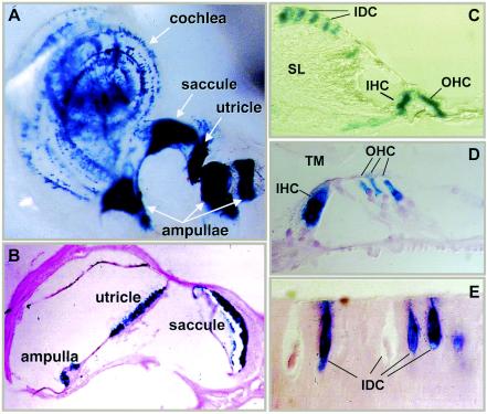
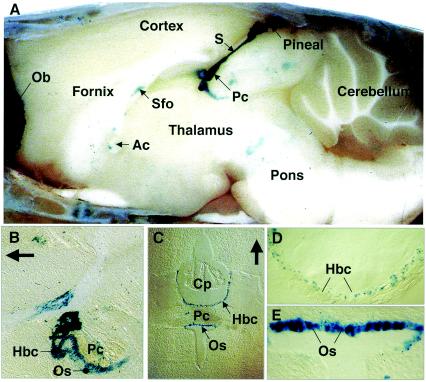
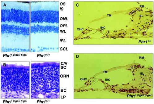
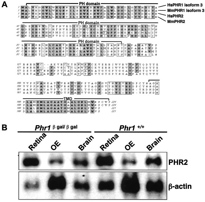
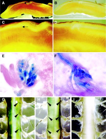
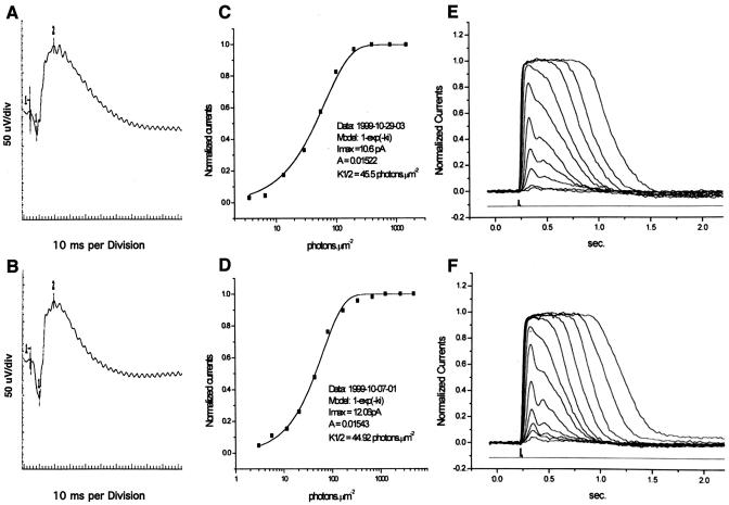
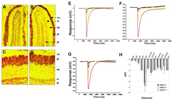
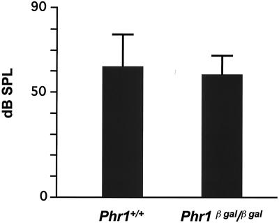
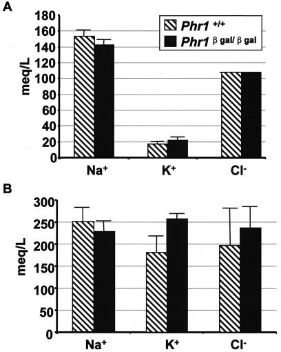
Previously, we identified PHR1 as an abundantly expressed gene in photoreceptors and showed that it encodes four isoforms, each with N-terminal pleckstrin homology (PH) and C-terminal transmembrane domains. To better understand PHR1 function and expression, we made a Phr1 null mouse by inserting a beta-galactosidase/neor cassette into exon 3. In addition to photoreceptors, we found abundant expression of specific Phr1 splice forms in olfactory receptor neurons and vestibular and cochlear hair cells. We also found Phr1 expression in cells with a possible sensory function, including peripheral retinal ganglion cells, cochlear interdental cells, and neurons of the circumventricular organ. Despite this discrete expression in known and putative sensory neurons, mice lacking PHR1 do not have overt sensory deficits.


Figures

FIG. 1.
FIG. 1.
Targeted disruption of Phr1. (A) The targeting construct (top) and targeted region of Phr1 (bottom) are shown. We inserted the β-Gal/neor cassette into exon 3 of Phr1 following codon L22. The neor cassette includes a 5′ PGK promoter and is transcribed with the same orientation as Phr1. The black ovals indicate the location of the 5′ photoreceptor-specific (Pr) and the internal, general (Pi) promoter of Phr1. The positions of the 5′ and 3′ probes used for Southern blot screening of potential recombinant ES cell clones are shown below the Phr1 structural gene as solid black lines. (B) Southern blot, hybridized with both the 5′ and 3′ probes, of BclI-digested genomic DNA from parents heterozygous for the targeted Phr1 allele and their offspring. The 5′ probe recognizes an 8.5-kb fragment in the targeted allele and a 9.5-kb fragment in the wild-type allele. The 3′ probe detects a 3.5-kb fragment in the targeted allele and a 9.5-kb fragment in the wild-type allele. The filled symbol indicates a homozygote for the targeted allele, a symbol with a central black dot indicates a heterozygote for the targeted allele, and an open symbol indicates a homozygote for the wild-type allele.
FIG. 2.
FIG. 2.
Expression of Phr1 as detected by β-Gal staining in adult mouse retina. Sections from comparable regions of retina from Phr1+/β-Gal (A) and Phr1+/+ (B) animals are shown following β-Gal staining. The retinal layers are indicated as follows: OS, photoreceptor outer segments; IS, photoreceptor inner segments; ONL, outer nuclear layer; OPL, outer plexiform layer; INL, inner nuclear layer; IPL, inner plexiform layer; GCL, ganglion cell layer. A lower power view of a retinal cross section (C) shows staining of cells in the peripheral (solid arrowheads) but not in the central (*) ganglion cell layer of the retina. (D to F) Higher-power views of the peripheral (D), medial (E), and central (F) retina. The arrowheads point to β-Gal-positive cells in the ganglion cell layer.
FIG. 3.
FIG. 3.
Expression of Phr1 as detected by β-Gal staining and immunohistochemistry of olfactory structures in Phr1+/β-Gal mice. (A) Mid-sagittal view of a whole mount of the nasal septum. The dashed lines indicate the location of the sections shown in panels G and H. (B) Higher-power view of a section through the OE showing β-Gal staining of the receptor neurons and their axons. Immunohistochemical localization of PHR1 is shown in coronal sections of the nasal septum stained with anti-PHR1 antiserum (C) or preimmune serum (D) and in higher-power cross-sectional views of OE stained with anti-PHR1 antiserum (E) or preimmune serum (F). (G) Localization of β-Gal staining in a cross-section of the septal organ. Note the prominent β-Gal staining of receptor neurons but not of the surrounding respiratory epithelium. (H) Cross-section of the vomeronasal organ with β-Gal staining of the receptor neurons. Abbreviations: MOE, major olfactory epithelium; OB, olfactory bulb; RE, respiratory epithelium; SO, septal organ of Masera; VNO, vomeronasal organ.
FIG. 4.
FIG. 4.
The Phr1β-Gal allele is a null. (A) Northern blot of total cellular RNA (5 μg/lane) isolated from retina, OE, and brain of mice of the indicated genotypes and probed with a full-length Phr1 cDNA. Note that Phr1 transcripts were undetectable in the Phr1β-Gal/β-Gal mice and that Phr1 was highly expressed in OE of Phr1+/+ mice with a transcript size (Phr1 transcripts 3 and 4) similar to that in the brain and smaller than those (Phr1 transcripts 1 and 2) in retina. A faintly hybridizing 1.5-kb transcript was detected in OE RNA isolated from mice of both genotypes. The bottom panel shows the same filter hybridized with a β-actin probe to control for RNA quality and quantity. (B) RT-PCR analysis using RNA isolated from the indicated tissues of Phr1β-Gal/β-Gal (here designated as Phr1/) and Phr1+/+ mice. In Phr1+/+ mice, a primer pair (mf0 and mr6) that amplifies only Phr1 transcripts 1 and 2 detected transcript 1 in retina but not in RNA from other tissues, while a primer pair (mf13 and mr4) that detects all four Phr1 splice forms detected transcripts in RNA from all three tissues. N designates a lane in which no RT was added to an RT-PCR with retinal RNA. The open rectangles on each side show the Phr1 exons contributing to the segment of the transcript amplified. In brain and OE, transcript 4 lacking exon 7 predominates. In retina, transcript 1 including exon 7 predominates. No Phr1 transcripts were detected by either primer pair in RNA isolated from Phr1β-Gal/β-Gal mice. (C) Immunoblot analysis of extracts (50 μg of protein) of retina, OE, and brain. Abundant PHR1 was present in OE extracts, with the same size as PHR1 in brain. The loading for the retinal lane was only 5 μg. No PHR1 was detected in the tissue extracts from Phr1β-Gal/β-Gal mice.
FIG. 5.
FIG. 5.
Expression of Phr1 as detected by β-Gal staining in the inner ear of Phr1+/β-Gal mice. (A) β-Gal staining in a whole mount of the membranous labyrinth of a 4-week-old mouse. (B) Section through the vestibular organ of a 6-week-old mouse counterstained with eosin. (C) β-Gal staining in a section through the spiral limbus and organ of Corti of a 4-week-old mouse. (D) Higher-power view of the organ of Corti from a 6-week-old mouse, counterstained with eosin. (E) High-power view of a section through the spiral limbus of a 6-week-old mouse, showing the interdental cells, some of which are positive for β-Gal staining while others are not. Abbreviations: IDC, interdental cell; IHC, inner hair cell; OHC, outer hair cell; SL, spiral limbus; TM, tectorial membrane.
FIG. 6.
FIG. 6.
Expression of Phr1 as detected by β-Gal staining in brain from a PHR1β-Gal/+ mouse. (A) Mid-sagittal view of a whole mount of the brain, showing dense staining of the pineal body and stalk (S). (B) Sagittal section through the region of the posterior commissure (Pc) and organus subcommisurale (Os). The arrow points rostrally. (C to E) Coronal sections through the CVO. Panels D and E are higher-power views of the structures shown in panel C with the same orientation. The arrow points dorsally. Abbreviations: Ac, anterior commissure; Cp, choroidal plexus; F, fornix; Hbc, habenular commissure; Ob, olfactory bulb; Os, organus subcommisurale; Pc, posterior commissure; Sfo, subfornical organ.
FIG. 7.
FIG. 7.
Histology of selected sensory organs. (A) Hematoxylin-eosin (HE)-stained sections of retina from 6-month-old mice of the indicated genotypes. Abbreviations: GCL, ganglion cell layer; INL, inner nuclear layer; IPL, inner plexiform layer; IS, inner segment; ONL, outer nuclear layer; OPL, outer plexiform layer; OS, outer segment. (B) HE-stained sections of OE from 4-week-old mice of the indicated genotypes. In review of multiple sections from multiple animals, we saw no consistent difference between wild-type and Phr1β-Gal/-β-Gal animals. Abbreviations: C/V, olfactory cilia and microvilli of supporting cells; SC, supporting cells; ORN, olfactory receptor neurons; BC, basal cells; LP, lamina propria. (C and D) HE-stained sections of the organ of Corti from 6-month-old mice of the indicated genotypes. The sections show the basal turn of the cochlea. Similar results were observed in sections of the middle turn (data not shown). Abbreviations: IHC, inner hair cells; OHC, outer hair cells; RM, Reissner's membrane; SL, spiral limbus; TM, tectorial membrane.
FIG. 8.
FIG. 8.
(A) Alignment of the amino acid sequences of human and murine PHR1 (isoform 3) and PHR2. Overall, murine PHR2 (isoform 3) has 38% amino acid identity with murine PHR1 isoform 3. The similarity is much higher in certain domains. For example, the N-terminal PH domains of PHR1 and PHR2 are 50% identical and 73% similar. Their C-terminal transmembrane domains (TMD) are 61% identical and 68% similar. Boxed bold or shaded letters are identical residues in three or four sequences, respectively. Boxed-only letters are similar residues. (B) Northern blot analysis of total cellular RNA (5 μg/lane) harvested from the indicated tissues from Phr1β-Gal/β-Gal and Phr1+/+ mice. A 3.2-kb Phr2 transcript is present in RNA from retina, OE, and brain, with levels in the following order: retina > brain > OE.
FIG. A1.
FIG. A1.
Expression of Phr1 as detected by β-Gal staining of the tongue and spinal cord. Low-power (A and B) and high-power (C and D) views of sagittal sections of the tongue of Phr1β-Gal/β-Gal (A and C) and Phr+/+ (B and D) mice. The arrow in panel C points to a stained vallate papilla. (E and F) High-power views of a taste bud (E) and of the stretch receptor in the tongue (F) of a Phr1β-Gal/β-Gal animal. (G to I) Segments of thoracic spinal cord from a Phr1β-Gal/β-Gal animal (dorsal view) (G), a Phr1β-Gal/β-Gal animal (ventral view) (H), and a Phr1+/+ animal (dorsal view) (I). The black arrows indicate the stained dorsal roots in panel G and the nonstaining ventral roots in panel H.
FIG. A2.
FIG. A2.
Retinal electrophysiology in Phr1β-Gal/β-Gal mice (A, C, and E) and Phr+/+ littermates (B, D, and F). (A and B) ERGs measured in 6-month-old animals. (C to F) Suction pipette recordings from single rod photoreceptors isolated from 2-month-old mice. Panels C and D show normalized responses of a rod (Phr1β-Gal/β-Gal [C] and Phr1+/+ [D]) to 500-nm flashes of increasing strength. Each trace is the averaged response from multiple flash trials (see Materials and Methods). Panels E and F show the relationship between the peak amplitude of flash response and the flash intensity for the rods shown in panels C and D.
FIG. A3.
FIG. A3.
Evaluation of olfaction in Phr1β-Gal/β-Gal mice. (A to D) Immunohistochemical localization of tyrosine hydroxylase in sections of OE from 6-week-old Phr1β-Gal/β-Gal mice (A and C) and normal littermates (B and D). Original magnification, ×50 (A and B) or ×100 (C and D). Abbreviations: ep, external plexiform layer; gl, glomerular layer; gr, granule cell layer; m, mitral cell layer; on, olfactory nerve layer. (E to G) EOG responses to 10−5(red), 10−4 (yellow), and 10−3 M (green) amyl acetate in Phr1β-Gal/β-Gal (E), Phr1β-Gal/+ (F), and Phr1+/+ (G) mice. The black curve shows the response to water as a negative control. (H) Peak responses to all odorants. AA6, AA5, AA4, and AA3 correspond to 10−6, 10−5, 10−4, and 10−3 M amyl acetate, respectively; carvone4, 10−4 M carvone; cineole4, 10−4 M cineole; hexanal4, 10−4 M hexanal; citrial4, 10−4 M citrial; heptanol4, 10−4 M heptanol.
FIG. A4.
FIG. A4.
Auditory function as measured by ABR recordings for 6-month-old Phr1β-Gal/β-Gal mice and Phr+/+ littermate controls. The bars indicate the mean (± 1 standard deviation) sound pressure level (decibels) detected in three animals of the indicated genotypes.
FIG. A5.
FIG. A5.
Electrolyte levels in serum (A) and urine (B) obtained from 7-month-old animals of the indicated genotypes. The heights of the bars indicate the means, and the thin lines indicate the ranges of determinations in three animals. The significance of the 43% increase in the mean urinary K+ concentration in the Phr1β-Gal/β-Gal animals is uncertain, particularly in light of the corresponding increase in serum K+.

Similar articles

Cited by

References

    1. Abe, S., T. Katagiri, A. Saito-Hisaminato, S.-I. Usami, Y. Inoue, T. Tsunoda, and Y. Nakamura. 2003. Identification of CRYM as a candidate responsible for nonsyndromic deafness, through cDNA microarray analysis of human cochlear and vestibular tissues. Am. J. Hum. Genet. 72:73-82. - PMC - PubMed
    1. Akazawa, C., M. Ishibashi, C. Shimizu, S. Nakanshi, and R. Kageyama. 1995. A mammalian helix-loop-helix factor structurally related to the product of Drosophila proneural gene atonal is a positive transcriptional regulator expressed in the developing nervous system. J. Biol. Chem. 270:8730-8738. - PubMed
    1. Andrews, K., P. Potdar, P. Nettesheim, and L. E. Ostrowski. 2000. KPL1, which encodes a novel PH domain-containing protein, is induced during ciliated cell differentiation of rat tracheal epithelial cells. Exp. Lung Res. 26:257-271. - PubMed
    1. Baker, H., D. M. Cummings, S. D. Munger, J. W. Margolis, L. Franzen, R. R. Reed, and F. L. Margolis. 1999. Targeted deletion of a cyclic nucleotide-gated channel subunit (OCNC1): biochemical and morphological consequences in adult mice. J. Neurosci. 19:9313-9321. - PMC - PubMed
    1. Berson, D. M., F. A. Dunn, and M. Takao. 2002. Phototransduction by retinal ganglion cells that set the circadian clock. Science 295:1070-1074. - PubMed

Publication types

MeSH terms