. 2004 Dec;15(12):5700-11.
doi: 10.1091/mbc.e04-08-0732. Epub 2004 Sep 29.

Construction, characterization, and complementation of a conditional-lethal DNA topoisomerase IIalpha mutant human cell line

Affiliations

Construction, characterization, and complementation of a conditional-lethal DNA topoisomerase IIalpha mutant human cell line

Adam J Carpenter et al. Mol Biol Cell. 2004 Dec.

Abstract

DNA Topoisomerase IIalpha (topoIIalpha) is a DNA decatenating enzyme, abundant constituent of mammalian mitotic chromosomes, and target of numerous antitumor drugs, but its exact role in chromosome structure and dynamics is unclear. In a powerful new approach to this important problem, with significant advantages over the use of topoII inhibitors or RNA interference, we have generated and characterized a human cell line (HTETOP) in which >99.5% topoIIalpha expression can be silenced in all cells by the addition of tetracycline. TopoIIalpha-depleted HTETOP cells enter mitosis and undergo chromosome condensation, albeit with delayed kinetics, but normal anaphases and cytokineses are completely prevented, and all cells die, some becoming polyploid in the process. Cells can be rescued by expression of topoIIalpha fused to green fluorescent protein (GFP), even when certain phosphorylation sites have been mutated, but not when the catalytic residue Y805 is mutated. Thus, in addition to validating GFP-tagged topoIIalpha as an indicator for endogenous topoIIalpha dynamics, our analyses provide new evidence that topoIIalpha plays a largely redundant role in chromosome condensation, but an essential catalytic role in chromosome segregation that cannot be complemented by topoIIbeta and does not require phosphorylation at serine residues 1106, 1247, 1354, or 1393.


Figures
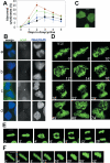
Figure 5.
Figure 5.
Abnormal chromosome segregation in topoIIα-depleted cells. (A) Frequency of abnormal cytokineses after addition of doxycycline. The samples were the same as those analyzed in Figure 3A: no further treatment (circles; green) or 7-h treatments with nocodazole (triangles; blue), caffeine (diamonds; brown), or nocodazole and caffeine (squares; black). (B) Fluorescence and immunofluorescence confocal microscopy of DNA (DAPI; blue) and phosphorylated Histone H3 (green), respectively, in normal (a) and abnormal (b–d) cytokinesis in cultures treated with doxycycline for 1 (a and b), 2 (c), or 3 (d) d. (C) Confocal fluorescence microscopy of an abnormal cytokinesis in HTETOP/H2B-GFP cells treated with doxycycine for 3 d. (D) Time-lapse confocal microscopy showing an abnormal cytokinesis arising from an abnormal anaphase in HTETOP/H2B-GFP cells treated with doxycycline for 1 d. (E) Time-lapse confocal microscopy showing normal anaphase in HTETOP/H2B-GFP cells not treated with doxycycline. (F) Time-lapse confocal microscopy showing abortive anaphase in HTETOP/H2B-GFP cells treated with doxycycline for 2 d. All bars, 10 μm. Images in D, E, and F were taken from videos (Fig 5video1.mov, Fig 5video2.mov and Fig 5video3.mov, respectively) that can been seen as online Supplementary Material.
Figure 1.
Figure 1.
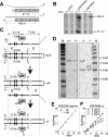
Construction and initial characterization of HTETOP cells. (A) Principal of RNAse protection assay use to detect endogenous (top) and exogenous (bottom) topoIIα transcripts. The 5′ ends of transcripts are shown as shaded boxes, numbered according to exons. The exogenous transcript has non-topoIIα-derived sequences at its 5′ end (unshaded box). Protected and unprotected regions of the RNA probe are shown above each transcript (solid and broken lines, respectively). (B) Products of RNase protection assay outlined in A were separated by denaturing PAGE. Protecting RNA was from the indicated cell lines grown in the presence or absence of doxycycline for 2 d. (C) Gene targeting strategy. Bold lines represent 5′ regions of TOPOIIα genes with exons 1–4 (left to right) shown as black boxes. Pairs of TOPOIIα alleles in HTETOPwt (+/+) HTETOPhet (-/+) and HTETOP (-/-) cells are shown with relevant sites for PstI (P), and the resulting fragment sizes are indicated. Linearized targeting constructs are shown with TOPOIIα DNA aligned to TOPOIIα target alleles, and the position (disrupting exon 4) and orientation (opposite to TOPOIIα) of their gpt and zeo selection cassettes are indicated; remaining plasmid DNA is shown as a thin line. The vertical dotted line indicates the limit of homology between targeting constructs and TOPOIIα alleles. The probe (Pr) used for Southern analysis (gray bar) and oligonucleotides (O1, O2, and O3) used for PCR assays (arrowheads) are shown aligned with homologous DNA. (D) Southern analysis. PstI-digested genomic DNA isolated from HTETOPwt (+/+), HTETOPhet (-/+), and HTETOP (-/-) cells, and labeled marker DNA (M), were separated, blotted, and probed with the probe shown in C. (E) HTETOPhet doubling times during continuous culturing (see Materials and Methods) with (open circles) or without (squares) added tetracycline at 1 μg/ml. (F) HTETOP doubling times, grown as in E but with tetracycline at 0 (squares), 1 (diamonds), or 2 (circles) ng/ml.
Figure 2.
Figure 2.
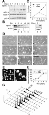
TopoIIα depletion is accompanied by loss of proliferation, cell death, abnormal cell and nuclear morphology, and increased ploidy. (A) Western analysis of HTETOP cells grown in doxycycline for the indicated number of days. Duplicate blots were probed with antibodies to topoIIα and actin or Ku80 and p53. (B) The indicated dilutions of lysates from untreated HTETOP cells (lanes 1–5) or undiluted lysates from HTETOP cells treated with doxycycline for 1 (lane 7) or 3 (lane 6) d, were probed with antibodies to topoIIα(top) or actin (bottom). (C) Growth curves for HTETOP cells after addition (open circles) or mock addition (squares) of tetracycline at day 0. (D) Phase contrast microscopy of HTETOP grown in doxycycline for the indicated number of days. Bar, 250 μm. (E) Fluorescence microscopy of DAPI-stained nuclei of HTETOP cells treated with doxycycline for the indicated number of days. Bars, 10 μm. (F) Frequency of abnormal (enlarged and/or distorted as in E) nuclei in HTETOP cells grown in doxycycline for the indicated times. Cultures were treated with (diamonds) or without (open circles) caffeine for 7 h before fixing. The experiment was done in triplicate, scoring ∼1000 cells for each; means and standard deviations are shown. (G) Flow cytometric analysis of DNA content in nuclei prepared from HTETOP at the indicated number of days after addition of doxycycline. x-axis, fluorescence (log scale); y-axis, frequency; and z-axis, time (days).
Figure 3.
Figure 3.
Mitosis after topoIIα-depletion. (A) Mitotic indices in HTETOP cells were determined at the indicated times after doxycycline addition. Cells were fixed for microscopic analysis after no further treatment (circles; green) or 7-h treatments with nocodazole (triangles; blue), caffeine (diamonds; brown), or nocodazole and caffeine (squares; black). The experiment was done in triplicate, scoring ∼1000 cells in each; means and standard deviations are shown. The same samples were used in Figures 2E, 3C, and 5A. (B) Fluorescent and immunofluorescent confocal microscopy of DNA (DAPI; blue) and topoIIα (fluorescein isothiocyanate; green), respectively, in mitotic HTETOP cells grown with (+) or without (-) doxycycline for 2 d. Bars, 10 μm. No topoIIα-positive cells were detected in many fields (representing >>1000 cells) of such doxycycline-treated cells. (C) Frequency of normal anaphases (open symbols) or anaphases with DNA bridges (closed symbols) in HTETOP cells after addition of doxycycline. Samples were treated with (diamonds; brown) or without (circles; green) caffeine for 7 h before harvesting and were the same as those describe in A. (D) Fluorescent confocal microscopy of mitotic HTETOP/H2B-GFP cells grown without (-) or with (+) doxycycline for 0 (a), 1 (b), or 3 (c) d. Bars, 10 μm.
Figure 4.
Figure 4.
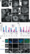
Chromosome spreads from HTETOP cells. (A) DAPI-stained spreads from untreated cells (a and b) or cells treated with doxycycline for 2–3 d (c–e), or ICRF-193 for 2 h (f–h). These were placed in the low (h and f), medium (b[lower spread], c, and g), or high (a, b[upper spread], d, and e) categories of chromosome condensation. (B) Frequencies of spreads with high, medium, or low levels of chromosome condensation (as defined in methods). Spreads were prepared from untreated cells or cells treated with colcemid (Colc) for 2 h, doxycycline (Dox) for 2–3 d, or ICRF-193 (ICRF) for 2 h, in the indicated combinations. The number of mitotic spreads categorized (n) and mitotic indices (MI) for each treatment are indicated. (C) As for B, but this experiment was done in triplicate and error bars show standard deviations. (D) Fluorescent and immunofluorescent microscopy of DNA and topoIIα, respectively, in chromososome spreads of cells grown with (+) or without (-) doxycycline for 3 d. Bars, 10 μm.
Figure 6.
Figure 6.
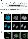
GFP-topoIIα rescues HTETOP from doxycycline sensitivity. (A) Western analysis of six puromycin-resistant HTETOP/GFP-topoIIα clones, treated with or without doxycycline for 2 d, with antibodies to topoIIα (top) and actin (bottom). All clones except clone I were doxycycline resistant. (B) Confocal fluorescence microscopy of GFP-topoIIα (green) and DNA (DAPI, blue) in fixed nuclei of clone B grown continuously in doxycycline. (C) Confocal fluorescence microscopy of GFP-topoIIα in nuclei of live clone B cells; a and b are separate mitosis; b′ is a single z-section of b.
Figure 7.
Figure 7.
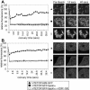
FRAP of GFP-topoIIα-rescued HTETOP cells shows topoIIα to be highly mobile. (A) Left. Relative fluorescence at 1.6-s intervals after photobleaching of mitotic chromosomes in HTETOP/H2B-GFP cells (triangles) or clone B (GFP-topoII-rescued HTETOP) cells untreated (closed squares) or treated (open squares) with ICRF-193 for 1 h before bleaching. Right, examples of images analyzed. Dotted white squares (7 μm2) indicate areas of analysis. (B) Analysis as in A but for nucleoplasmic fluorescence of interphase cells.

References

    1. Adachi, N., Suzuki, H., Iiizumi, S., and Koyama, H. (2003). Hypersensitivity of nonhomologous DNA end-joining mutants to VP-16 and ICRF- 193, implications for the repair of topoisomerase II-mediated DNA damage. J. Biol. Chem. 278, 35897-35902. - PubMed
    1. Adachi, Y., Luke, M., and Laemmli, U.K. (1991). Chromosome assembly in vitro: topoisomerase II is required for condensation. Cell 64, 137-148. - PubMed
    1. Agostinho, M., Rino, J., Braga, J., Ferreira, F., Steffensen, S., and Ferreira, J. (2004). Human topoisomerase IIalpha: targeting to subchromosomal sites of activity during interphase and mitosis. Mol. Biol. Cell 15, 2388-2400. - PMC - PubMed
    1. Akimitsu, N., Adachi, N., Hirai, H., Hossain, M.S., Hamamoto, H., Kobayashi, M., Aratani, Y., Koyama, H., and Sekimizu, K. (2003). Enforced cytokinesis without complete nuclear division in embryonic cells depleting the activity of DNA topoisomerase IIalpha. Genes Cells 8, 393-402. - PubMed
    1. Andersen, C.L., Wandall, A., Kjeldsen, E., Mielke, C., and Koch, J. (2002). Active, but not inactive, human centromeres display topoisomerase II activity in vivo. Chromosome Res. 10, 305-312. - PubMed

MeSH terms

LinkOut - more resources