. 2004 Oct 12;101(41):14776-81.
doi: 10.1073/pnas.0405175101. Epub 2004 Sep 29.

De novo design of conformationally flexible transmembrane peptides driving membrane fusion

Affiliations

De novo design of conformationally flexible transmembrane peptides driving membrane fusion

Mathias W Hofmann et al. Proc Natl Acad Sci U S A. .

Abstract

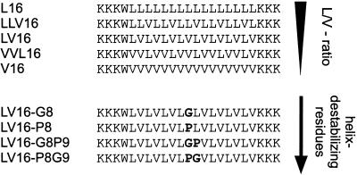
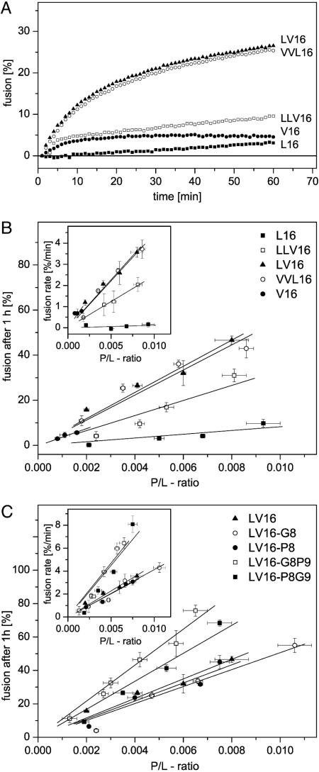
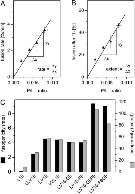
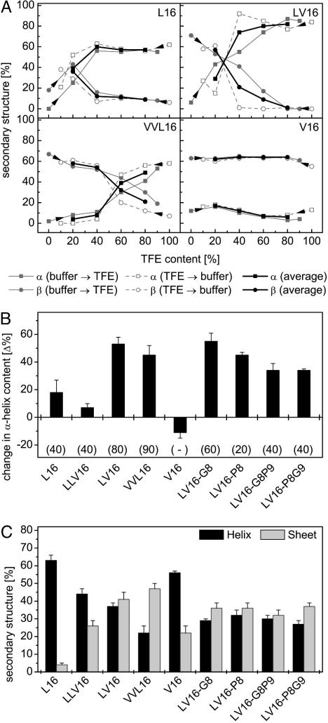
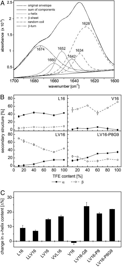
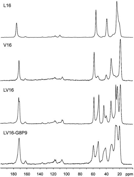
Fusion of biological membranes is mediated by distinct integral membrane proteins, e.g., soluble N-ethylmaleimide-sensitive factor attachment protein receptors and viral fusion proteins. Previous work has indicated that the transmembrane segments (TMSs) of such integral membrane proteins play an important role in fusion. Furthermore, peptide mimics of the transmembrane part can drive the fusion of liposomes, and evidence had been obtained that fusogenicity depends on their conformational flexibility. To test this hypothesis, we present a series of unnatural TMSs that were designed de novo based on the structural properties of hydrophobic residues. We find that the fusogenicity of these peptides depends on the ratio of alpha-helix-promoting Leu and beta-sheet-promoting Val residues and is enhanced by helix-destabilizing Pro and Gly residues within their hydrophobic cores. The ability of these peptides to refold from an alpha-helical state to a beta-sheet conformation and backwards was determined under different conditions. Membrane fusogenic peptides with mixed Leu/Val sequences tend to switch more readily between different conformations than a nonfusogenic peptide with an oligo-Leu core. We propose that structural flexibility of these TMSs is a prerequisite of fusogenicity.


Figures

Fig. 1.
Fig. 1.
Design of fusogenic peptides. Peptides contain hydrophobic core sequences based on different Leu/Val (L/V) ratios and helix-destabilizing Pro and Gly residues (in bold).
Fig. 2.
Fig. 2.
Fusogenic activities of TMS peptides. (A) Typical fusion kinetics reveal that fusogenicity depends on the Leu/Val ratio of the hydrophobic peptide core sequences. P/L ratios were from 0.0035 to 0.005, except for peptide V16, which was incorporated up to a P/L ratio of only 0.0011. (B) Quantitative comparison of fusogenicities of the different aliphatic core sequences by plotting fusion extents, as seen after 1 h, or initial rates (Insets) as a function of P/L ratio. (C) Fusogenicities of peptides containing Gly and/or Pro relative to the parental LV16. Data points represent means ± SE (n = 4–18 independent experiments).
Fig. 3.
Fig. 3.
Comparison of fusogenicities. (A) Taking LV16 as an example, the schematic depicts how the slopes of the relationships between P/L ratio and initial fusion rate (Left) or fusion extent (Right) were derived for the comparison shown in B. V16 was omitted here because of its inefficient membrane incorporation.
Fig. 4.
Fig. 4.
Structural plasticity of LV peptides determined by CD spectroscopy. (A) Contents of α-helical and β-sheet structure are shown for some peptides (0.1 mg/ml) upon titrating from either aqueous buffer or from TFE solution as indicated by arrowheads. Bold lines represent the averaged values of both types of titrations. (B) Conformational change of peptides as revealed by differences in average helix content seen at 20% and 80% TFE, respectively. Gains in secondary structure upon decreasing polarity result in positive values. Note that the change in helix content tended to be larger with the more fusogenic peptides. Data are shown as means ± SE (n = 4–5). (C) Contents of α-helical and β-sheet structure in inverse sodium bis(2-ethylhexyl)sulfosuccinate/water/iso-octane micelles. Data are shown as means ± SE (n = 8–10).
Fig. 5.
Fig. 5.
Structural plasticity of LV peptides determined by FTIR spectroscopy. (A) Original spectrum of LV16 in 100% TFE and decomposition of the amide-I band into individual bands according to ref. ; the superposition of the original envelope with the sum of components demonstrates the quality of the fit. (B) α-Helix and β-sheet contents of some peptides (1.5 mg/ml) upon titration from TFE solution. (C) Conformational change of peptides as revealed by differences in helix content seen at 10% and 100% TFE, respectively. Gains in secondary structure upon decreasing polarity result in positive values. Note that the changes in helix contents tended to be larger with the more fusogenic peptides. Data are shown as means ± SE (n = 3); error bars were omitted when they were smaller than the symbols. Results obtained with peptide LV16-G8P9 are not shown because it adhered to the cuvette surface with unknown effects on its secondary structure.
Fig. 6.
Fig. 6.
One-dimensional 13C-natural abundance CP-MAS solid-state NMR spectra of lyophilized peptides. Please note the different 13C-carbonyl resonances for L16 and V16. The resonances from 20 ppm to 60 ppm correspond to Cα, Cβ, and side-chain carbons.

References

    1. Jahn, R., Lang, T. & Südhof, T. C. (2003) Cell 112, 519–533. - PubMed
    1. Tamm, L. K., Crane, J. & Kiessling, V. (2003) Curr. Opin. Struct. Biol. 13, 453–466. - PubMed
    1. Weber, T., Zemelman, B. V., McNew, J. A., Westermann, B., Gmachl, M., Parlati, F., Söllner, T. H. & Rothman, J. E. (1998) Cell 92, 759–772. - PubMed
    1. Schuette, C. G., Hatsuzawa, K., Margittai, M., Stein, A., Riedel, D., Küster, P., König, M., Seidel, C. & Jahn, R. (2004) Proc. Natl. Acad. Sci. USA 101, 2858–2863. - PMC - PubMed
    1. Tucker, W. C., Weber, T. & Chapman, E. R. (2004) Science 304, 435–438. - PubMed

Publication types

LinkOut - more resources