

Crystal structure of human GGA1 GAT domain complexed with the GAT-binding domain of Rabaptin5
- PMID: 15457209
- PMCID: PMC524345
- DOI: 10.1038/sj.emboj.7600411
Crystal structure of human GGA1 GAT domain complexed with the GAT-binding domain of Rabaptin5
Abstract
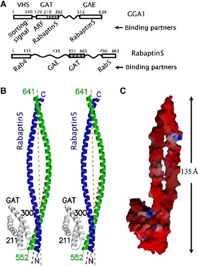
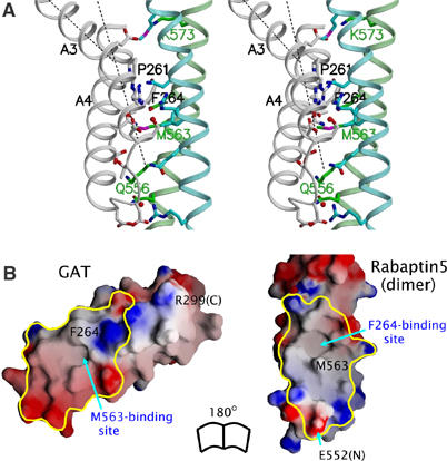
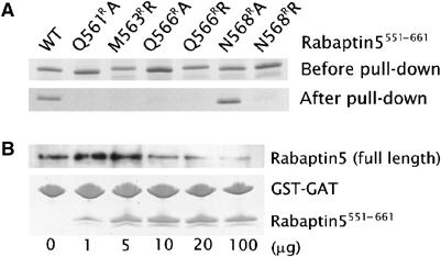
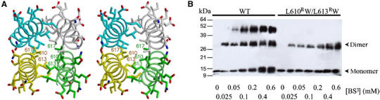
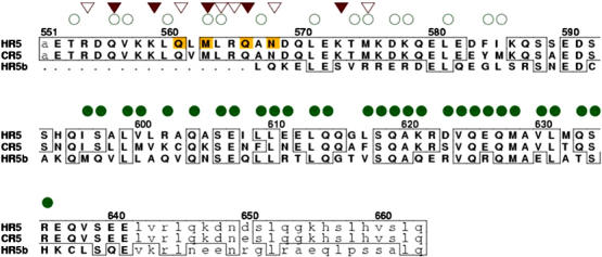
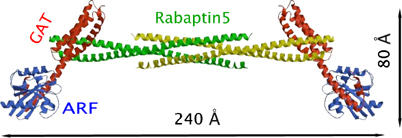
GGA proteins coordinate the intracellular trafficking of clathrin-coated vesicles through their interaction with several other proteins. The GAT domain of GGA proteins interacts with ARF, ubiquitin, and Rabaptin5. The GGA-Rabaptin5 interaction is believed to function in the fusion of trans-Golgi-derived vesicles to endosomes. We determined the crystal structure of a human GGA1 GAT domain fragment in complex with the Rabaptin5 GAT-binding domain. In this structure, the Rabaptin5 domain is a 90-residue-long helix. At the N-terminal end, it forms a parallel coiled-coil homodimer, which binds one GAT domain of GGA1. In the C-terminal region, it further assembles into a four-helix bundle tetramer. The Rabaptin5-binding motif of the GGA1 GAT domain consists of a three-helix bundle. Thus, the binding between Rabaptin5 and GGA1 GAT domain is based on a helix bundle-helix bundle interaction. The current structural observation is consistent with previously reported mutagenesis data, and its biological relevance is further confirmed by new mutagenesis studies and affinity analysis. The four-helix bundle structure of Rabaptin5 suggests a functional role in tethering organelles.
Figures
Similar articles
-
Structural mechanism for ubiquitinated-cargo recognition by the Golgi-localized, gamma-ear-containing, ADP-ribosylation-factor-binding proteins.Proc Natl Acad Sci U S A. 2005 Feb 15;102(7):2334-9. doi: 10.1073/pnas.0500118102. Epub 2005 Feb 8. Proc Natl Acad Sci U S A. 2005. PMID: 15701688 Free PMC article.
-
Crystal structure of the human GGA1 GAT domain.Biochemistry. 2003 Jun 3;42(21):6392-9. doi: 10.1021/bi034334n. Biochemistry. 2003. PMID: 12767220
-
The interaction of the human GGA1 GAT domain with rabaptin-5 is mediated by residues on its three-helix bundle.Biochemistry. 2003 Dec 2;42(47):13901-8. doi: 10.1021/bi035392b. Biochemistry. 2003. PMID: 14636058
-
Membrane recruitment of effector proteins by Arf and Rab GTPases.Curr Opin Struct Biol. 2005 Dec;15(6):681-9. doi: 10.1016/j.sbi.2005.10.015. Epub 2005 Nov 9. Curr Opin Struct Biol. 2005. PMID: 16289847 Review.
-
The GGA proteins: adaptors on the move.Nat Rev Mol Cell Biol. 2004 Jan;5(1):23-32. doi: 10.1038/nrm1279. Nat Rev Mol Cell Biol. 2004. PMID: 14708007 Review.
Cited by
-
Structural mechanism for ubiquitinated-cargo recognition by the Golgi-localized, gamma-ear-containing, ADP-ribosylation-factor-binding proteins.Proc Natl Acad Sci U S A. 2005 Feb 15;102(7):2334-9. doi: 10.1073/pnas.0500118102. Epub 2005 Feb 8. Proc Natl Acad Sci U S A. 2005. PMID: 15701688 Free PMC article.
-
Application of Gaussia luciferase in bicistronic and non-conventional secretion reporter constructs.BMC Biochem. 2014 Jul 9;15:14. doi: 10.1186/1471-2091-15-14. BMC Biochem. 2014. PMID: 25007711 Free PMC article.
-
Roles of RabGEF1/Rabex-5 domains in regulating Fc epsilon RI surface expression and Fc epsilon RI-dependent responses in mast cells.Blood. 2007 Jun 15;109(12):5308-17. doi: 10.1182/blood-2007-01-067363. Epub 2007 Mar 6. Blood. 2007. PMID: 17341663 Free PMC article.
-
Rabaptin-5-independent membrane targeting and Rab5 activation by Rabex-5 in the cell.Mol Biol Cell. 2007 Oct;18(10):4119-28. doi: 10.1091/mbc.e07-02-0100. Epub 2007 Aug 15. Mol Biol Cell. 2007. PMID: 17699593 Free PMC article.
-
Rabaptin5 targets autophagy to damaged endosomes and Salmonella vacuoles via FIP200 and ATG16L1.EMBO Rep. 2022 Jan 5;23(1):e53429. doi: 10.15252/embr.202153429. Epub 2021 Oct 26. EMBO Rep. 2022. PMID: 34704340 Free PMC article.
References
-
- Boman AL (2001) GGA proteins: new players in the sorting game. J Cell Sci 114: 3413–3418 - PubMed
-
- Brunger AT, Adams PD, Clore GM, DeLano WL, Gros P, Grosse-Kunstleve RW, Jiang JS, Kuszewski J, Nilges M, Pannu NS, Read RJ, Rice LM, Simonson T, Warren GL (1998) Crystallography & NMR system: a new software suite for macromolecular structure determination. Acta Crystallogr D 54: 905–921 - PubMed
-
- Collins BM, Watson PJ, Owen DJ (2003) The structure of the GGA1-GAT domain reveals the molecular basis for ARF binding and membrane association of GGAs. Dev Cell 4: 321–332 - PubMed
Publication types
MeSH terms
Substances
Grants and funding
LinkOut - more resources
Full Text Sources
Other Literature Sources
Molecular Biology Databases
