. 2004 Oct 27;23(21):4146-55.
doi: 10.1038/sj.emboj.7600428. Epub 2004 Sep 30.

GTP/GDP exchange by Sec12p enables COPII vesicle bud formation on synthetic liposomes

Affiliations

GTP/GDP exchange by Sec12p enables COPII vesicle bud formation on synthetic liposomes

Eugene Futai et al. EMBO J. .

Abstract

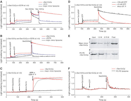
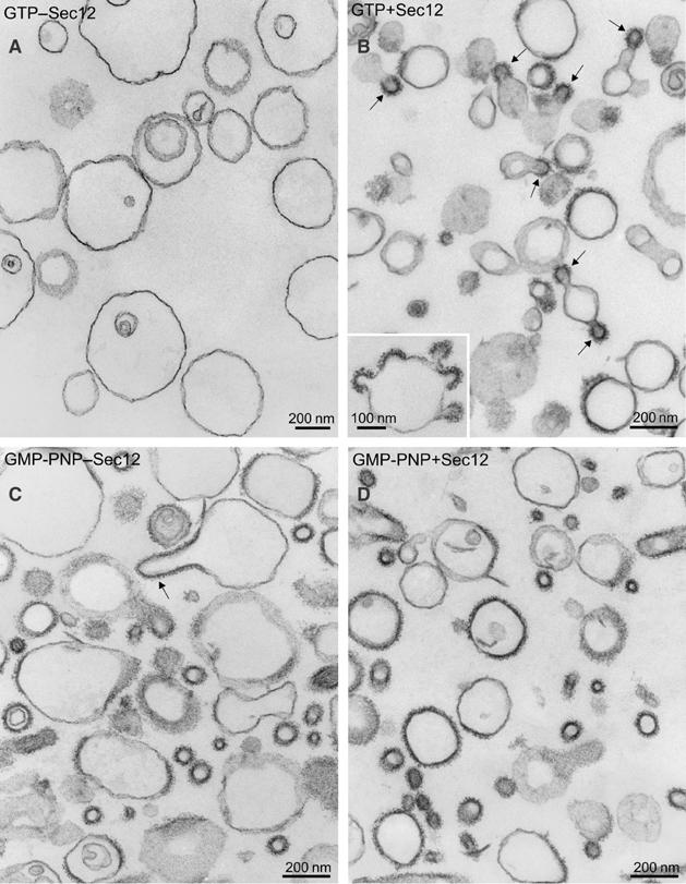
The generation of COPII vesicles from synthetic liposome membranes requires the minimum coat components Sar1p, Sec23/24p, Sec13/31p, and a nonhydrolyzable GTP analog such as GMP-PNP. However, in the presence of GTP and the full complement of coat subunits, nucleotide hydrolysis by Sar1p renders the coat insufficiently stable to sustain vesicle budding. In order to recapitulate a more authentic, GTP-dependent budding event, we introduced the Sar1p nucleotide exchange catalyst, Sec12p, and evaluated the dynamics of coat assembly and disassembly by light scattering and tryptophan fluorescence measurements. The catalytic, cytoplasmic domain of Sec12p (Sec12DeltaCp) activated Sar1p with a turnover 10-fold higher than the GAP activity of Sec23p stimulated by the full coat. COPII assembly was stabilized on liposomes incubated with Sec12DeltaCp and GTP. Numerous COPII budding profiles were visualized on membranes, whereas a parallel reaction conducted in the absence of Sec12DeltaCp produced no such profiles. We suggest that Sec12p participates actively in the growth of COPII vesicles by charging new Sar1p-GTP molecules that insert at the boundary between a bud and the surrounding endoplasmic reticulum membrane.


Figures

Figure 1
Figure 1
Sec12ΔCp stabilizes GTP-driven COPII coat assembly on liposomes. (A–D) The light scattering of a suspension of major–minor mix liposomes (100 μg ml−1) was continuously monitored upon the addition of 950 nM Sar1p GDP, 160 nM Sec23/24p, 260 nM Sec13/31p, and 100 μM GTP or GMP-PNP, at specific time points as indicated by arrows. (A, B) The initial mixture contained 50 nM Sec12ΔCp (red trace), or 2 mM EDTA (blue traces) in HKM buffer (20 mM Hepes-K, pH 6.8, 160 mM KOAc, 1 mM MgCl2), or buffer alone (black trace). EDTA decreases the concentration of free Mg2+ from 1 mM to 1 μM, and accelerates GDP–GTP exchange by Sar1p. Sec13/31p was added to the initial mixture in (B). (C) Sar1p-GDP was activated by addition of GTP (bold trace) or GMP-PNP (thin trace). The initial mixture contained 50 nM Sec12ΔCp in HKM buffer (red trace), or buffer alone (black trace). (D) Different levels of GTP were added to the reaction mixture: 100 μM GTP (black trace), 10 μM GTP (red trace), or without GTP (gray trace). The initial mixture contained Sec13/31p and 50 nM Sec12ΔCp. (E) Sec12ΔCp binding to liposomes. Major–minor or PC/PE liposomes (400 μg ml−1) were incubated with Sec12ΔCp (500 nM) and separated by flotation centrifugation through sucrose step gradients (1.0, 0.75, and 0 M sucrose in HKM buffer). Proteins recovered in fractions (input (1.0 M sucrose), 1.0 or 0.75 M sucrose, and float fractions (without sucrose)) were analyzed by SDS–PAGE. (F) Light scattering of a suspension of PC/PE liposomes was monitored upon addition of Sar1p-GDP, GTP, and Sec23/24p. The initial mixture contained Sec13/31p and 50 nM Sec12ΔCp (red trace) or HKM buffer alone (black trace).
Figure 2
Figure 2
Exchange activity of Sec12ΔCp on Sar1p is ∼10-fold higher than GAP activity of Sec23/24p complex with saturating Sec13/31p. (A) The GEF activity of Sec12ΔCp and the GAP activity of Sec23/24p were measured by tryptophan fluorescence of Sar1p. A large tryptophan fluorescence change accompanies a conformational change by Sar1p from the GDP-bound to the GTP-bound state. Sar1p-GDP (2 μM) was added to major–minor liposomes (300 μg ml−1) and activated by the addition of GTP. After 6 min, Sec23/24p (55 nM) was added to activate GTP hydrolysis. The initial mixture contained 50 nM Sec12ΔCp (red trace), 2 mM EDTA (blue traces) in HKM buffer, or buffer alone (black trace) and 90 nM Sec13/31p, which stimulates GAP activity at saturation. EDTA decreases the concentration of free Mg2+ from 1 mM to 1 μM, and accelerates GDP–GTP exchange by Sar1p. (B) The rate constants for Sar1p activation were plotted against Sec12ΔCp concentration (left) to determine the nucleotide exchange activity (kactivation=kspontaneous+kexchange [Sec12ΔCp]). The specific nucleotide exchange activity (kexchange/[Sec12ΔCp]) corresponds to the rate constant of the activation of Sar1p normalized to the concentration of Sec12ΔCp (see Materials and methods). The rate constant for Sar1p inactivation by Sec23/24p with saturating Sec13/31p was plotted against Sec23/24p concentration (right) to determine catalytic activity (kinactivation=kcatalyze [Sec23/24p]). (C) Light scattering assays with the indicated concentration of Sec12ΔCp (0–200 nM). The experimental conditions were as in Figure 1. (D) In the experiments (C), light scattering signal decays instantly after Sec23/24p addition. The intensity of signals after instant decay was measured at 400 s. Percentage of the maximal amplitude was calculated, and plotted against Sec12ΔCp concentration.
Figure 3
Figure 3
Morphology of major–minor liposomes incubated with COPII proteins and Sec12ΔCp. (A–D) The reaction contains major–minor liposomes (100 μg ml−1), 950 nM Sar1p, 160 nM Sec23/24p, and 260 nM Sec13/31p. The reaction was initiated by the addition of 300 μM GTP and incubated at 27°C. After 15 min, the samples were processed for EM. On thin-section electron micrographs, characteristic profiles observed without (A) or with (B) 50 nM Sec12ΔCp are shown. The inset in (B) depicts liposome with multiple coated buds. Comparison of COPII-like vesicles formed with GMP-PNP in the absence (C) or presence (D) of Sec12ΔCp is shown. Scale bars, 200 nm. The arrows point to coated buds (B, C).
Figure 4
Figure 4
Exchange activity of Sec12ΔCp mutants on Sar1p. (A) For each mutant, experiments similar to that shown in Figure 2A were performed to determine value ±s.e. of the specific exchange activity (kexchange/[Sec12ΔCp]). Except for [N40A] Sec12ΔCp, the mutants were assayed at a concentration up to 20 nM. A very weak stimulation of Sar1p activation as detected by varying concentrations of [N40A] Sec12ΔCp up to 600 nM. (B) Sar1p binding to GSTSec12ΔCp wild type or mutants. GSTSec12ΔCp (3 μM) was incubated with Sar1p-GDP (3 μM) and GMP-PNP (300 μM) (left), GDP (300 μM) (middle), or EDTA (2 mM) with alkaline phosphatase (calf intestine, Promega) (right) in HKM buffer containing 0.2% octylglucoside. The incubation with EDTA and alkaline phosphatase renders Sar1p nucleotide free. GSTSec12ΔCp was recovered with glutathione agarose and washed extensively with HKM buffer (0.2% octylglucoside) containing GMP-PNP (left), GDP (middle), or EDTA (right). Sar1p bound to the beads was analyzed by SDS–PAGE and quantified by Sypro-Ruby staining. (C) Light scattering assays with Sec12ΔCp wild type or mutants. The experimental conditions were as in Figure 1. Sec12ΔCp (50 nM) wild-type or mutant proteins were added to the initial mixture.
Figure 5
Figure 5
Sorting of Sec12TMp and cargo proteins into COPII vesicles. (A) Proteoliposomes (100 μg ml−1) with Sec12TMp, Bet1p, Bos1p, and GST-Ufe1p were incubated for 30 min at 27°C with COPII proteins (130 μg ml−1 Sec23/24p, 150 μg ml−1 sec13/31p, and 80 mg ml−1 Sar1p) and nucleotide (GMP-PNP or GDP). COPII vesicles were separated from donor proteoliposomes by sucrose density gradient sedimentation (2.2–0.2 M sucrose). After separation, lipid recovery was monitored by fluorescence of Texas red-PE. (B) Bet1p, Bos1p, Sec12TMp, and GST-Ufe1p in each fraction were detected by immunoblot. (C) From the blot in (B), recovery of Bet1p and Sec12TMp was quantified using 35S-labeled anti-IgG and a phosphorimager. The recovery in the fractions for liposomes (Fr 1–9) or COPII vesicles (Fr 10–15) was calculated. Bos1 and Bet1 were enriched in the COPII vesicles, but Sec12TM and Ufe1 were neither enriched nor excluded in the vesicle fractions.
Figure 6
Figure 6
Coordination of COPII coating and uncoating by localized action of Sec12 GEF.

References

    1. Antonny B, Gounon P, Schekman R, Orci L (2003) Self-assembly of minimal COPII cages. EMBO Reports 4: 419–424 - PMC - PubMed
    1. Antonny B, Madden D, Hamamoto S, Orci L, Schekman R (2001) Dynamics of the COPII coat with GTP and stable analogues. Nat Cell Biol 3: 531–537 - PubMed
    1. Barlowe C, Orci L, Yeung T, Hosobuchi M, Hamamoto S, Salama N, Rexach MF, Ravazzola M, Amherdt M, Schekman R (1994) COPII: a membrane coat formed by Sec proteins that drive vesicle budding from the endoplasmic reticulum. Cell 77: 895–907 - PubMed
    1. Barlowe C, Schekman R (1993) SEC12 encodes a guanine nucleotide exchange factor essential for transport vesicle budding from ER. Nature 365: 347–349 - PubMed
    1. Beraud-Dufour S, Robineau S, Chardin P, Paris S, Chabre M, Cherfils J, Antonny B (1998) A glutamic finger in the guanine nucleotide exchange factor ARNO displaces Mg2+ and the beta-phosphate to destabilize GDP on ARF1. EMBO J 17: 3651–3659 - PMC - PubMed

Publication types

MeSH terms