. 2004 Oct 27;23(21):4127-33.
doi: 10.1038/sj.emboj.7600429. Epub 2004 Sep 30.

Variable LH2 stoichiometry and core clustering in native membranes of Rhodospirillum photometricum

Affiliations

Variable LH2 stoichiometry and core clustering in native membranes of Rhodospirillum photometricum

Simon Scheuring et al. EMBO J. .

Abstract

The individual components of the photosynthetic unit (PSU), the light-harvesting complexes (LH2 and LH1) and the reaction center (RC), are structurally and functionally known in great detail. An important current challenge is the study of their assembly within native membranes. Here, we present AFM topographs at 12 A resolution of native membranes containing all constituents of the PSU from Rhodospirillum photometricum. Besides the major technical advance represented by the acquisition of such highly resolved data of a complex membrane, the images give new insights into the organization of this energy generating apparatus in Rsp. photometricum: (i) there is a variable stoichiometry of LH2, (ii) the RC is completely encircled by a closed LH1 assembly, (iii) the LH1 assembly around the RC forms an ellipse, (iv) the PSU proteins cluster together segregating out of protein free lipid bilayers, (v) core complexes cluster although enough LH2 are present to prevent core-core contacts, and (vi) there is no cytochrome bc1 complex visible in close proximity to the RCs. The functional significance of all these findings is discussed.


Figures
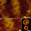
Figure 1
Figure 1
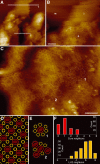
High-resolution AFM of native photosynthetic membranes of Rsp. photometricum. (A) Large native membrane patch (128 nm) at submolecular resolution (scale bar: 20 nm; full color scale: 6 nm). (B) Close-up view of an assembly of multiple core complexes and LH2s. Topography substructure can be seen on the individual LH2 and RC molecules (scale bar: 10 nm; full color scale: 6 nm). (C) Averages of the core complexes (top, 15 Å resolution) and the LH2 (bottom, 12 Å resolution) calculated from two hand-picked families of 235 and 1301 particles, respectively (scale bar: 20 nm; full color scale: 2 nm).
Figure 2
Figure 2
Diversity of LH complexes. (A) A large ∼80 Å diameter (arrow L) light-harvesting complex, (B, C) small ∼40 Å diameter (arrow S) light-harvesting complexes, and (D) a tiny (arrow T) particle in assembly with LH2s and core complexes (all scale bars: 5 nm; full color scale: 4 nm). (E) Graph reporting the abundance of different-sized LH complexes. The analysis of the diameter of the complexes represents a Gaussian distribution peaking at 50 Å. A small fraction of LH complexes with diameters up to ∼85 Å are detected. (F) Averages of the LH components calculated after classification of their rotational power spectra (as described in Materials and methods). From the most abundant complexes with diameters around 50 Å, three classes with 8-, 9-, and 10-fold symmetry could be calculated. A representative complex with a diameter of ∼80 Å is shown surrounded by six ‘normal' LH2s. Judged from its size, we estimate this complex to consist of ∼14 LH subunits. These complexes were rare and strongly variable in size inhibiting the calculation of an average (scale bars: 5 nm; full color scale: 2 nm). (G) Gel exclusion chromatography analysis of the complex size heterogeneity of LH2. In initial elution (black line), the fraction size was 100 μl. Re-chromatography of three fractions eluting at 6.0 ml (dark gray line), 6.5 ml (medium gray line), and 7.0 ml (light gray line). The red line displays the near-infrared absorption maximum of the elution fractions (right red Y-axis). The larger complexes absorb at 841 nm and the latest eluting smallest complexes at 838.5 nm.
Figure 3
Figure 3
Observation of particular complex assemblies. (A) Core in which the RC is capped by additional topography interpreted as a cytochrome c2 (arrow c2) neighbored by three ‘normal' cores (one of them encircled). (B) Section analysis of the c2 capped (top) and ‘normal' (bottom) core complexes shown in (A). Right panel: Section analysis profiles along the dashed white lines in the left panel. While the RC surface of the ‘normal' core complex reveals similar height as the LH1 units around, the c2 capped RC protrudes significantly stronger (horizontal scale bar: 5 nm; vertical scale bar: 1 nm). (C) A moon-shaped assembly of probably four LH2 subunits (arrow M) and a core in which the LH1 assembly reveals an ∼20 Å gap (arrow D). While the cytochrome c2 capped core represents an intermediate state in the photosynthetic cycle, the complexes depicted in the right panel represent most certainly defective molecules (scale bars: 5 nm; full color scale: 4 nm).
Figure 4
Figure 4
Arrangement of the PSU. (A) Overview topograph of a native membrane patch constituted of densely packed and pure lipid membrane parts. Section analysis along the two white lines (1 and 2) resulted in thickness measures of ∼4 nm (line 1) and ∼8 nm (line 2), respectively (scale bars: 50 nm; full color scale: 15 nm). (B) Boarder delineating the area containing the PSU proteins (p) from protein-free lipid bilayer (l). The smooth area in the top left is the mica AFM support (m) (scale bars: 20 nm; full color scale: 12 nm) (C) Medium-resolution topograph allowing the molecular arrangement of the proteins of the PSU to be studied (scale bars: 20 nm; full color scale: 10 nm). (D) Model membrane packing based on a PSU constituted of eight LH2s around one core (dashed line; Papiz et al, 1996). In such a system, each core has eight LH2 neighbors, zero contacts with other cores, while the average ratio of LH2–core complexes is 4. (E) Two examples (depicted in (C) by dashed contour line) of core–neighbor assemblies: (1) core in contact with seven LH2s and zero cores and (2) core in contact with four LH2s and three cores. (F) Graphs displaying numbers of neighboring LH2s and neighboring cores to a core. Top panel: Number of neighboring cores—‘only' 27% of the cores have zero direct contact with another core, 44% have one core contact, and the remaining 29% make two or maximally three contacts with neighboring cores (see example 2 in C and E). Bottom panel: Number of neighboring LH2s—the assembly of eight LH2s surrounding a core is rarely (9%) found; in these cases, at least one of the LH2s does not make physical contact with the core. Most cores are connected with seven LH2s (30%). We never found a core with less than three direct LH2 neighbors. The average ratio of LH2/core in the membranes analyzed is ∼5.

References

    1. Allen JP, Feher G, Yeates TO, Komiya H, Rees DC (1987) Structure of the reaction center from Rhodobacter sphaeroides R-26: the protein subunits. Proc Natl Acad Sci USA 84: 6162–6166 - PMC - PubMed
    1. Bahatyrova S, Frese RN, Van Der Werf KO, Otto C, Hunter CN, Olsen JD (2004) Flexibility and size heterogeneity of the LH1 light harvesting complex revealed by atomic force microscopy: functional significance for bacterial photosynthesis. J Biol Chem 279: 21327–21333 - PubMed
    1. Creighton TE (1993) Proteins. WH Freeman & Company: New York, NY
    1. Deisenhofer J, Epp O, Miki K, Huber R, Michel H (1984) X-ray structure analysis of a membrane protein complex. J Mol Biol 180: 385. - PubMed
    1. Deisenhofer J, Epp O, Miki K, Huber R, Michel H (1985) Structure of the protein subunits in the photosynthetic reaction centre of Rhodopseudomonas viridis at 3 Å resolution. Nature 318: 618–624 - PubMed

Publication types

MeSH terms

Substances