

Replication and gene expression of mutant hepatitis B virus in a transgenic mouse containing the complete viral genome with mutant s gene
- PMID: 15457560
- PMCID: PMC4611258
- DOI: 10.3748/wjg.v10.i21.3141
Replication and gene expression of mutant hepatitis B virus in a transgenic mouse containing the complete viral genome with mutant s gene
Abstract
Aim: To establish the transgenic mouse line harbouring complete hepatitis B virus (HBV) genome with mutant s gene (adr subtype).
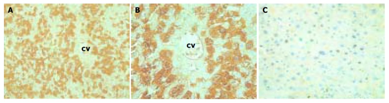
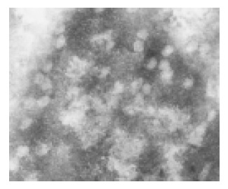
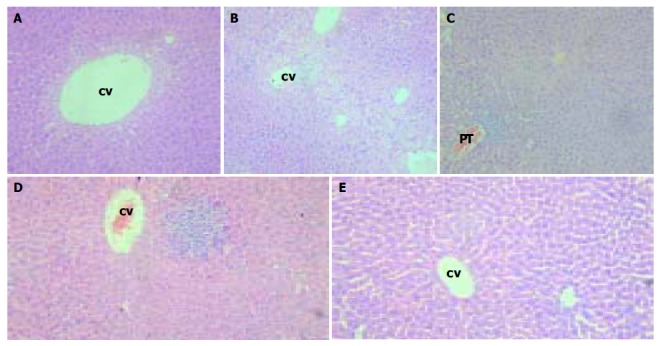
Methods: Transgenic mice were generated by microinjecting HBV genome into fertilized eggs. Integration, expression, replication of HBV gene and histological changes in transgenic mice were estimated by genomic DNA PCR, serum DNA PCR, Southern blot, ELISA, HE staining, immunohistochemistry and transmission electron microscopy. Transgenic mice with HBsAg positive in serum were bred and analyzed.
Results: A total of 288 eggs survived from microinjections were transplanted into the oviducts of 13 pseudopregnant mice and 49 pups were produced. Twenty-six mice were identified to have the integrated HBV gene. Serum HBsAg and HBeAg were detected in 2 of 43 mice. HBsAg and HBcAg in cytoplasm or nuclei of hepatocytes were detected in 10 mice. Founders with HBsAg in serum were named lineages G145R-15 and G145R-18. Of the 16 F1 offsprings generated by G145R-15 founder, 12 were positive for HBV genome with PCR, 10 were positive for HBsAg and HBcAg with immunohistochemistry and 7 were positive for HBsAg and HBeAg with ELISA. Only 1 of 8 F1 offsprings generated by G145R-18 founder was survived and it was detected positive for HBV genome, HBsAg, HBcAg and HBeAg. Both of the two lineages had some pathological characteristics of mild chronic hepatitis B in the liver, such as swelling of hepatocytes and focal hepatocellular necrosis and parenchymal lymphomononuclear cell infiltrate.
Conclusion: Transgenic mice harbouring HBV with mutant s gene can be generated. The HBV genes are integrated in the transgenic mice genome and can be expressed, replicated, packaged and excreted. HBV DNA can be stably transmitted in the transgenic mice.
Figures

Similar articles
-
Stable transmission and expression of the hepatitis B virus total genome in hybrid transgenic mice until F10 generation.J Exp Zool A Comp Exp Biol. 2006 May 1;305(5):420-7. doi: 10.1002/jez.a.277. J Exp Zool A Comp Exp Biol. 2006. PMID: 16489557
-
[Establishment and identification of highly expressing and replicating hepatitis B virus genome transgenic mouse models].Zhonghua Gan Zang Bing Za Zhi. 2003 Jun;11(6):338-40. Zhonghua Gan Zang Bing Za Zhi. 2003. PMID: 12837210 Chinese.
-
Gene expression profile of transgenic mouse kidney reveals pathogenesis of hepatitis B virus associated nephropathy.J Med Virol. 2006 May;78(5):551-60. doi: 10.1002/jmv.20575. J Med Virol. 2006. PMID: 16555286
-
Hepatitis B virus transgenic mice: models of viral immunobiology and pathogenesis.Curr Top Microbiol Immunol. 1996;206:149-73. doi: 10.1007/978-3-642-85208-4_9. Curr Top Microbiol Immunol. 1996. PMID: 8608715 Review.
-
Virion Secretion of Hepatitis B Virus Naturally Occurring Core Antigen Variants.Cells. 2020 Dec 30;10(1):43. doi: 10.3390/cells10010043. Cells. 2020. PMID: 33396864 Free PMC article. Review.
Cited by
-
TREM2 promotes Aβ phagocytosis by upregulating C/EBPα-dependent CD36 expression in microglia.Sci Rep. 2017 Sep 11;7(1):11118. doi: 10.1038/s41598-017-11634-x. Sci Rep. 2017. PMID: 28894284 Free PMC article.
References
-
- Merican I, Guan R, Amarapuka D, Alexander MJ, Chutaputti A, Chien RN, Hasnian SS, Leung N, Lesmana L, Phiet PH, et al. Chronic hepatitis B virus infection in Asian countries. J Gastroenterol Hepatol. 2000;15:1356–1361. - PubMed
-
- Birrer RB, Birrer D, Klavins JV. Hepatocellular carcinoma and hepatitis virus. Ann Clin Lab Sci. 2003;33:39–54. - PubMed
-
- Rabe C, Cheng B, Caselmann WH. Molecular mechanisms of hepatitis B virus-associated liver cancer. Dig Dis. 2001;19:279–287. - PubMed
-
- Bréchot C, Gozuacik D, Murakami Y, Paterlini-Bréchot P. Molecular bases for the development of hepatitis B virus (HBV)-related hepatocellular carcinoma (HCC) Semin Cancer Biol. 2000;10:211–231. - PubMed
Publication types
MeSH terms
Substances
LinkOut - more resources
Full Text Sources
Medical
Molecular Biology Databases
Research Materials
