. 2004 Dec 17;279(51):53828-39.
doi: 10.1074/jbc.M408472200. Epub 2004 Sep 30.

A naturally occurring mutation of the opsin gene (T4R) in dogs affects glycosylation and stability of the G protein-coupled receptor

Affiliations

A naturally occurring mutation of the opsin gene (T4R) in dogs affects glycosylation and stability of the G protein-coupled receptor

Li Zhu et al. J Biol Chem. .

Abstract

Rho (rhodopsin; opsin plus 11-cis-retinal) is a prototypical G protein-coupled receptor responsible for the capture of a photon in retinal photoreceptor cells. A large number of mutations in the opsin gene associated with autosomal dominant retinitis pigmentosa have been identified. The naturally occurring T4R opsin mutation in the English mastiff dog leads to a progressive retinal degeneration that closely resembles human retinitis pigmentosa caused by the T4K mutation in the opsin gene. Using genetic approaches and biochemical assays, we explored the properties of the T4R mutant protein. Employing immunoaffinity-purified Rho from affected RHO(T4R/T4R) dog retina, we found that the mutation abolished glycosylation at Asn(2), whereas glycosylation at Asn(15) was unaffected, and the mutant opsin localized normally to the rod outer segments. Moreover, we found that T4R Rho(*) lost its chromophore faster as measured by the decay of meta-rhodopsin II and that it was less resistant to heat denaturation. Detergent-solubilized T4R opsin regenerated poorly and interacted abnormally with the G protein transducin (G(t)). Structurally, the mutation affected mainly the "plug" at the intradiscal (extracellular) side of Rho, which is possibly responsible for protecting the chromophore from the access of bulk water. The T4R mutation may represent a novel molecular mechanism of degeneration where the unliganded form of the mutant opsin exerts a detrimental effect by losing its structural integrity.


Figures

F<sc>ig</sc>. 1
Fig. 1
Models of WT and T4R Rho. A, three-dimensional models of WT (upper panel) and T4R mutant (middle panel) Rho embedded in phospholipid bilayers. The model of oligomeric WT Rho in the membranes was described previously (41), and it is based on the crystal structure of bovine Rho (8). Only the first shell of phospholipids is shown. The electrostatic potential is mapped onto the rhodopsin surface. Red denotes negatively charged areas, and blue denotes positively charged areas. The T4R mutation changes the charge distribution around Asn2, making its surroundings positive and preventing glycosylation at this site. The electrostatic potential is nearly unaffected at the Asn15 site. Lower panel, detailed view of the intradiscal domain of T4R Rho. Arg4 likely forms an ion pair with Glu5. B, two-dimensional model of Rho for bovine (black), human (green), mouse (blue), and dog (red). Mutations associated with autosomal dominant RP (ADRP) are on a black background, whereas those associated with autosomal recessive RP (ARRP) are on a gray background. The T4R and T4K point mutations are shown on a red background. Note that T4R Rho found in dogs (22) resembles RP mutation T4K in man (16), possibly affecting glycosylation at position 2.
F<sc>ig</sc>. 2
Fig. 2
Fundus photography and retinal morphology of affected dogs. A1, photomontage of the interior posterior surface of the eyeball, including the retina, optic disc, macula, and posterior pole (fundus) of the left eye of an 8-month-old affected RHOT4R/+ dog. The fundus is normal. A2, retinal morphology of the central superior (tapetal) region of the retina of the right eye of the same dog. A3, retinal morphology of the peripheral region of the same dog. The retina is uniformly normal morphologically. B1, fundus photomontage of the left eye of a 6.5-month-old RHOT4R/+ RPE65−/− double mutant dog. There is hyperreflectivity of the tapetal fundus (indicative of retinal thinning) and moderate vascular attenuation, changes typical of mid-stage progressive retinal atrophy. B2 and B3, superior and inferior retinas, respectively, of a 6.5-month-old RHOT4R/+ RPE65−/− double mutant dog. Mid-stage outer retinal degeneration is present uniformly throughout the double mutant retina. Scale bars = 5.0 mm (A1 and B1) and 50 μm (A2, A3, B2, and B3).
F<sc>ig</sc>. 3
Fig. 3
Localization and quantification of Rho in the retina of aRHOT4R/T4R mutant dog and retinoid analysis. A, retinal immunocytochemistry of a frozen section from the right eye of a 3-month-old homozygous affected RHOT4R/T4R dog. Opsin is shown in green, and the retinal pigment epithelium protein RPE65 is in red. RPE, retinal pigment epithelium; OS, outer segment; IS, inner segment; ONL, outer nuclear layer; OPL, outer plexiform layer; INL, inner nuclear layer; IPL, inner plexiform layer. Nuclei were visualized by staining with Hoechst 33342 dye (blue). Scale bar = 50 μm. B, UV-visible absorption spectra (left panel) and silver-stained SDS-polyacrylamide gel (right panel) of immunoaffinity-purified WT and T4R Rho. The asterisks indicate SDS-induced oligomers of Rho and the mutant. Inset, the amount of Rho solubilized in 1% DM, 150 mm NaCl, and 10 mm bis-tris propane (pH 7.5) from one retina of a WT, RHOT4R/+, or RHOT4R/T4R dog. The amounts of Rho were measured in three independent experiments from which the standard deviations were calculated. The retinas were harvested from 3-month-old dogs for all three genetic backgrounds. Au, absorbance units. C, retinoid composition of WT, RHOT4R/+, and RHOT4R/T4R dog retinas in the superior central region (one-sixth of the eye surface) analyzed by HPLC. Peak 1, all-trans-retinyl ester; peak 2, syn-11-cis-retinal oxime; peak 3, syn-all-trans-retinal oxime; peak 4, syn-9-cis-retinal oxime; peak 5, syn-13-cis-retinal oxime; peak 6, 11-cis-retinol; peak 7, all-trans-retinol; peak 2′ and 3′, anti-isomers of the corresponding compounds, respectively. The asterisk indicates influence from change of the solvent (for details, see “Experimental Procedures”). Proportional amounts of retinoid between genetic backgrounds were found in 3-mm retina punches or in extracts from whole eyes. mAu, milli-absorbance units.
F<sc>ig</sc>. 4
Fig. 4
Glycosylation status of T4R Rho. A, glycosylation status of extracted and purified Rho from dogs of different genetic backgrounds with respect to Rho mutation. The Coomassie Blue-stained SDS-polyacrylamide gel shows supernatant solubilized in 1% DM (left) and immunoaffinity-purified Rho (right) from WT, RHOT4R/+, and RHOT4R/T4R dog retinas. B, immunoblot of immunoaffinity-purified Rho. Rho from WT, RHOT4R/+, and RHOT4R/T4R dog retinas before and after deglycosylation with peptide N-glycosidase F (PNGase F) was detected with anti-Rho C terminus antibody 1D4. C, immunoblot of Rho from different quadrants of WT, RHOT4R/+, and RHOT4R/T4R dog retinas. Similar results were obtained in three to five independent experiments. SN, superior nasal; ST, superior temporal; IN, inferior nasal; IT, inferior temporal.
F<sc>ig</sc>. 5
Fig. 5
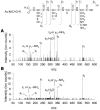
LC-MS/MS analysis of tryptic N-terminal peptides. Shown are the results of analysis using the standard synthetic peptide Ac-MNGREGPNGYV (A) and immunoaffinity-purified T4R Rho (B). The expected product, Ac-MNGR, [M + H]+ at m/z 519.2354, was not observed, but instead, its deaminated product, Ac-MDGR, [M + H]+ at m/z 520.2184, was detected in both cases. Both MS/MS spectra of Ac-MDGR at m/z 520.2310 from the standard (A) and at m/z 520.1900 from T4R Rho (B) were nearly identical. The characteristic fragments for calculated m/z values are as follows: y4 , 520.2184; y3, 347.1673; y2, 232.1404; y1, 175.1189; z3- or y3-NH 3, 330.1408; z1- or y1-NH3, 158.0924; a3-H2O, 300.0970; and a1, 146.0640. The structure at the top shows the fragmentation patterns of this tetrapeptide. The analysis was reproduced in two independent experiments. The deamidated residue is shown in gray.
F<sc>ig</sc>. 6
Fig. 6
UV-visible spectra of WT and T4R Rho after bleaching. Immunoaffinity-purified WT Rho (A and C) and T4R Rho (B and D) were bleached for the indicated times without (A and B) or with (C and D) 20 mm neutral NH2OH. In A and B, the pH of the samples was adjusted to 1.9 with H2SO4 to protonate the Schiff base. The bleaching of WT and T4R Rho was similar, whereas the amount of acid-trapped retinylidene-opsin was lower in the Rho mutant. Similar spectra were obtained in three independent experiments.
F<sc>ig</sc>. 7
Fig. 7
Meta II decay of WT and T4R Rho. A, shown are the fluorescence emission spectra of WT Rho (left panel) and T4R Rho (right panel) before (black trace), immediately after (dark gray trace), and after thorough (light gray trace) photobleaching. B, the decay of WT Rho (black trace) and T4R Rho (red trace) Meta II was recorded with (lower panel) or without (upper panel)10mm NH2OH at pH 6.0. Similar results were obtained in five independent experiments. C, the data from B without NH2OH were fitted to a first-order reaction, which gives the relaxation times (τ) of WT Rho (left panel) and T4R Rho (right panel) Meta II as 33.3 and 12.2 min, respectively. Similar results were obtained in five independent experiments.
F<sc>ig</sc>. 8
Fig. 8
Stability of WT and T4R Rho. The stability of Rho was measured as a change in the UV-visible absorption spectra at its absorption maximum at 37 °C without (A) or with (B) 20 mm neutral NH2OH. The results were plotted assuming 100% absorption at the initial point. Bovine Rho was used as a control for the NH2OH sample. Similar results were obtained in three independent experiments.
F<sc>ig</sc>. 9
Fig. 9
Regeneration of WT and T4R Rho in digitonin. 11-cis-Retinal was added in 50-fold excess to the purified opsins solubilized in 2% digitonin, 10 mm bis-tris propane, and 500 mm NaCl (pH 7.5). UV-visible spectra were recorded as a function of time, and the data were then fitted to a pseudo first-order reaction: ln(A0X) = −kt + C, where X is the absorbance at 500 nm at any given time and A0 is the absorbance at 500 nm before Rho was bleached. WT opsin regenerated at a rate ∼30 times faster than the T4R mutant. Regeneration reached the level of ∼70% compared with the starting amount of Rho.
F<sc>ig</sc>. 10
Fig. 10
Limited proteolysis of WT and T4R Rho. At a 200:1 molar ratio of Rho to trypsin, immunoaffinity-purified WT (A–C) or T4R (D–F) opsin (second through sixth lanes) or Rho (seventh through tenth lanes) was trypsinized for the indicated times (0, 5, 15, 30, and 45 min) at room temperature. Opsin was generated from Rho by bleaching with a Fiber-Lite illuminator for 5 min at room temperature in the presence of 20 mm neutral NH2OH. A and D, silver-stained SDS-polyacrylamide gels; B and E, anti-Rho N terminus antibody B6-30N immunoblots; C and F, anti-Rho C terminus antibody 1D4 immunoblots. The arrows indicates the trypsin band. Similar digestion patterns were obtained in three independent experiments.
F<sc>ig</sc>. 11
Fig. 11
Gt activation assay with WT and T4R Rho*. A and B, Gt incubated with Rho* (see “Experimental Procedures”) was submitted to size exclusion chromatography, and the resulting fractions were examined by immunoblotting with anti-Rho C terminus antibody 1D4 (upper panels) or anti-Gα and anti-Gβt antibodies (lower panels). Fraction numbers are indicated. Similar results were obtained in three independent experiments. In B, GTPγS was present in the samples before loading onto the column. In A and B, the arrows inside the box represent a molecular mass marker of 38.5 kDa. The asterisks denote the fractions with the highest protein content. C, the intrinsic fluorescence Gt assay was performed with WT Rho at pH 6.0 (black trace) and pH 7.5 (blue trace) and with T4R Rho at pH 6.0 (red trace) and pH 7.5 (green trace) and upon incubation 800 s before addition of GTPγS at pH 7.5 (gray trace) at a 1:10 molar ratio of Rho to Gt. The apparent initial rates after curve fitting were calculated as follows: WT Rho at pH 7.5, k0 = 0.0026 s−1; WT Rho at pH 6.0, k0 = 0.0019 s−1; T4R Rho at pH 6.0, k0 = 0.0013 s−1; T4R Rho at pH 7.5, k0 = 0.0005 s−1; and T4R Rho at 800 s > τMeta II at pH 7.5, k0 = 0.0003 s−1. The initial Gt activation rates were calculated from three independent experiments under each condition.

Similar articles

Cited by

References

    1. Mirzadegan T, Benko G, Filipek S, Palczewski K. Biochemistry. 2003;42:2759–2767. - PMC - PubMed
    1. Pierce KL, Premont RT, Lefkowitz RJ. Nat. Rev. Mol. Cell. Biol. 2002;3:639–650. - PubMed
    1. Filipek S, Stenkamp RE, Teller DC, Palczewski K. Annu. Rev. Physiol. 2003;65:851–879. - PMC - PubMed
    1. Ridge KD, Abdulaev NG, Sousa M, Palczewski K. Trends Biochem. Sci. 2003;28:479–487. - PubMed
    1. Okada T, Ernst OP, Palczewski K, Hofmann KP. Trends Biochem. Sci. 2001;26:318–324. - PubMed

Publication types

MeSH terms