. 2004 Nov 15;561(Pt 1):53-64.
doi: 10.1113/jphysiol.2004.069005. Epub 2004 Sep 30.

NMDA receptors induce somatodendritic secretion in hypothalamic neurones of lactating female rats

Affiliations

NMDA receptors induce somatodendritic secretion in hypothalamic neurones of lactating female rats

Christiaan P J de Kock et al. J Physiol. .

Abstract

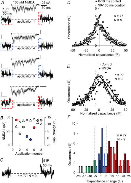
Many neurones in the mammalian brain are known to release the content of their vesicles from somatodendritic locations. These vesicles usually contain retrograde messengers that modulate network properties. The back-propagating action potential is thought to be the principal physiological stimulus that evokes somatodendritic release. In contrast, here we show that calcium influx through NMDA receptor (NMDAR) channels, in the absence of postsynaptic cell firing, is also able to induce vesicle fusion from non-synaptic sites in nucleated outside-out patches of dorsomedial supraoptic nucleus (SON) neurones of adult female rats, in particular during their reproductive stages. The physiological significance of this mechanism was characterized in intact brain slices, where NMDAR-mediated release of oxytocin was shown to retrogradely inhibit presynaptic GABA release, in the absence of postsynaptic cell firing. This implies that glutamatergic synaptic input in itself is sufficient to elicit the release of oxytocin, which in turn acts as a retrograde messenger leading to the depression of nearby GABA synapses. In addition, we found that during lactation, when oxytocin demand is high, NMDA-induced oxytocin release is up-regulated compared to that in non-reproductive rats. Thus, in the hypothalamus, local signalling back and forth between pre- and postsynaptic compartments and between different synapses may occur independently of the firing activity of the postsynaptic neurone.


Figures

Figure 1
Figure 1. Action potentials induce exocytosis from somatic nucleated (i.e. ‘giant’) outside-out patches
A: upper panel, image of acute coronal brain slice containing supraoptic nucleus in close apposition to the optic chiasm; lower image, giant nucleated outside-out patch from dorsomedial neurone. B, inward currents during a single and a short train of action potentials (upper traces) measured under whole-cell voltage clamp in slices from adult lactating female rats (lactating days (L)7–9, average amplitude of inward current 1920.2 ± 252.5 pA, average cell capacitance 29.2 ± 1.8 pF, n = 8). C, current trace, corresponding membrane capacitance and membrane conductance during a single action potential in voltage clamp recording from nucleated outside-out patches in neurones from adult lactating females (average amplitude inward current 130.2 ± 7.0 pA, average patch size 2.3 ± 0.3 pF, n = 8). Capacitance changes were observed in 2 out of 8 patches tested. The lowest trace shows average capacitance change in these two experiments (average capacitance increase 12.1 fF, n = 2). D, same as C, but during a short train of action potentials in voltage clamp. Action potential template as in B. Capacitance changes were observed in 4 out of 8 patches tested (average capacitance increase 29.0 ± 16.3 fF, n = 4). Calibration in A: 20 μm in both panels.
Figure 2
Figure 2. NMDAR activation induces exocytosis without action potential firing of postsynaptic compartment
A, selection of example traces (applications 1, 4–6 shown in B) of NMDA-induced current and corresponding membrane capacitance changes during single 200 ms NMDA (100 μm) applications recorded from the same nucleated outside-out patch (L7–9 females, voltage clamped at −70 mV). Note that in the second and third measurements, the capacitance changes are < 5 fF. Areas marked before and after application of NMDA were used for further analysis of the capacitance trace. Colour coding refers to data in B. B, amplitudes of NMDA current responses (open triangles) and capacitance changes (filled circles, colour coding refers to applications shown in A), respectively, of the applications from the experiment shown in A. C, overall average capacitance change obtained by averaging all responses to NMDA applications (n = 77) in all experiments (N = 9 animals). D, pooled all-points histogram of membrane capacitance during two 10 ms episodes taken with an interval of 80 ms during control recording (i.e. before application of NMDA, n = 77 measurements from N = 9 animals). E, pooled all-points histogram of membrane capacitance during 10 ms after (•) compared to 100 ms before NMDA application (○, n = 77 measurements from N = 9 animals). After NMDA application, the relative membrane capacitance was significantly increased (from 0.12 ± 0.29 to 5.75 ± 0.89 fF, Kolmogorov-Smirnov, P < 0.00001). F, frequency distribution of capacitance responses from all responses (n = 77) on nucleated patches from N = 9 L7–9 females in 5 mm extracellular calcium. Dashed lines indicate separators to define exocytosis (> 5 fF), failures (> −5 fF but < 5 fF) or endocytosis (< −5 fF).
Figure 3
Figure 3. NMDAR-mediated exocytosis in nucleated patches is calcium dependent
A, NMDA-induced current and corresponding membrane capacitance change and membrane conductance measurement during 200 ms NMDA (100 μm) application recorded from a nucleated outside-out patch from L7–9 females voltage clamped at −70 mV (average amplitude of NMDA-induced current 35.9 ± 4.3 pA, average capacitance increase 14.2 ± 1.9 fF, n = 6, N = 1). The lowest trace shows average capacitance change obtained by averaging all > 5 fF responses from all experiments normalized to average capacitance change (N = 9). B, same as in A, but with decreased extracellular calcium concentration, i.e. 2.4 mm Ca2+ (average amplitude of NMDA-induced current 33.0 ± 7.4 pA, n = 6, average capacitance increase 16.7 ± 3.8 fF, n = 2, N = 1). Note that there are fewer > 5 fF responses per recording. C and D, frequency distribution of capacitance responses from all experiments on nucleated patches from lactating females in 5 and 2.4 mm extracellular calcium, respectively (numbers are given). Dashed lines indicate separators to classify responses in exocytosis (> 5 fF), failures (> −5 fF but < 5 fF) and endocytosis (< −5 fF), respectively. E, summary of the probability of exocytotic, failure-like and/or endocytotic responses upon NMDAR activation for the two conditions. Endocytosis probability increased (P < 0.01), failure rate was unaltered, while exocytosis probability was decreased (P < 0.01) upon lowering the extracellular calcium concentration (unpaired t tests).
Figure 4
Figure 4. Somatic vesicle release induced by NMDAR activation is reduced before the reproductive cycle
A, NMDA-induced current and corresponding membrane capacitance changes during 200 ms NMDA (100 μm) application recorded from nucleated outside-out patches from adult virgin animals, voltage clamped at −70 mV in 5 mm extracellular calcium (average amplitude of NMDA-induced current 36.3 ± 6.9 pA, average capacitance increase 14.5 ± 3.1 fF, n = 7). We have averaged the three types of capacitance responses within this experiment using the −5 fF to 5 fF ‘failure’ range as classification criterium. B, probability of endocytosis-like capacitance changes at virgin stage is increased compared to females at L7–9 (unpaired t test P < 0.01) whereas exocytosis rate is reduced at virgin stage (P < 0.01). C and D, frequency distribution of pooled capacitance responses from all experiments on nucleated patches from lactating females and adult virgin animals (77 versus 141 NMDA applications) in 5 mm extracellular calcium. Note: this extracellular concentration was used to fortify the probability of exocytosis; no capacitance responses > 5 fF were observed, N = 3 virgin animals at 5 mm (n = 25) (not shown).
Figure 5
Figure 5. Postsynaptic NMDA receptor activation inhibits GABAergic transmission
A, voltage protocol for NMDAR activation by endogenous glutamate. Experiments were performed in the presence of CNQX (10 μm), L-CGG-III (10 μm) and CPPG (30 nm). B, time course of the frequency of spontaneous GABAergic IPSCs recorded from oxytocin neurones in slices of adult lactating female rats at −30 mV. Frequency is normalized to that during first 10 s at −30 mV. Open bar is an average of the last 5 points (n = 14). C and D, example traces of IPSCs at start (C, shown by left box in A) and at the end (D, right box in A) at −30 mV. The insets show IPSCs at higher time resolution (calibration 40 pA, 50 ms). E, at −30 mV the specific NMDA antagonist APV (50 μm) significantly reduced the rapid decline in sIPSC frequency (residual sIPSC frequency 87.5 ± 7.2%, n = 7, 2-way ANOVA, P < 0.01). F, summary of the effects of specific blockers on the frequency of GABAergic sIPSCs at −30 mV. d(CH2)5-OVT ([des-glycinamide9,d(CH2),O-Me-Tyr2,Thr4,Orn8]-vasotocin; 1 μm) partly but significantly reduced the retrograde effect (n = 6, ANOVA P < 0.01), indicating that oxytocin receptors were involved (*P < 0.01; **P < 0.001).
Figure 6
Figure 6. NMDAR activation induces release of retrograde messengers to inhibit GABAergic synaptic transmission
a, glutamatergic synaptic transmission activates NMDA receptors, which induce calcium influx. b, in turn, oxytocin is released from non-synaptic locations to act as a retrograde messenger, reducing GABAergic synaptic transmission, via both a presynaptic (c) and a postsynaptic (d) mechanism.

Similar articles

Cited by

References

    1. Al-Ghoul WM, Meeker RB, Greenwood RS. Differential expression of five N-methyl-D-aspartate receptor subunit mRNAs in vasopressin and oxytocin neuroendocrine cells. Brain Res Mol Brain Res. 1997;44:262–272. 10.1016/S0169-328X(96)00205-7. - DOI - PubMed
    1. Artalejo CR, Elhamdani A, Palfrey HC. Sustained stimulation shifts the mechanism of endocytosis from dynamin-1-dependent rapid endocytosis to clathrin- and dynamin-2-mediated slow endocytosis in chromaffin cells. Proc Natl Acad Sci U S A. 2002;99:6358–6363. 10.1073/pnas.082658499. - DOI - PMC - PubMed
    1. Bekkers JM. Properties of voltage-gated potassium currents in nucleated patches from large layer 5 cortical pyramidal neurones of the rat. J Physiol. 2000;525:593–609. - PMC - PubMed
    1. Brussaard AB, Devay P, Leyting-Vermeulen JL, Kits KS. Changes in properties and neurosteroid regulation of GABAergic synapses in the supraoptic nucleus during the mammalian female reproductive cycle. J Physiol. 1999;516:513–524. - PMC - PubMed
    1. Brussaard AB, Herbison AE. Long-term plasticity of postsynaptic GABAA-receptor function in the adult brain: insights from the oxytocin neurone. Trends Neurosci. 2000;23:190–195. - PubMed

MeSH terms

LinkOut - more resources