

Interhemispheric interaction between human dorsal premotor and contralateral primary motor cortex
- PMID: 15459244
- PMCID: PMC1665328
- DOI: 10.1113/jphysiol.2004.072843
Interhemispheric interaction between human dorsal premotor and contralateral primary motor cortex
Abstract
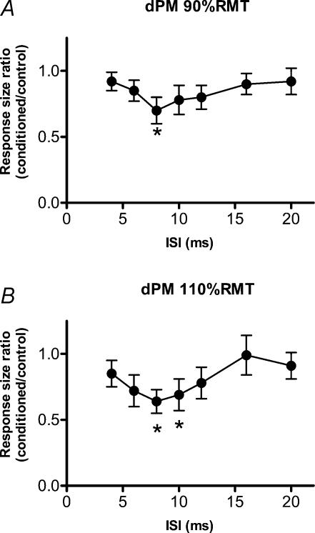
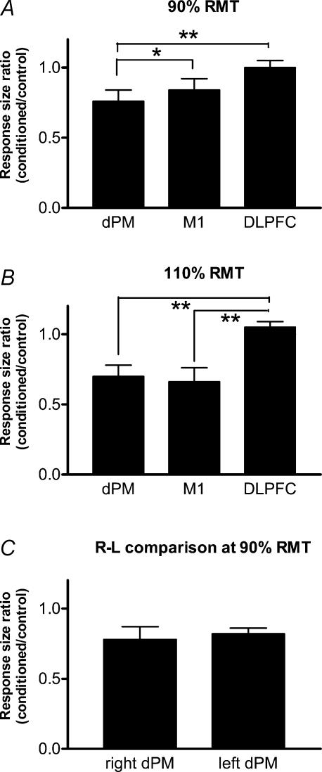
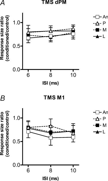
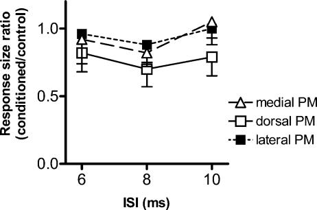
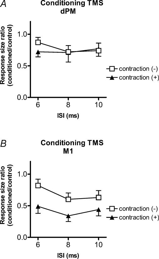
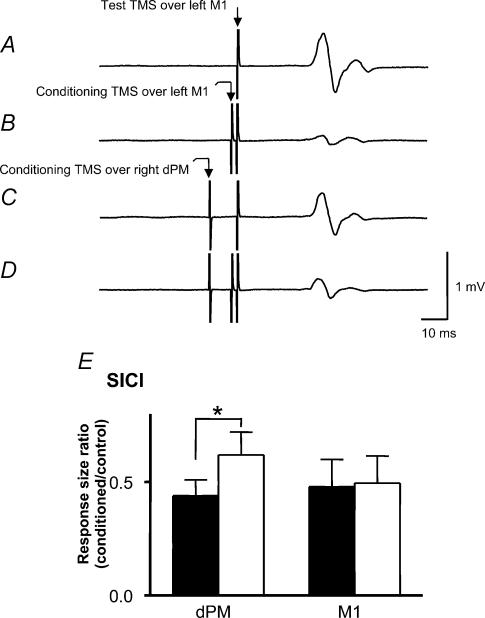
We used transcranial magnetic stimulation (TMS) in a paired pulse protocol to investigate interhemispheric interactions between the right dorsal premotor (dPM) and left primary motor cortex (M1) using interstimulus intervals of 4, 6, 8, 10, 12, 16 and 20 ms in ten healthy subjects. A conditioning stimulus over right dPM at an intensity of either 90 or 110% resting motor threshold (RMT) suppressed motor-evoked potentials (MEPs) evoked in the first dorsal interosseous (FDI) muscle by stimulation of left M1. Maximum effects occurred for interstimulus intervals (ISIs) of 8-10 ms. There was no effect if the conditioning stimulus was applied 2.5 cm lateral, anterior or medial to dPM. The effect differed from previously described M1 interhemispheric inhibition in that the threshold for the latter was greater than 90% RMT, whereas stimulation of the dPM at the same intensity led to significant inhibition. In addition, voluntary contraction of the left FDI (i.e. contralateral to the conditioning TMS) enhanced interhemispheric inhibition from right M1 but had no effect on the inhibition from right dPM. Finally, conditioning to right dPM at 90% RMT reduced short-interval intracortical inhibition (SICI; at ISI = 2 ms) in left M1 whilst there was no effect if the conditioning stimulus was applied to right M1. We conclude that conditioning TMS over dPM has effects that differ from the previous pattern of interhemispheric inhibition described between bilateral M1s. This may reflect the existence of commissural fibres between dPM and contralateral M1 that may play a role in bimanual coordination.
Figures
Similar articles
-
Organization of ipsilateral excitatory and inhibitory pathways in the human motor cortex.J Neurophysiol. 2003 Mar;89(3):1256-64. doi: 10.1152/jn.00950.2002. Epub 2002 Oct 30. J Neurophysiol. 2003. PMID: 12611955
-
Further evidence for excitability changes in human primary motor cortex during ipsilateral voluntary contractions.Neurosci Lett. 2008 Mar 12;433(2):135-40. doi: 10.1016/j.neulet.2007.12.058. Epub 2008 Jan 10. Neurosci Lett. 2008. PMID: 18261851
-
Effect of low-frequency repetitive transcranial magnetic stimulation on interhemispheric inhibition.J Neurophysiol. 2005 Sep;94(3):1668-75. doi: 10.1152/jn.01306.2004. Epub 2005 May 4. J Neurophysiol. 2005. PMID: 15872061
-
Triad TMS of the human motor cortex.Neurosci Res. 2020 Jul;156:245-249. doi: 10.1016/j.neures.2019.11.005. Epub 2019 Nov 12. Neurosci Res. 2020. PMID: 31730779 Review.
-
PMd and action preparation: bridging insights between TMS and single neuron research.Trends Cogn Sci. 2023 Aug;27(8):759-772. doi: 10.1016/j.tics.2023.05.001. Epub 2023 May 25. Trends Cogn Sci. 2023. PMID: 37244800 Review.
Cited by
-
Increased primary motor cortical excitability by a single-pulse transcranial magnetic stimulation over the supplementary motor area.Exp Brain Res. 2012 Jun;219(3):339-49. doi: 10.1007/s00221-012-3095-7. Epub 2012 Apr 25. Exp Brain Res. 2012. PMID: 22532164
-
Recovery of motor function after stroke.Dev Psychobiol. 2012 Apr;54(3):254-62. doi: 10.1002/dev.20508. Epub 2010 Nov 17. Dev Psychobiol. 2012. PMID: 22415914 Free PMC article. Review.
-
Motor and premotor cortices in subcortical stroke: proton magnetic resonance spectroscopy measures and arm motor impairment.Neurorehabil Neural Repair. 2013 Jun;27(5):411-20. doi: 10.1177/1545968312469835. Epub 2013 Jan 8. Neurorehabil Neural Repair. 2013. PMID: 23300210 Free PMC article.
-
Transcranial magnetic stimulation of the brain: What is stimulated? - A consensus and critical position paper.Clin Neurophysiol. 2022 Aug;140:59-97. doi: 10.1016/j.clinph.2022.04.022. Epub 2022 May 18. Clin Neurophysiol. 2022. PMID: 35738037 Free PMC article. Review.
-
Activity of right premotor-parietal regions dependent upon imagined force level: an fMRI study.Front Hum Neurosci. 2014 Oct 8;8:810. doi: 10.3389/fnhum.2014.00810. eCollection 2014. Front Hum Neurosci. 2014. PMID: 25339893 Free PMC article.
References
-
- Chen R, Yung D, Li J. Organization of ipsilateral excitatory and inhibitory pathways in the human motor cortex. J Neurophysiol. 2003;89:1256–1264. - PubMed
-
- Chouinard PA, Van Der Werf YD, Leonard G, Paus T. Modulating neural networks with transcranial magnetic stimulation applied over the dorsal premotor and primary motor cortices. J Neurophysiol. 2003;90:1071–1083. - PubMed
-
- Civardi C, Cantello R, Asselman P, Rothwell JC. Transcranial magnetic stimulation can be used to test connections to primary motor areas from frontal and medial cortex in humans. Neuroimage. 2001;14:1444–1453. - PubMed
-
- Daskalakis ZJ, Christensen BK, Fitzgerald PB, Roshan L, Chen R. The mechanisms of interhemispheric inhibition in the human motor cortex. J Physiol. 2002;543:317–326. 10.1113/jphysiol.2002.017673. - DOI - PMC - PubMed
-
- De Gennaro L, Bertini M, Pauri F, Cristiani R, Curcio G, Ferrara M, et al. Callosal effects of transcranial magnetic stimulation (TMS): the influence of gender and stimulus parameters. Neurosci Res. 2004;48:129–137. 10.1016/j.neures.2003.10.004. - DOI - PubMed
MeSH terms
LinkOut - more resources
Full Text Sources