. 2004 Oct 12;101(41):14901-6.
doi: 10.1073/pnas.0406196101. Epub 2004 Sep 30.

Mediation of Af4 protein function in the cerebellum by Siah proteins

Affiliations

Mediation of Af4 protein function in the cerebellum by Siah proteins

Peter L Oliver et al. Proc Natl Acad Sci U S A. .

Abstract

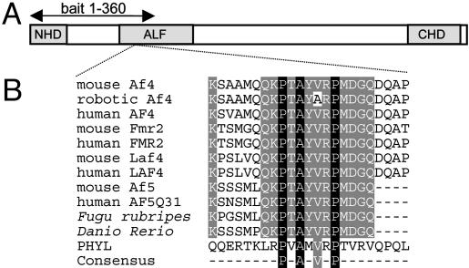
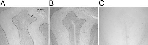
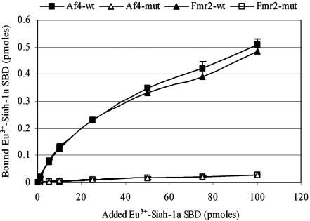
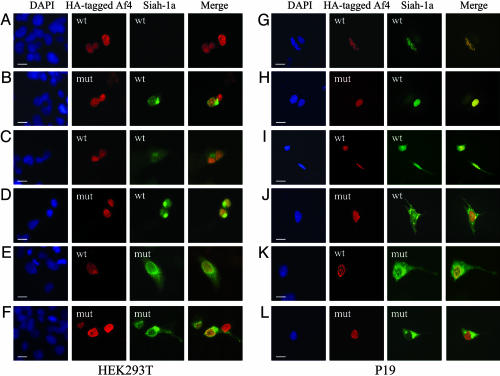
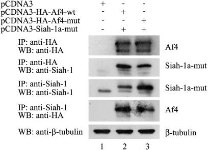
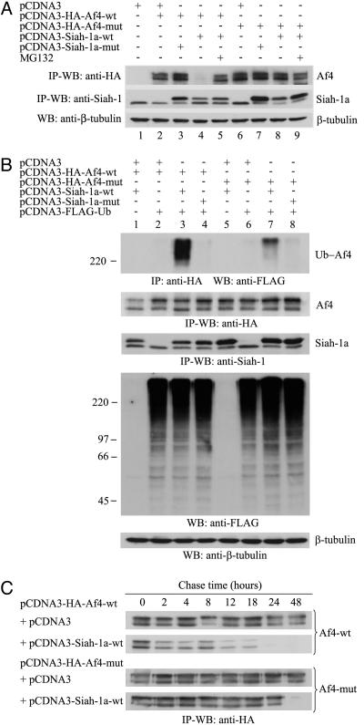
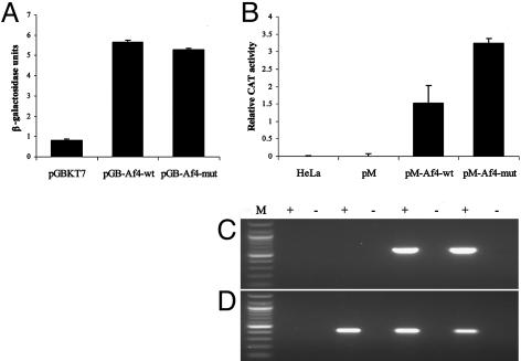
We have established that the gene AF4, which had long been recognized as disrupted in childhood leukemia, also plays a role in the CNS. Af4 is mutated in the robotic mouse that is characterized by ataxia and Purkinje cell loss. To determine the molecular basis of this mutation, we carried out a yeast two-hybrid screen and show that Af4 binds the E3 ubiquitin ligases Drosophila seven in absentia (sina) homologues (Siah)-1a and Siah-2 in the brain. Siah-1a and Af4 are expressed in Purkinje cells and colocalize in the nucleus of human embryonic kidney 293T and P19 cells. In vitro binding assays and coimmunoprecipitation reveal a significant reduction in affinity between Siah-1a and robotic mutant Af4 compared with wild-type, which correlates with the almost complete abolition of mutant Af4 degradation by Siah-1a. These data strongly suggest that an accumulation of mutant Af4 occurs in the robotic mouse due to a reduction in its normal turnover by the proteasome. A significant increase in the transcriptional activity of mutant Af4 relative to wild-type was obtained in mammalian cells, suggesting that the activity of Af4 is controlled through Siah-mediated degradation. Another member of the Af4 family, Fmr2, which is involved in mental handicap in humans, binds Siah proteins in a similar manner. These results provide evidence that a common regulatory mechanism exists that controls levels of the Af4/Fmr2 protein family. The robotic mouse thus provides a unique opportunity to understand how these proteins play a role in disorders as diverse as leukemia, mental retardation, and neurodegenerative disease.


Figures

Fig. 1.
Fig. 1.
ALF family protein structure. (A) The conserved N- and C-terminal homology (NHD/CHD) and ALF domains are shown and the yeast two-hybrid bait region is marked. (B) Multiple amino acid alignment of the ALF family including the 23-mer Af4 peptides used for the Siah-1a dissociation-enhanced lanthanide fluorescence immunoassay. GenBank accession nos. from top to bottom: NP_598680, AY749168, NP_005926, NP_032058, NP_002016, XP_283603, NP_002276, NP_291043, and NP_055238; Fugu rubripes and Danio rerio sequences were taken from ENSEMBL database-predicted proteins SINFRUP00000169375 and ENSDARP0000009290, respectively, which can be accessed at www.ensembl.org. The Siah-binding sequence of PHYL (NP_725394) is shown, in addition to the consensus motif (18).
Fig. 2.
Fig. 2.
Coexpression of Af4 and Siah-1a in mouse cerebellum. In situ hybridization was performed from wild-type mouse brain sections by using antisense riboprobes for Af4 (A) and Siah-1a (B). Expression of both genes occurs in the Purkinje cell layer (PCL). (C) A negative control sense probe for Siah-1a is shown. Lobe II of the cerebellum is shown.
Fig. 3.
Fig. 3.
In vitro binding assay of Siah-1a to wild-type and mutant Af4 and Fmr2 peptides. Biotinylated Af4/Fmr2 wild-type and mutant peptides were incubated in the presence of increasing quantities of Eu3+-labeled recombinant Siah-1a SBD. The amount of bound protein was determined by converting the fluorescence count values to that obtained for the Eu3+ standard (0.1 pmol). Data are presented as means + SD for three independent experiments.
Fig. 4.
Fig. 4.
Siah-1a and Af4 colocalization in mammalian cells. HEK293T cells (A-F) and differentiated P19 cells (G-L) were cotransfected with equal amounts of pCDNA3-HA-Af4-wt (A, C, E, G, I, and K) or pCDNA3-HA-Af4-mut (B, D, F, H, J, and L) and pCDNA3-Siah-1a-wt (A-D and G-J) or pCDNA3-Siah-1a-mut (E, F, K, and L) constructs. Cells were cultured for 7 h in the presence (C, D, I, and J) or absence (A, B, E, F, K, and L) of MG132. Cells were labeled with mouse anti-HA and goat anti-Siah-1 primary antibodies followed by Alexa Fluor 594 anti-mouse and Alexa Fluor 488 anti-goat secondary antibodies. Preparations were counterstained with 4′,6-diamidino-2-phenylindole (DAPI) to visualize the nuclei (blue); regions of overlap between Af4 (red) and Siah-1a (green) appear in yellow in the merged images. (Scale bars, 10 μm.)
Fig. 5.
Fig. 5.
Siah-1a interacts with Af4 in mammalian cells. HEK293T cells were cotransfected with the indicated constructs. Total cell lysates were immunoprecipitated (IP), resolved by SDS/PAGE, and analyzed by Western blot (WB). To verify equal inputs, 20 μg of each total lysate was analyzed by Western blot by using an anti-β-tubulin antibody.
Fig. 6.
Fig. 6.
Siah-1a mediates Af4 degradation through the Ub-proteasome pathway. (A-C) HEK293T cells were cotransfected with the indicated pCDNA3 constructs. Unless otherwise stated, cells were cultured for 7 h in the presence of MG132. In C, cells were subsequently washed and chased for the indicated times in the presence of cycloheximide. Total cell lysates were analyzed as in Fig. 5. In B, FLAG-Ub-conjugated proteins were detected by Western blot from 20 μg of each lysate using an anti-FLAG antibody.
Fig. 7.
Fig. 7.
Effect of the robotic mutation on the transactivation potential of Af4. (A) Yeast o-nitrophenyl-β-d-galactopyranoside activation assay. pGBKT7 fusions were transformed into Y187 cells and β-galactosidase activity was assayed. One unit of β-galactosidase is defined as the amount that hydrolyses 1 mmol of o-nitrophenyl-β-d-galactopyranoside to o-nitrophenol and d-galactose per minute. Results are shown + SD. (B) Mammalian cell activation assay. pM fusions were cotransfected into HeLa cells with reporter vector pG5CAT. After transfection, CAT activity was measured by ELISA. Results are shown + SD. (C and D) RT-PCR analysis of mammalian activation assay. Primers specific for pM-Af4-wt/mut (C) or pG5CAT (D) were used to amplify cDNA from the corresponding cotransfections in B. + and -, reactions with and without the presence of reverse transcriptase in the cDNA synthesis step. Reactions were run with a 100-bp ladder (lane M) (New England Biolabs).

References

    1. Heintz, N. & Zoghbi, H. (2000) Annu. Rev. Physiol. 62, 779-802. - PubMed
    1. Patil, N., Cox, D. R., Bhat, D., Faham, M., Myers, R. M. & Peterson, A. S. (1995) Nat. Genet. 2, 126-129. - PubMed
    1. Zuo, J., De Jager, P. L., Takahashi, K. A., Jiang, W., Linden, D. J. & Heintz, N. (1997) Nature 388, 769-773. - PubMed
    1. Burright, E. N., Orr, H. T. & Clark, H. B. (1997) Brain Pathol. 7, 965-977. - PMC - PubMed
    1. Isaacs, A. M., Oliver, P. L., Jones, E. L., Jeans, A., Potter, A., Hovik, B. H., Nolan, P. M., Vizor, L., Glenister, P., Simon, A. K., et al. (2003) J. Neurosci. 23, 1631-1637. - PMC - PubMed

MeSH terms

Associated data

LinkOut - more resources