

Review
doi: 10.1038/nrm1494.
Imaging gene expression in single living cells
Affiliations
- PMID: 15459666
- PMCID: PMC4942131
- DOI: 10.1038/nrm1494
Item in Clipboard
Review
Imaging gene expression in single living cells
Nat Rev Mol Cell Biol.
2004 Oct.
Abstract
Technical advances in the field of live-cell imaging have introduced the cell biologist to a new, dynamic, subcellular world. The static world of molecules in fixed cells has now been extended to the time dimension. This allows the visualization and quantification of gene expression and intracellular trafficking events of the studied molecules and the associated enzymatic processes in individual cells, in real time.
Figures
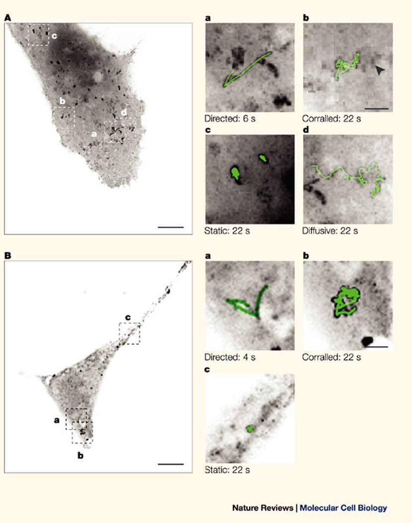
A | An mRNA transcript that contains the coding sequence of lacZ, 24 MS2 stem–loops and the 3' untranslated region (UTR) of the human growth-hormone gene (hGH) was transiently expressed in COS cells together with the green fluorescent protein (GFP)–MS2 fusion protein. GFP–MS2-tagged RNA particles were followed, and different types of motility were detected in single living cells: directed (Aa), corralled (Ab), static (Ac) and diffusive (Ad). The image in A is a maximum-intensity image projection of 200 time frames. Bar, 10 µm. Panels Aa–Ad are magnified sections of A that show an mRNA track (in green) superimposed on an enlargement from each of the indicated boxed areas. The arrowhead points to a 'static' particle in the vicinity of a 'corralled' particle. Bar, 2 µm. B | An mRNA transcript that contains the coding sequence of lacZ, 24 MS2 stem–loops and the 3' UTR of SV40 was transiently expressed in COS cells together with the GFP–MS2 protein and analysed as above. Reproduced with permission from Ref.
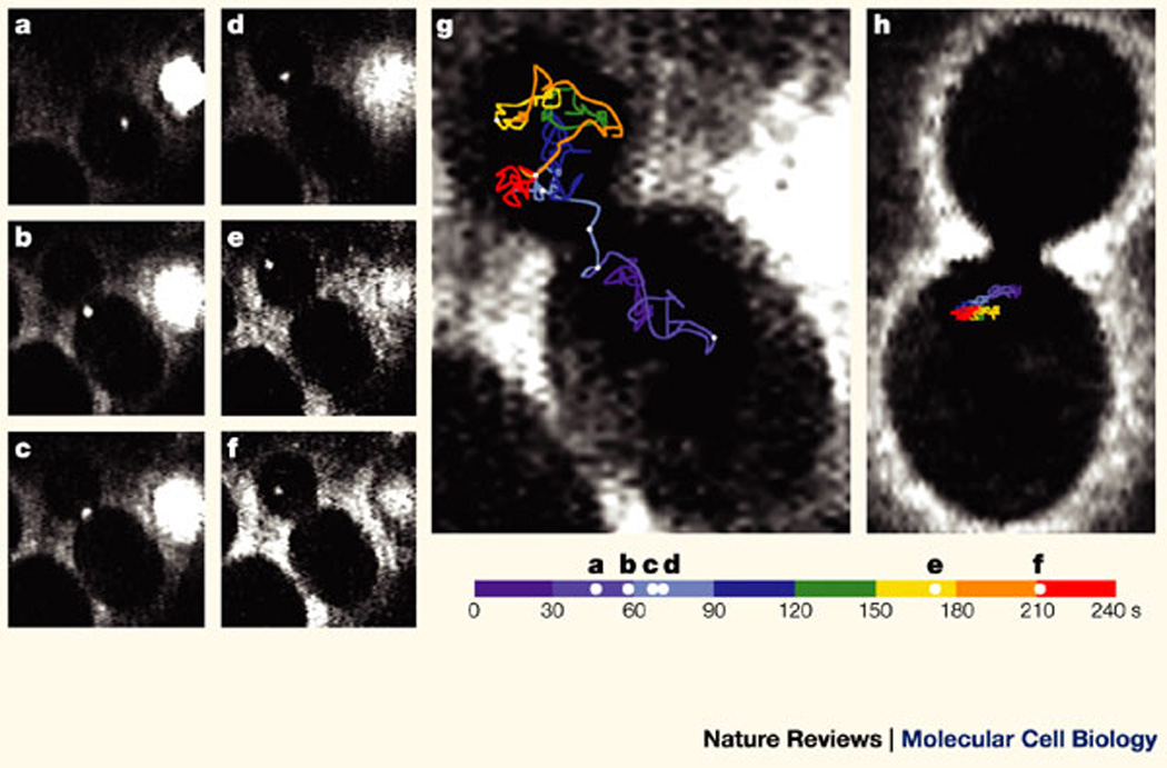
Yeast that expresses both the ASH1 (asymmetric synthesis of HO endonuclease) reporter gene, which contains MS2 stem–loops, and the green fluorescent protein (GFP)–MS2 protein were observed using epifluorescence and bright-field microscopy. Movement of the GFP–MS2-coated ASH1 mRNA particle was recorded and followed over time. Images are presented at indicated intervals. a–f | Movement of the particle from the mother cell to the bud (total distance: 23 µm in 128 s). g | The diagram shows the total path of the particle movement (43 µm in 240 s). Intervals of 30 s each are represented by different colours and images a–f are indicated by white dots on the travel line. The particle spends 180 out of 240 s in the bud and ~ 60 s localized at or near the bud tip. h | A yeast strain that contains a deletion of she1 (a microfilament motor that is required for the localization of ASH1 to the bud tip) and was analysed by the same approach showed significantly less net displacement and stayed within the mother cell, never localizing to the bud tip. Bar, 2 µm. Reproduced with permission from Ref.
References
-
- Gerlich D, Ellenberg J. 4D imaging to assay complex dynamics in live specimens. Nature Cell Biol. 2003;5:S14–S19. - PubMed
-
- Zimmermann T, Rietdorf J, Pepperkok R. Spectral imaging and its applications in live cell microscopy. FEBS Lett. 2003;546:87–92. - PubMed
-
- Patterson G, Day RN, Piston D. Fluorescent protein spectra. J. Cell Sci. 2001;114:837–838. - PubMed
-
- Peercy PS. The drive to miniaturization. Nature. 2000;406:1023–1026. - PubMed
-
- Carrington WA, Fogarty K, Fay FS. In: 3D fluorescence imaging of single cells using image restoration. Foskett K, Grinstein S, editors. New York: Wiley–Liss Inc.; 1990.
Publication types
MeSH terms
Substances
Grants and funding
LinkOut - more resources
Full Text Sources
Other Literature Sources
Medical
Molecular Biology Databases
