. 2004 Dec;87(6):3775-85.
doi: 10.1529/biophysj.104.050955. Epub 2004 Oct 1.

Filtering of calcium transients by the endoplasmic reticulum in pancreatic beta-cells

Affiliations

Filtering of calcium transients by the endoplasmic reticulum in pancreatic beta-cells

Richard Bertram et al. Biophys J. 2004 Dec.

Abstract

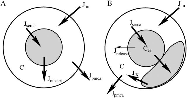
Calcium handling in pancreatic beta-cells is important for intracellular signaling, the control of electrical activity, and insulin secretion. The endoplasmic reticulum (ER) is a key organelle involved in the storage and release of intracellular Ca2+. Using mathematical modeling, we analyze the filtering properties of the ER and clarify the dual role that it plays as both a Ca2+ source and a Ca2+ sink. We demonstrate that recent time-dependent data on the free Ca2+ concentration in pancreatic islets and beta-cell clusters can be explained with a model that uses a passive ER that takes up Ca2+ when the cell is depolarized and the cytosolic Ca2+ concentration is elevated, and releases Ca2+ when the cell is repolarized and the cytosolic Ca2+ is at a lower concentration. We find that Ca2+-induced Ca2+ release is not necessary to explain the data, and indeed the model is inconsistent with the data if Ca2+-induced Ca2+ release is a dominating factor. Finally, we show that a three-compartment model that includes a subspace compartment between the ER and the plasma membrane provides the best agreement with the experimental Ca2+ data.


Figures

FIGURE 1
FIGURE 1
Illustration of the Ca2+ fluxes incorporated in (A) the two-compartment model and (B) the three-compartment subspace model. The thin arrow in B represents a small flux from the ER to the bulk cytosol.
FIGURE 2
FIGURE 2
(A) Simulated voltage protocol mimicking the biphasic response to an increase in glucose concentration. (B) Cytosolic Ca2+ response with SERCA pumps enabled (solid line) and disabled (dashed line). During each imposed oscillation, the nadir is defined as the lowest value of C during the repolarized phase. The amplitude is the difference between the peak and the nadir. (C) The ER Ca2+ concentration. No driving force is established between the ER and the cytosol when SERCA pumps are disabled (dashed line).
FIGURE 3
FIGURE 3
(A) Doubling the rate of Ca2+ release from the ER, per, has a transient effect on the cytosolic Ca2+ response (dashed line) to the voltage protocol, but no long-term effect. (B) Cer adapts to the doubling in per (dashed line) by declining to a value one-half that of the control.
FIGURE 4
FIGURE 4
(A) Reducing the rate of pumping across the plasma membrane by half (dashed line) slightly increases the amplitude of the Ca2+ response to depolarization and greatly increases the Ca2+ nadir. (B) The increase in the Ca2+ nadir is due to an increase in Cer.
FIGURE 5
FIGURE 5
(A) An increase in the glucose concentration is mimicked (at arrow) by increasing the depolarized phase duration from 8 s to 12 s, while keeping the oscillation period fixed at 24 s. (B) In the control simulation (solid line), increasing the duration of depolarization results in an increase in the Ca2+ nadir, but not in the amplitude, whereas there is no effect when SERCA pumps are disabled (dashed line). (C) In the control simulation, an increase in the ER Ca2+ concentration is responsible for the increased cytosolic Ca2+ nadir.
FIGURE 6
FIGURE 6
(A) When the Ca2+ channel conductance, gCa, is doubled (at arrow), both the Ca2+ amplitude and nadir are increased. This is true with SERCA pumps enabled (solid line) or disabled (dashed line), although the nadir increase in the latter case is quite small. (B) As in Fig. 5, the nadir increase is due primarily to an increase in the ER Ca2+ concentration, although increased Ca2+ entry also plays a role.
FIGURE 7
FIGURE 7
(A) Voltage protocol for compound bursting. (B) In the control system the Ca2+ nadir rises during a compound burst and slowly falls between (solid line). This does not occur when the SERCA pumps are disabled (dashed line). (C) The rise and fall of the nadir reflects the dynamics of ER Ca2+.
FIGURE 8
FIGURE 8
Demonstration that the slow rise and fall of the cytosolic Ca2+ nadir depends critically on the speed at which the ER takes up and releases Ca2+. (A) The speed of the ER is increased by setting fer = 0.1 (solid line), or decreased by setting fer = 0.0002 (dashed line). (B) There are no slow Cer dynamics when fer = 0.1, and there is only a small increase in Cer during a compound burst when fer = 0.0002.
FIGURE 9
FIGURE 9
Ca2+ response to a sequence of depolarizations when CICR dominates release from the ER. (A) Sequence of brief depolarizations. (B) Cytosolic Ca2+ rises during depolarization and falls during repolarization. (C) The ER releases Ca2+ during the depolarization due to CICR, and refills during the repolarization.
FIGURE 10
FIGURE 10
(A) Biphasic voltage protocol applied to the model in which CICR dominates. (B) Even with active SERCA pumps the Ca2+ nadir is not elevated and the amplitude is large (solid line), so that blocking SERCA pumps has only a small effect (dashed line). (C) The cytosolic Ca2+ nadir is not elevated despite the fact that the ER is filled.
FIGURE 11
FIGURE 11
Ca2+ concentrations during a sustained depolarization from −70 mV to −30 mV with the three-compartment subspace model. (A) The weighted average of the bulk cytosolic Ca2+ and the subspace Ca2+ concentrations is lower when SERCA pumps are inhibited (dashed line) than in the control system (solid line). The ER Ca2+ concentration (B) and the subspace Ca2+ concentration (D) are lower when SERCA pumps are inhibited. (C) The bulk cytosolic concentration satisfies Eq. 16, so its steady state is independent of the Cer or ER parameters.

Similar articles

Cited by

References

    1. Ämmälä, C., P.-O. Larsson, P.-O. Berggren, K. Bokvist, L. Juntti-Berggren, H. Kindmark, and P. Rorsman. 1991. Inositol trisphosphate-dependent periodic activation of a Ca2+-activated K+ conductance in glucose-stimulated pancreatic β-cells. Nature. 353:849–852. - PubMed
    1. Arredouani, A., Y. Guiot, J.-C. Jonas, L. H. Liu, M. Nenquin, J. A. Pertusa, J. Rahier, J.-F. Rolland, G. E. Shull, M. Stevens, F. Wuytack, J. C. Henquin, and P. Gilon. 2002a. SERCA3 ablation does not impair insulin secretion but suggests distinct roles of different sarcoendoplasmic reticulum Ca2+ pumps for Ca2+ homeostasis in pancreatic β-cells. Diabetes. 51:3245–3253. - PubMed
    1. Arredouani, A., J.-C. Henquin, and P. Gilon. 2002b. Contribution of the endoplasmic reticulum to the glucose-induced [Ca2+]c response in mouse pancreatic islets. Am. J. Physiol. 282:E982–E991. - PubMed
    1. Ashcroft, F. M., and P. Rorsman. 1989. Electrophysiology of the pancreatic β-cell. Prog. Biophys. Mol. Biol. 54:87–143. - PubMed
    1. Barbosa, R. M., A. M. Silva, A. R. Tomé, J. A. Stamford, R. M. Santos, and L. M. Rosário. 1998. Control of pulsatile 5-HT/insulin secretion from single mouse pancreatic islets by intracellular calcium dynamics. J. Physiol. 510:135–143. - PMC - PubMed

Publication types