. 2004 Oct 12;101(41):14931-6.
doi: 10.1073/pnas.0404416101. Epub 2004 Oct 1.

Conditional mutagenesis using site-specific recombination in Plasmodium berghei

Affiliations

Conditional mutagenesis using site-specific recombination in Plasmodium berghei

Teresa Gil Carvalho et al. Proc Natl Acad Sci U S A. .

Abstract

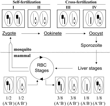
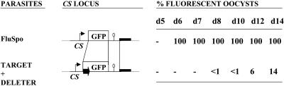
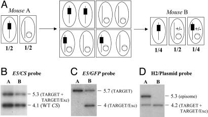
Reverse genetics in Plasmodium, the genus of parasites that cause malaria, still faces major limitations. Only red blood cell stages of this haploid parasite can be transfected. Consequently, the function of many essential genes in these and subsequent stages, including those encoding vaccine candidates, cannot be addressed genetically. Here, we establish conditional mutagenesis in Plasmodium by using site-specific recombination and the Flp/FRT system of yeast. Site-specific recombination is induced after cross-fertilization in the mosquito vector of two clones containing either the target sequence flanked by two FRT sites or the Flp recombinase. Parasites that have undergone recombination are recognized in the cross progeny through the expression of a fluorescence marker. This approach should permit to dissect the function of any essential gene of Plasmodium during the haploid phase of its life, i.e., during infection of salivary glands in the mosquito and infection of both the liver and red blood cells in the mammal.


Figures

Fig. 1.
Fig. 1.
Strategy for conditional mutagenesis in Plasmodium. The life cycle of Plasmodium takes place in a mammalian and a mosquito host. The haploid, RBC stages of the parasite generate gametocytes (parallel lines). In the lumen of the mosquito midgut, released gametocytes transform into gametes, which fertilize to create a zygote. Zygotes transform into ookinetes, which differentiate into oocysts. These three parasite stages (underlined) contain the two parental genomes; oocysts contain thousands of copies of the genomes generated by meiotic reduction in the diploid zygote. Sporozoites bud off from the multinucleate oocysts, traverse mosquito salivary glands, and invade mammalian hepatocytes, where they generate RBC-infecting stages. Sporozoites, liver stages, and RBC stages are uninucleate, haploid stages of the parasite. Shown around the life cycle is a parasite cross for conditional mutagenesis; boxes and ellipses indicate parasitic cells and nuclei, respectively. Two parasite clones are mixed, each carrying one marker (black or open rectangles, symbolizing a flirted sequence and the Flp-encoding locus, respectively) on distinct chromosomes (thin or thick lines, respectively). Self-fertilization propagates the parental genotypes, A+/B- and A-/B+, in type I and II cells, respectively. Cross-fertilization either regenerates the parental genotypes within type III cells or creates new, hybrid genotypes (A-/B- and A+/B+) in type IV cells. Assuming similar frequencies of self- and cross-fertilizations, as well as random chromosome segregation during meiosis in the zygotes, then 1/8 of the sporozoites, and subsequent liver and RBC stages, should contain both markers in their haploid genome.
Fig. 2.
Fig. 2.
Construction of the P. berghei TARGET and DELETER clones. (A)(Left) Schematic representation of the WT CS locus and the TARGET recombinant locus generated by homologous integration of plasmid pTARGET at the CS locus of WT P. berghei NK65 (not drawn to scale). Plasmid pTARGET contained 1.3 kb of CS upstream region (thin arrow), the hDHFR selectable marker (M, 1.7 kb, including its own expression sequences) flanked on either side by a FRT sequence (solid arrows), the GFP gene (0.7 kb) followed by 0.3 kb of CS downstream region (ellipse), and a pUC plasmid backbone (thick line). Plasmid pTARGET integrated via the CS promoter region, which is thus duplicated in the TARGET locus. The predicted size (in kilobases) of restriction fragments generated by digestion with EcoRV (E5) or AflII (A2) in the WT CS or the TARGET locus is shown. (Right) Southern hybridization of genomic DNA of the WT and TARGET P. berghei by using a CS internal probe. Mouse A and B correspond to parasite RBC stages collected before and after cycling through mosquitoes, respectively. (B) Schematic representation of the TARGET/Exc locus created by Flp-mediated SSR at the TARGET locus. In the TARGET/Exc locus, the 5′ promoter region of the CS gene is WT to the ATG start codon. The start codon is immediately followed by the CTTAAGGC sequence (AflII restriction site underlined), the FRT site (boxed, two inverted repeats in italics flanking the central spacer region), a CTTAAG sequence (AflII restriction site), and the full-length GFP sequence. The TARGET/Exc locus therefore encodes a GFP protein possessing the N-terminal extension MLKAKFLFSRKYRNFLK. (C) (Left) Schematic representation of the WT TRAP locus and the DELETER recombinant locus generated by homologous integration of the plasmid pDELETER at the TRAP locus of WT P. berghei NK65 (not drawn to scale). The plasmid pDELETER contained 1.5 kb of TRAP promoter region (thin arrow), the Flp gene (1.3 kb), 0.6 kb of TRAP downstream region (ellipse), the pUC backbone (thick line), and the hDHFR selectable marker (M, 1.7 kb). The plasmid pDELETER integrated via the TRAP promoter region, so that in the DELETER locus, expression of both Flp and TRAP is controlled by TRAP expression sequences. The predicted size (in kilobases) of restriction fragments generated by digestion with BamHI (B) or HincII (H2) in the WT TRAP or the DELETER locus is shown. (Right) Southern hybridization of genomic DNA of the WT and DELETER P. berghei by using a TRAP internal probe.
Fig. 3.
Fig. 3.
Progeny of a TARGET × DELETER cross. (A) The TARGET and DELETER clones were mixed in equal proportions in the same mouse and transmitted to 100 A. stephensi female mosquitoes. The percentage of fluorescent sporozoites (spz) observed in the salivary glands of infected mosquitoes at days 15 and 18 postinfection is ≈25% on average (mean of three experiments). Bars represent standard deviation values. (B) Southern hybridization of genomic DNA of the TARGET + DELETER mixture or the TARGET clone alone collected from mouse A (before mosquito infection) and mouse B (after mosquito infection). The predicted size (in kilobases) of restriction fragments generated by digestion with EcoRV (E5) at the CS, TARGET, and TARGET/Exc loci are shown. The CS probe shows a similar intensity of the EcoRV fragments corresponding to the WT CS (4.1 kb) and the TARGET + TARGET/Exc loci (5.3 kb) in both mouse A and mouse B. The GFP probe shows a similar intensity of the EcoRV fragments corresponding to the TARGET (5.7 kb) and the TARGET/Exc (4 kb) loci in mouse B. Therefore, ≈25% of all RBC stages from mouse B after the cross have a TARGET/Exc locus. The 4-kb band is not detected in mouse B when the TARGET clone is cycled alone.
Fig. 4.
Fig. 4.
Analysis of clones from of a TARGET × DELETER cross. The RBC stages of mouse B after the TARGET × DELETER cross were cloned by limiting dilution, and their CS and TRAP loci were analyzed by Southern hybridization after HindIII (H3) digestion by using a mix of Flp and GFP internal probes. The predicted sizes (in kilobases) of restriction fragments generated by digestion with HindIII at the TARGET, TARGET/Exc, and DELETER loci are shown on the right. A clone carrying a TARGET/Exc locus without the Flp gene at the TRAP locus is shown in lane 1. Clones carrying a TARGET locus and a WT TRAP, a WT CS and DELETER locus, and a WT CS and a WT TRAP are shown in lanes 2, 3, and 4, respectively.
Fig. 5.
Fig. 5.
Stage specificity of recombinase expression. The percentage of fluorescent oocysts of the FluSpo clone and of the progeny of a TARGET × DELETER parasite cross was estimated at various days postinfection. GFP expression controlled by the CS regulatory sequences, as in the FluSpo recombinant locus, results in 100% fluorescent oocysts from day 6 onward. When a similar locus is created upon SSR controlled by the TRAP regulatory sequences (note that the residual FRT site in the TARGET/Exc locus is located inside the GFP coding sequence, not in the 5′ CS upstream region), the first fluorescent oocysts are detected in significant numbers only from day 12 onward.
Fig. 6.
Fig. 6.
Progeny of a TARGET × DELETER-EPI cross. (A) Schematics of the parasite cross. In mouse A, the two clones are mixed in equal proportions, one having the integrated TARGET locus (black box) and the other carrying the Flp gene on an episome (open box on a circle). After transmission of the parasite mixture to mosquitoes, nuclei fusion during cross-fertilization should transmit the episome to the nuclei containing the TARGET locus. Assuming similar frequencies of self- and cross-fertilizations and stable maintenance of the episome in parasites resulting from cross-fertilizations, then 1/4 of the emerging sporozoites, as well as RBC stages in mouse B, are expected to carry both the TARGET locus and the episome-borne Flp. Episomes can be lost (+/-) during parasite multiplication in the oocyst, liver, and RBC in mouse B. Boxes indicate parasitic cells, and ellipses indicate nuclei. (B) Southern hybridization of the parasite mixture TARGET + DELETER-EPI collected from mouse A and mouse B by using a CS probe. The probe shows a similar intensity in mice A and B of the EcoRV (E5) fragments corresponding to the WT CS (4.1 kb) and the TARGET + TARGET/Exc loci (5.3 kb) (Fig. 3B). (C) Southern hybridization of the parasite mixture TARGET + DELETER-EPI collected from mouse A and mouse B by using a GFP probe. The probe shows a similar intensity in mice A and B of the EcoRV (E5) fragments corresponding to the TARGET (5.7 kb) and the TARGET/Exc (4 kb) loci (Fig. 3B). (D) Southern hybridization of the parasite mixture TARGET + DELETER-EPI collected from mouse A and mouse B by using a plasmid pUC probe. The probe detects a HincII (H2) fragment of 4.2 kb, corresponding to the TARGET or TARGET/Exc locus, in mouse A and mouse B. In contrast, the probe detects a HincII fragment of 5.3 kb (corresponding to the episome, not shown) in mouse A that is absent in RBC stages from mouse B.

Similar articles

Cited by

References

    1. Wu, Y., Sifri, C. D., Lei, H. H., Su, X. Z. & Wellems, T. E. (1995) Proc. Natl. Acad. Sci. USA 92, 973-977. - PMC - PubMed
    1. van Dijk, M. R., Waters, A. P. & Janse, C. J. (1995) Science 268, 1358-1362. - PubMed
    1. Wu, Y., Kirkman, L. A. & Wellems, T. E. (1996) Proc. Natl. Acad. Sci. USA 93, 1130-1134. - PMC - PubMed
    1. van Dijk, M. R., Janse, C. J. & Waters, A. P. (1996) Science 271, 662-665. - PubMed
    1. Crabb, B. S. & Cowman, A. F. (1996) Proc. Natl. Acad. Sci. USA 93, 7289-7294. - PMC - PubMed

Publication types

Substances