. 2004 Oct;186(20):6983-98.
doi: 10.1128/JB.186.20.6983-6998.2004.

ParB of Pseudomonas aeruginosa: interactions with its partner ParA and its target parS and specific effects on bacterial growth

Affiliations

ParB of Pseudomonas aeruginosa: interactions with its partner ParA and its target parS and specific effects on bacterial growth

Aneta A Bartosik et al. J Bacteriol. 2004 Oct.

Abstract

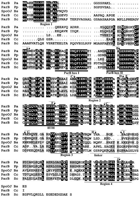
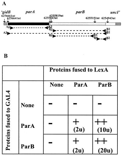
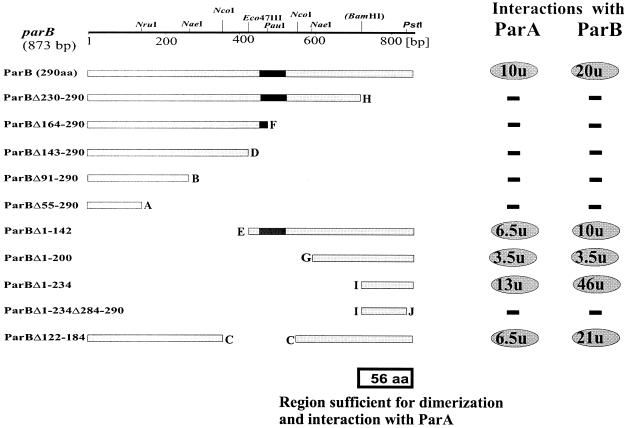
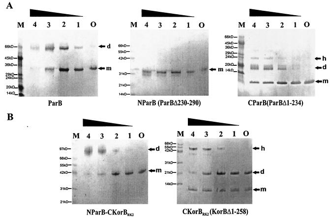
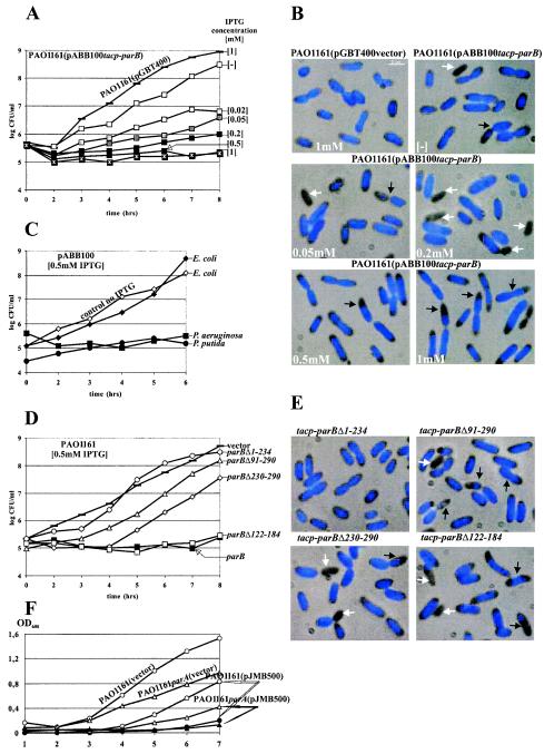
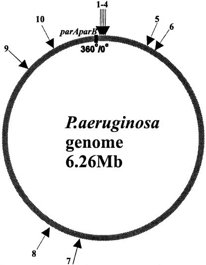
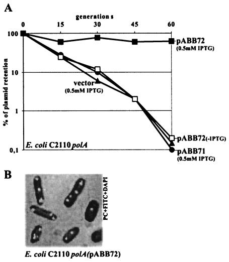
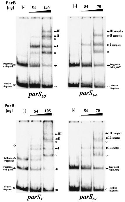
The par genes of Pseudomonas aeruginosa have been studied to increase the understanding of their mechanism of action and role in the bacterial cell. Key properties of the ParB protein have been identified and are associated with different parts of the protein. The ParB- ParB interaction domain was mapped in vivo and in vitro to the C-terminal 56 amino acids (aa); 7 aa at the C terminus play an important role. The dimerization domain of P. aeruginosa ParB is interchangeable with the dimerization domain of KorB from plasmid RK2 (IncP1 group). The C-terminal part of ParB is also involved in ParB-ParA interactions. Purified ParB binds specifically to DNA containing a putative parS sequence based on the consensus sequence found in the chromosomes of Bacillus subtilis, Pseudomonas putida, and Streptomyces coelicolor. The overproduction of ParB was shown to inhibit the function of genes placed near parS. This "silencing" was dependent on the parS sequence and its orientation. The overproduction of P. aeruginosa ParB or its N-terminal part also causes inhibition of the growth of P. aeruginosa and P. putida but not Escherichia coli cells. Since this inhibitory determinant is located well away from ParB segments required for dimerization or interaction with the ParA counterpart, this result may suggest a role for the N terminus of P. aeruginosa ParB in interactions with host cell components.


Figures

FIG. 1.
FIG. 1.
Comparison of P. aeruginosa ParB with the other well-studied chromosomal members of the ParB family (P. aeruginosa, Pa; P. putida, Pp; B. subtilis, Bs; C. crescentus, Cc; S. coelicolor, Sc). A black background indicates homology in all five proteins, and a gray background indicates homology in three or four proteins. Regions of conservation are underlined; a thick line corresponds to previously identified blocks (54), and a thin line corresponds to blocks identified here (for the first time). A broken line indicates the proposed linker region referred to in the text. Positions defining the ends of the deletions (see Fig. 3) are marked with coordinate numbers and letters for cross-referencing.
FIG. 2.
FIG. 2.
Detection of ParA-ParA, ParA-ParB, and ParB-ParB interactions with the yeast two-hybrid system (YTH). (A) Region of the genome used as the template in PCRs; genome coordinates define the primer sites. Coordinates shown correspond to P. aeruginosa genome coordinates. Primer A3 includes the ribosome-binding site for parA. Primers B3 and B4 introduce a BamHI site into the DNA sequence without changing the protein sequence. (B) Summary of results of the YTH assay. S. cerevisiae strain L40 was transformed with two compatible plasmids expressing only Gal4AD, LexA, or products of translational fusions between Gal4AD or LexA and ParA or ParB (parA was amplified with primers A1 and A2; parB was amplified with primers B1 and B2). −, no interactions (β-galactosidase activity of <0.2 U); +, weak interactions (β-galactosidase activity of 1 to 3 U); ++, strong interactions (β-galactosidase activity of >10 U).
FIG. 3.
FIG. 3.
Mapping of ParB-ParB and ParB-ParA interaction domains with the yeast two-hybrid system. The linear map at the top shows the restriction sites used for manipulations. The BamHI site was engineered by PCR (Fig. 2A). The putative H-T-H motif in ParB is indicated by black box. Lettering at the ends of deletions is the same as in Fig. 1. Interactions between deletion derivatives of ParB fused to LexA (pBTM derivatives) and intact ParA or ParB fused to Gal4AD (pGAD424 derivatives) were monitored. The two panels on the right summarize β-galactosidase activity: −, no interactions (β- galactosidase activity of <0.2 U); ovals, positive interactions (numbers in ovals correspond to mean units of β-galactosidase activity averaged from at least three assays).
FIG. 4.
FIG. 4.
Cross-linking of purified ParB and its truncated derivatives by glutaraldehyde. Proteins at a concentration of 0.1 mg/ml were incubated with various amounts of glutaraldehyde (GA). The samples were separated on homogeneous gels by SDS-12.5 or 20% PAGE and visualized by Coomassie blue staining (PHAST gel system). Lane O corresponds to the purified protein without GA; lanes 1 to 4 correspond to GA concentrations of 0.001, 0.002, 0.005, and 0.01% (wt/vol), respectively. m, monomer; d, dimer; h, higher-order complex. Lane M contains protein molecular weight markers; different molecular markers were used—one for the gels with ParB and C-ParB and another for the gels with N-ParB, N-ParB-C-KorB, and C-KorB.
FIG.5.
FIG.5.
Inhibition of host growth by the expression of parB. Overnight bacterial cultures carrying the plasmids indicated and grown under selection were diluted 104-fold into fresh selective medium without or with IPTG over a range of concentrations. Every hour, culture samples were serially diluted, and 20-μl aliquots were spotted on L agar and L agar-streptomycin plates. No plasmid loss was observed. The data shown are based on CFU from L agar plates. After approximately 4 h, samples were stained with DAPI and studied. (A) Effect on the growth of PAO1161 of increasing ParB expression from pABB100. Bacteria with empty vector pGBT400 and grown with 1 mM IPTG were used as the negative control. (B) Fluorescent images of samples from cultures monitored in panel A. White arrows indicate anucleate cells; black arrows indicate dislocalized, condensed chromosomes. (C) Effect of ParB production on the growth of E. coli DH5α and P. putida KT2442 carrying pABB100. PAO1161(pABB100) was used as a control. The data for all cultures grown without IPTG looked very similar and are represented by DH5α(pABB100) as the control (no IPTG). (D) Effect of overproduction of truncated forms of ParB on P. aeruginosa growth. PAO1161(pGBT400) not producing ParB and PAO1161(pABB100) producing wild-type ParB were used as controls. No difference in growth was observed for uninduced cultures. Only data for the induced cultures are presented. Expression plasmids with different derivatives of parB are listed in Table 1. (E) Fluorescent images of PAO1161 transformants overproducing truncated forms of ParB. The cultures were sampled after 4 h of growth in the presence of 0.5 mM IPTG. White arrows indicate anucleate cells; black arrows indicate cells with condensed chromosomes. (F) Effect of ParB overproduction on the growth of the PAO1161 parA::smh strain. The broad-host-range plasmid pBBR1MCS1 and its derivative pJMB500 carrying lacIq and a tacp- parB transcriptional fusion were used to transform PAO1161 parA and PAO1161. The cultures of the transformants were diluted 100-fold into L broth with 0.5 mM IPTG and without IPTG. Growth was monitored as the OD600. The growth of strains with vector pBBR1MCS1 is shown only for cultures with 0.5 mM IPTG.
FIG. 6.
FIG. 6.
Circular map of P. aeruginosa with localization of the putative ParB-binding sequences. The arrows indicate the putative ParB-binding sites; the numbers correspond to the positions of these sites on the circular map of the chromosome. The black box indicates the position of parAB less than 150 kb counterclockwise from oriC (position 0). The nucleotide sequences and exact locations of identified sites are listed in Table 2.
FIG. 7.
FIG. 7.
Segregational stabilization of an unstable plasmid in E. coli C2110 polA by parABS of P. aeruginosa. (A) Rate of loss of vector pOG04 and its derivatives with parS2/3 only (pABB71) and with a tacp-parAB transcriptional fusion and parS2/3 (pABB72), determined as described in Material and Methods. The values presented were calculated relative to the plasmids present in the initial overnight cultures. These values were about 30% for pOG04 and all of its derivatives, with the exception of pABB72, for which 50% plasmid retention occured. No effect of IPTG was observed for all plasmids, with the exception of pABB72. To simplify the diagram, data for cultures without IPTG are shown only for pABB72. Data for pABB70, carrying the tacp-parAB transcriptional fusion but lacking a parS site, were the same as for pOG04 and pABB71 (data not shown). (B) E. coli C2110 polA (pABB72 parS tacp-parAB lacIq) from a logarithmic culture grown without selection for 3 h. The cells were stained with DAPI and treated with primary anti-ParB antibodies and secondary FITC-conjugated anti-rabbit IgG antibodies. Only cells with ParB foci are presented.
FIG. 8.
FIG. 8.
DNA-binding activity of ParB. A radioactively labeled 284-nt EcoRI-NdeI DNA fragment of pUC18 with different parS sequences inserted between SalI and HindIII sites was further cut with PvuI to produce 93- and 191-nt fragments, the latter with the putative parS sequence. Purified N-His6-ParB was used in mobility shift experiments at the amounts shown above the lanes. Lanes marked with minus signs indicate DNA in binding buffer, with no protein included. Different retarded species are indicated by arrows: black arrows for complexes formed with parS and white diamonds for nonspecific complexes.

References

    1. Arnold, F. H. 1991. Metal-affinity separations: a new dimension in protein processing. Bio/Technology 9:151-156. - PubMed
    1. Autret, S., R. Nair, and J. Errington. 2001. Genetic analysis of the chromosome segregation protein Spo0J of Bacillus subtilis: evidence for separate domains involved in DNA binding and interactions with Soj protein. Mol. Microbiol. 41:743-755. - PubMed
    1. Bignell, C. R., A. S. Haines, D. Khare, and C. M. Thomas. 1999. Effect of growth rate and incC mutation on symmetric plasmid distribution by the IncP-1 partitioning apparatus. Mol. Microbiol. 34:205-216. - PubMed
    1. Bignell, C. R., and C. M. Thomas. 2001. The bacterial ParA-ParB partitioning proteins. J. Biotechnol. 91:1-34. - PubMed
    1. Bouet, J. V., and B. E. Funnell. 1999. P1 ParA interacts with the P1 partition complex at parS and an ATP-ADP switch controls ParA activities. EMBO J. 18:1415-1424. - PMC - PubMed

Publication types

MeSH terms

Substances

LinkOut - more resources