

Case Reports
A review of two cases of fenestrated internal jugular veins as seen by CT angiography
Affiliations
- PMID: 15466347
- PMCID: PMC7975450
Item in Clipboard
Case Reports
A review of two cases of fenestrated internal jugular veins as seen by CT angiography
AJNR Am J Neuroradiol.
2004 Sep.
Abstract
Venous fenestrations are a rarely seen entity in the neck. Although their clinical significance is questionable, their importance in presurgical planning may be considerable. We present a report of two cases of internal jugular vein fenestration.
Figures
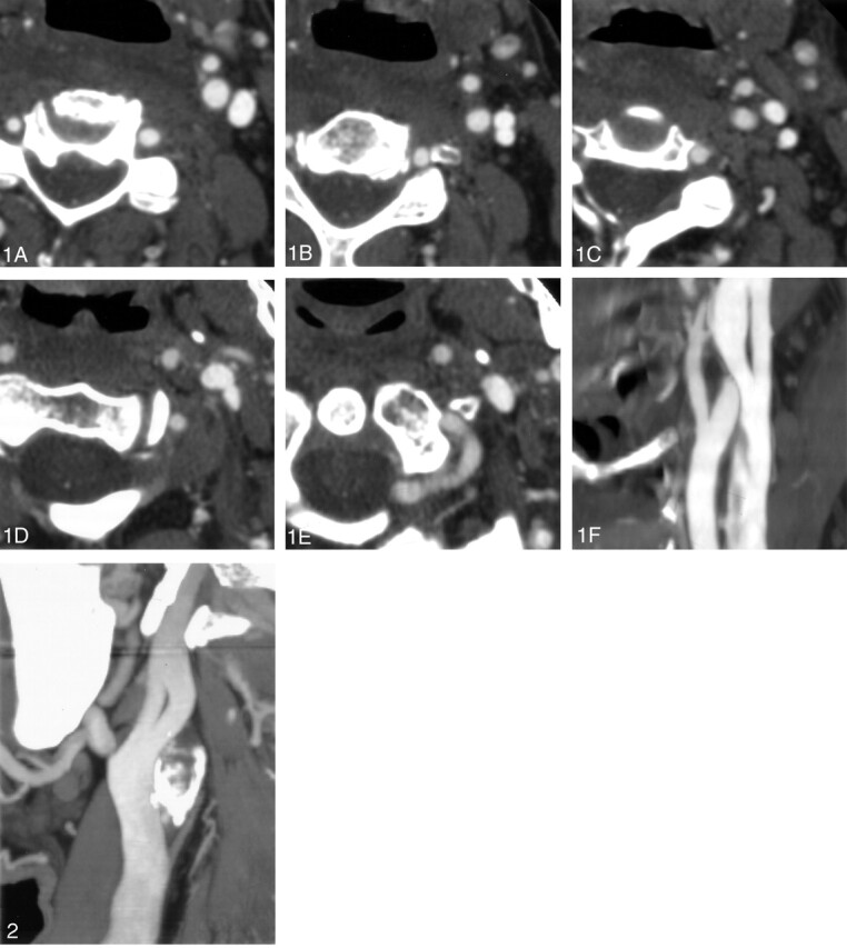
The appearance of an internal jugular vein on CTA images is characteristic. The vein begins its course with a single lumen (A, B). It then bifurcates forming two distinct lumena (C). As the vein continues caudally, it once again becomes a single vessel (D, E). Panel F shows the fenestrated internal jugular vein reformatted in a single sagittal, oblique plane.
The fenestrated internal jugular vein in our second patient is seen in a single oblique plane.
References
-
- Wollschlaeger G, Wollschlaeger PB, Lucas FV, Lopez VF. Experience and result with postmortem cerebral angiography performed as a routine procedure of the autopsy. AJR Am J Roentgenol 1967;101:68–87 - PubMed
-
- Prades JM, Timoshenko A, Dumollard JM, et al. High duplication of the internal jugular vein: clinical incidence in the adult and surgical consequences, a report of three clinical cases. Surg Radiol Anat 2002;24:129–132 - PubMed
-
- Som PM, Shugar JMA, Sacher M, Lanzieri CF. Internal jugular vein phlebectasia and duplication: CT features. J Comput Assist Tomogr 1985;9:390–392 - PubMed
-
- Rossi A, Tortoni-Donati P. Internal jugular vein phlebectasia and duplication: case report with magnetic resonance angiography features. Pediatr Radiol 2001;31:134. - PubMed
-
- Sylaidis P, Bardsley A, Montgomery P. Duplication of the internal jugular vein [letter]. Arch Otolaryngol Head Neck Surg 1997;123:1358. - PubMed
Publication types
MeSH terms
LinkOut - more resources
Full Text Sources
Medical