. 2004 Dec 3;279(49):51075-81.
doi: 10.1074/jbc.M408607200. Epub 2004 Oct 1.

Glycogen synthase kinase 3beta is a negative regulator of growth factor-induced activation of the c-Jun N-terminal kinase

Affiliations

Glycogen synthase kinase 3beta is a negative regulator of growth factor-induced activation of the c-Jun N-terminal kinase

Shuying Liu et al. J Biol Chem. .

Abstract

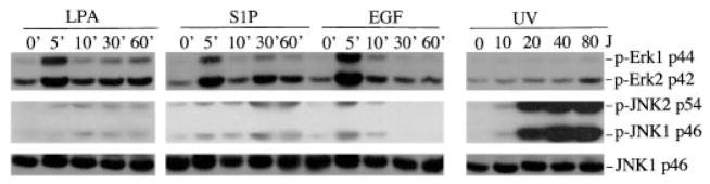
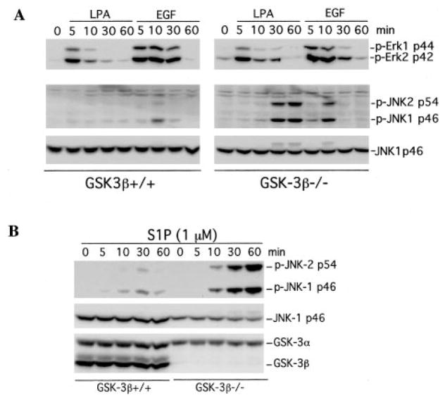
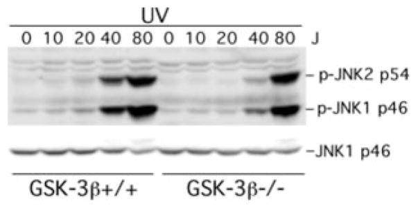
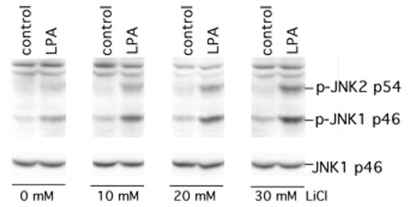
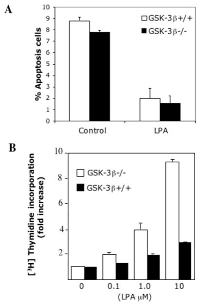
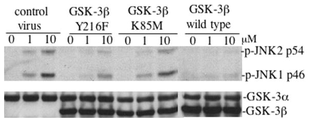
The c-Jun N-terminal kinase (JNK)/stress activated protein kinase is preferentially activated by stress stimuli. Growth factors, particularly ligands for G protein-coupled receptors, usually induce only modest JNK activation, although they may trigger marked activation of the related extracellular signal-regulated kinase. In the present study, we demonstrated that homozygous disruption of glycogen synthase kinase 3beta (GSK-3beta) dramatically sensitized mouse embryonic fibroblasts (MEFs) to JNK activation induced by lysophosphatidic acid (LPA) and sphingosine-1-phosphate, two prototype ligands for G protein-coupled receptors. To a lesser degree, a lack of GSK-3beta also potentiated JNK activation in response to epidermal growth factor. In contrast, the absence of GSK-3beta decreased UV light-induced JNK activation. The increased JNK activation induced by LPA in GSK-3beta null MEFs was insufficient to trigger apoptotic cell death or growth inhibition. Instead, the increased JNK activation observed in GSK-3beta-/- MEFs was associated with an increased proliferative response to LPA, which was reduced by the inhibition of JNK. Ectopic expression of GSK-3beta in GSK-3beta-negative MEFs restrained LPA-triggered JNK phosphorylation and induced a concomitant decrease in the mitogenic response to LPA compatible with GSK-3beta through the inhibition of JNK activation, thus limiting LPA-induced cell proliferation. Mutation analysis indicated that GSK-3beta kinase activity was required for GSK-3beta to optimally inhibit LPA-stimulated JNK activation. Thus GSK-3beta serves as a physiological switch to specifically repress JNK activation in response to LPA, sphingosine-1-phosphate, or the epidermal growth factor. These results reveal a novel role for GSK-3beta in signal transduction and cellular responses to growth factors.


Figures

Fig. 1
Fig. 1. JNK and ERK are differentially regulated by growth factors
Swiss 3T3 cells were starved in serum-free medium and incubated with LPA (1 μM), S1P (1 μM), or EGF (20 ng/ml). Cells were lysed in SDS sample buffer at 0, 5, 10, 30, and 60 min post-treatment and analyzed by immunoblotting for JNK and ERK phosphorylation using phospho-specific (p-Erk and p-JNK) antibodies. Starved Swiss 3T3 were subjected to UV light irradiation using an UV cross-linker at 10, 20, 40, and 80 J/m2 (J). UV light-irradiated cells were re-fed serum-free medium and returned to a CO2 incubator for 30 min before the cells were lysed and analyzed for JNK and ERK phosphorylation. Reprobing with anti-JNK1 antibody was included to show similar levels of loading among samples. For this and all other illustrations in the paper, similar results were obtained from three independent experiments.
Fig. 2
Fig. 2. Homozygous disruption of GSK-3β sensitizes cells to JNK phosphorylation induced by LPA, S1P, or EGF
GSK-3β knock-out (GSK-3β−/−) and wild type MEFs (GSK-3β+/+) were starved in a serum-free medium and stimulated with 1 μM LPA (A), 1 μM S1P (B), or 20 ng/ml EGF (A) for the indicated periods of time (minutes). The stimulated cells were lysed in SDS sample buffer and analyzed by immunoblotting for JNK and ERK phosphorylation using the corresponding phospho-specific (p-Erk and p-JNK) antibodies. Reprobing with an anti-JNK1 antibody was included to show equal loading among samples. The absence of GSK-3β in the knock-out MEFs was confirmed by reprobing with an anti-GSK-3α/β antibody (B).
Fig. 3
Fig. 3. Lack of GSK-3β does not enhance UV-induced JNK phosphorylation
GSK-3β−/− and GSK-3β+/+ MEFs were subjected to UV light radiation at the indicated doses (J, J/m2) using a UV light cross-linker as described under “Experimental Procedures.” Following UV light exposure, the cells were re-fed serum-free medium and returned to a CO2 incubator for 30 min. Cells were then lysed in SDS sample buffer and analyzed for JNK phosphorylation by immunoblotting with a JNK phospho-specific (p-JNK) antibody. Reprobing with anti-JNK1 antibody was included to confirm comparable levels of loading among samples.
Fig. 4
Fig. 4. Inhibition of GSK-3β activity by LiCl potentiates LPA-induced JNK phosphorylation
GSK-3β+/+ MEFs were starved and stimulated without (control) or with LPA (1 μM) for 30 min in the presence of lithium chloride at the indicated concentrations. LiCl was added to medium 75 min before LPA stimulation. Cell lysates were prepared and analyzed for JNK phosphorylation with a JNK phospho-specific (p-JNK) antibody. Reprobing with anti-JNK1 antibody was included to confirm comparable levels of loading among samples.
Fig. 5
Fig. 5. Increased JNK activation in GSK-3β−/− cells does not induce apoptosis and is associated with an increase in mitogenic response to LPA
A, GSK-3β−/− and GSK-3β+/+ cells were starved and incubated without or with LPA (10 μM). After 24 h, cells (including floating and adherent cells) were harvested, fixed, and stained via terminal deoxynucleotidyltransferase-mediated dUTP nick end-labeling (TUNEL) using ApopTag (Chemicon International). The percentages of TUNEL-stained positive apoptotic cells were determined by flow cytometry. The results are presented as means ± S.D. of duplicate data from three independent experiments. B, the mitogenic activity of LPA in GSK-3β−/− and GSK-3β+/+ cells was measured by [3H]thymidine incorporation as described under “Experimental Procedures.” The data were presented as fold increases (means ± S.D. of triplicate assays) with the activity in unstimulated control cells defined as one.
Fig. 6
Fig. 6. GSK-3β inhibits LPA-induced JNK phosphorylation
A, the effect of stable re-expression of GSK-3β. GSK-3β −/− cells were co-transfected with pcDNA3-HA-GSK-3β and pcDNA3.1/GS (containing the Zeocin resistance gene). Stable clones (G28 and G60) that expressed HA-GSK-3β were identified by immunoblotting with an antibody against GSK-3α/β and GSK-3β, with wild type MEFs included as a positive control (top). G28, G60, and empty vector control clone C9 were starved and treated with LPA (1 μM) for the indicated periods of time (min) (bottom). Cell lysates were prepared and analyzed by immunoblotting for JNK phosphorylation with a JNK phospho-specific (p-JNK) antibody (bottom). B, effect of the inducible expression of GSK-3β. The EC1214A-GSK3β inducible system involves a single vector carrying both the tetracycline-responsive transcriptional activator (tTA) and a tetracycline responsive element (TRE). The plasmid produces the tTA under the control of the cytomegalovirus promoter, and then tTA binds to TRE, leading to transcriptional activation of GSK3β in the absence of tetracycline. TK, thymidine kinase; BGH, bovine growth hormone; pA, poly(A). A stable clone with minimum expression of GSK-3β in the presence of tetracycline was identified and incubated in the presence (Tet 2 μg/ml) and absence of tetracycline (Tet 0 μg/ml) for 36 h. The cells were stimulated without or with LPA (1 μM) for 30 min and analyzed by immunoblotting for JNK phosphorylation with a JNK phospho-specific (p-JNK) antibody. Induction of GSK-3β expression in the inducible clone following removal of tetracycline was confirmed by immunoblotting with antibody against GSK-3α/β (bottom).
Fig. 7
Fig. 7. Re-expression of GSK-3β inhibits LPA-induced cell proliferation
GSK-3β−/− cells were infected with the HA-GSK-3β retro-virus or the control virus as detailed under “Experimental Procedures.” GFP-positive cells from HA-GSK-3β retrovirus-infected cells (GSK-3β infected) and control virus-infected cells (Vector control) were sorted, replated, and cultured for growth response to LPA. A, cell cycle analysis of the GSK-3β-infected, vector control, and GSK-3β+/+ cells. The cells were starved for 16 h and incubated with 10 μM LPA for 24 h before collection by trypsinization. Cells were stained with propidium iodide for flow cytometric analysis of cell cycle progression. B, growth curves of the GSK-3β-infected, vector control, and GSK-3β+/+ cells. Cells were seeded in 6-well plates (5 × 10⁴ cells/well). After attachment, cells were starved for 16 h and cultured in serum-free medium containing 10 μM LPA. Cell numbers were determined at 24-h intervals by counting in a hemocytometer. For both panel A and panel B data are presented as means ± S.D. of duplicate assays from three independent experiments. Statistical differences between GSK-3β-infected cells and vector control cells are indicated by an asterisk (p < 0.01).
Fig. 8
Fig. 8. Inhibition of JNK activation suppresses the increased proliferative response to LPA in GSK3β-negative cells
A, the effect of the JNK inhibitor SP600125 on LPA-induced c-Jun phosphorylation. GSK-3β-negative cells were starved and incubated with SP600125 at the indicated concentrations. One hour later the cells were stimulated with 10 μM LPA for 30 min. Cells were lysed in SDS sample buffer and analyzed by immunoblotting for c-Jun phosphorylation using phospho-specific (p-c-Jun) antibodies. GAPDH, glyceraldehyde-3-phosphate dehydrogenase. B, the effect of the JNK inhibitor on LPA-mediated cell proliferation. GSK3β-negative MEFs were prepared and plated as described for Fig. 7B. After attachment and starvation, cells were incubated with LPA (10 μM) in the presence of the indicated concentration of SP600125 that was added 1 h before the addition of LPA. Cell numbers were determined at 24-h intervals by counting in a hemocytometer. The data are presented as means ± S.D. of duplicate assays from three independent experiments. Statistical differences between cells treated with vehicle (Me2SO) and SP600125 are indicated by an asterisk (p < 0.01).
Fig. 9
Fig. 9. GSK-3β inhibition of LPA-induced JNK activation requires kinase activity
Wild type and mutant (Y216F and K85M) forms of GSK-3β were introduced into GSK-3β−/− cells by retrovirus-mediated gene transfer as detailed under “Experimental Procedures.” Enriched populations of GFP-positive cells were isolated by flow cytometry and replated in culture. After starvation in serum-free medium, the cells were incubated with LPA (1 and 10 μM) for 30 min before being lysed in SDS sample buffer and analyzed by immunoblotting for JNK phosphorylation with a JNK phospho-specific (p-JNK) antibody. Reprobing with an anti-GSK-3α/β antibody was included to serve as a loading control and to verify re-expression of GSK-3β in retrovirus-transduced cells. Similar results were obtained in three independent experiments.

References

    1. Hughes K, Nikolakaki E, Plyte SE, Totty NF, Woodgett JR. EMBO J. 1993;12:803–808. - PMC - PubMed
    1. Stambolic V, Woodgett JR. Biochem J. 1994;303:701–704. - PMC - PubMed
    1. Sutherland C, Leighton IA, Cohen P. Biochem J. 1993;296:15–19. - PMC - PubMed
    1. Cross DAE, Alessi DR, Cohen P, Andjelkovich M, Hemmings BA. Nature. 1995;378:785–789. - PubMed
    1. Fang X, Yu S, Lu Y, Bast RC, Jr, Woodgett JR, Mills GB. Proc Natl Acad Sci U S A. 2000;97:11960–11965. - PMC - PubMed

Publication types

MeSH terms