. 2004 Oct 11;167(1):35-41.
doi: 10.1083/jcb.200406136. Epub 2004 Oct 4.

XBP1: a link between the unfolded protein response, lipid biosynthesis, and biogenesis of the endoplasmic reticulum

Affiliations

XBP1: a link between the unfolded protein response, lipid biosynthesis, and biogenesis of the endoplasmic reticulum

Rungtawan Sriburi et al. J Cell Biol. .

Abstract

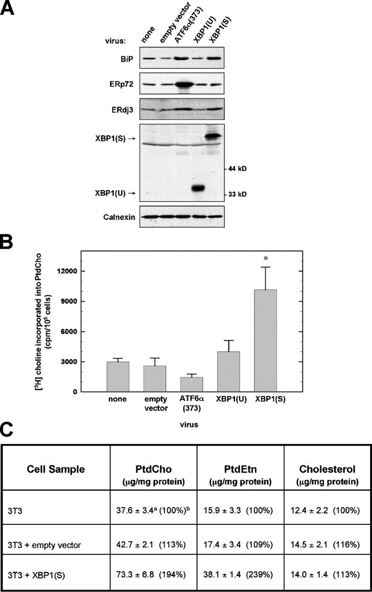
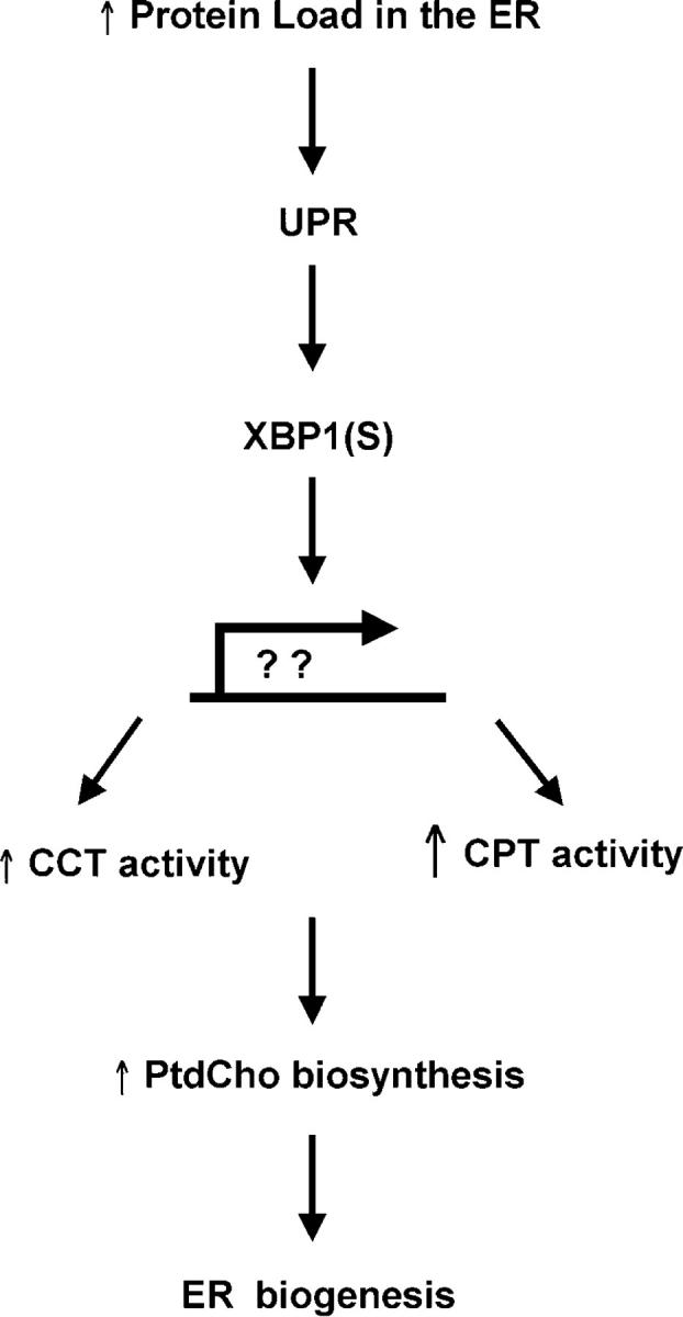
When the protein folding capacity of the endoplasmic reticulum (ER) is challenged, the unfolded protein response (UPR) maintains ER homeostasis by regulating protein synthesis and enhancing expression of resident ER proteins that facilitate protein maturation and degradation. Here, we report that enforced expression of XBP1(S), the active form of the XBP1 transcription factor generated by UPR-mediated splicing of XBP1 mRNA, is sufficient to induce synthesis of phosphatidylcholine, the primary phospholipid of the ER membrane. Cells overexpressing XBP1(S) exhibit elevated levels of membrane phospholipids, increased surface area and volume of rough ER, and enhanced activity of the cytidine diphosphocholine pathway of phosphatidylcholine biosynthesis. These data suggest that XBP1(S) links the mammalian UPR to phospholipid biosynthesis and ER biogenesis.


Figures

Figure 1.
Figure 1.
Phospholipid synthesis in NIH-3T3 fibroblasts retrovirally transduced with UPR transcriptional activators. NIH-3T3 cells were transduced with the indicated pBMN-I-GFP retroviral vectors. (A) At 48 h after transduction, cells were harvested and equivalent amounts of cell lysate protein were resolved by standard SDS-PAGE under reducing conditions. Chemiluminescent immunoblotting was performed. A nonspecific band just below the position of pXBP1(S) was detected by the anti-XBP1 antibody. Calnexin served as a loading control. (B) Cells were metabolically labeled with [3H]choline for 2 h, a labeling interval in which [3H]choline is incorporated exclusively into PtdCho. The amount of radiolabel present in cellular lipid extracts was normalized as cpm/105 cells and was plotted as the mean ± SD (n = 3; asterisk denotes P = 0.002 for comparison of XBP1(S) to empty vector). (C) Cells were harvested and total cellular mass of PtdCho, PtdEtn, and cholesterol was determined by flame ionization. Superscript a: average of triplicate determinations from two independent experiments (n = 6) ± SD. Superscript b: control NIH-3T3 values set at 100% for comparison.
Figure 2.
Figure 2.
Microscopy analysis of the ER. (A) At 48 h after transduction with the indicated retroviral vectors, thin sections were prepared from NIH-3T3 cells and examined by transmission EM. Representative micrographs of increasing magnification (from top to bottom) are shown with scale bar (1 μm) in top right corner: middle panels, arrow indicates representative ER; bottom panels, arrow indicates membrane-bound ribosomes. N, nucleus. (B) Stereological analysis of RER volume (top) and RER surface area (bottom) was performed on electron micrographs and the mean ± SEM are plotted (n = 4 stereological sets with 2–16 micrographs per set; *, P < 0.01). (C) Immunofluorescence analysis (using anti-KDEL mAb) and confocal microscopy was performed on NIH-3T3 cells grown on coverslips and transduced with the indicated retroviral vectors for 48 h.
Figure 3.
Figure 3.
Enzymatic activities in the CDP-choline pathway of PtdCho synthesis. The relative activities of CK, CCT, and CPT, which constitute the CDP-choline pathway of PtdCho biosynthesis (right side of figure), were determined using lysates or microsomes prepared from NIH-3T3 cells (▪), or NIH-3T3 cells transduced with XBP1(S) (•) or empty vector (○). The rates of production of phosphocholine (P-Choline), CDP-Choline, and PtdCho were compared as a function of total protein in each assay. (A) Data for CK are averaged from triplicate determinations and are representative of two independent experiments. (B) Data for CCT are averaged from five determinations obtained in two independent experiments. (C) Data for CPT are averaged from duplicate determinations and are representative of three independent experiments.
Figure 4.
Figure 4.
Expression of enzymes that function in the CDP-choline pathway of PtdCho synthesis. Total RNA was prepared from NIH-3T3 fibroblasts harvested at 24 and 48 h after transduction with the indicated retroviral vectors. (A) The relative levels of expression of the CCTα, β2, and β3 isoform cDNAs at the 48-h interval were measured by quantitative real-time PCR. The amount of target RNA was normalized to the endogenous GAPDH reference and related to the amount of target CCTα in NIH-3T3, which was set as the calibrator at 1.0 (superscript a). The mean ± SD (superscript b) of triplicate determinations is shown. (B) Northern blot analysis using 32P-labeled cDNA probes specific for CEPT1 and CPT1, the two genes encoding enzymes possessing CPT activity; ERdj3, a known XBP1 target as a positive control; and γ-actin as a loading control.
Figure 5.
Figure 5.
A model for how XBP1 links the UPR to ER biogenesis. In response to increased demand on the protein folding capacity of the ER, UPR-mediated splicing of XBP1 mRNA yields the XBP1(S) transcriptional activator. We propose that XBP1(S) induces expression of unknown gene products that lead to an increase in CCT activity and a large increase in CPT activity, thereby augmenting synthesis of PtdCho. Increased production of PtdCho, the major phospholipid of the ER membrane, is critical for expansion of the ER compartment.

Comment in

References

    1. Bligh, E.G., and W.J. Dyer. 1959. A rapid method of total lipid extraction and purification. Can. J. Med. Sci. 37:911–917. - PubMed
    1. Brewer, J.W., and J.A. Diehl. 2000. PERK mediates cell-cycle exit during the mammalian unfolded protein response. Proc. Natl. Acad. Sci. USA. 97:12625–12630. - PMC - PubMed
    1. Calfon, M., H. Zeng, F. Urano, J.H. Till, S.R. Hubbard, H.P. Harding, S.G. Clark, and D. Ron. 2002. IRE1 couples endoplasmic reticulum load to secretory capacity by processing the XBP-1 mRNA. Nature. 415:92–96. - PubMed
    1. Cornell, R.B., and I.C. Northwood. 2000. Regulation of CTP:phosphocholine cytidylyltransferase by amphitropism and relocalization. Trends Biochem. Sci. 25:441–447. - PubMed
    1. Cox, J.S., R.E. Chapman, and P. Walter. 1997. The unfolded protein response coordinates the production of endoplasmic reticulum protein and endoplasmic reticulum membrane. Mol. Biol. Cell. 8:1805–1814. - PMC - PubMed

Publication types

MeSH terms