. 2004 Oct 5;32(17):5249-59.
doi: 10.1093/nar/gkh842. Print 2004.

DNA double strand break repair in human bladder cancer is error prone and involves microhomology-associated end-joining

Affiliations

DNA double strand break repair in human bladder cancer is error prone and involves microhomology-associated end-joining

Johanne Bentley et al. Nucleic Acids Res. .

Abstract

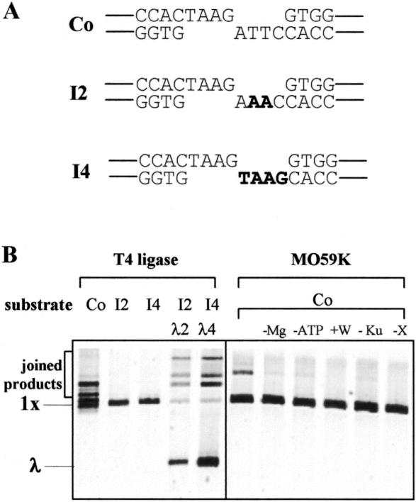
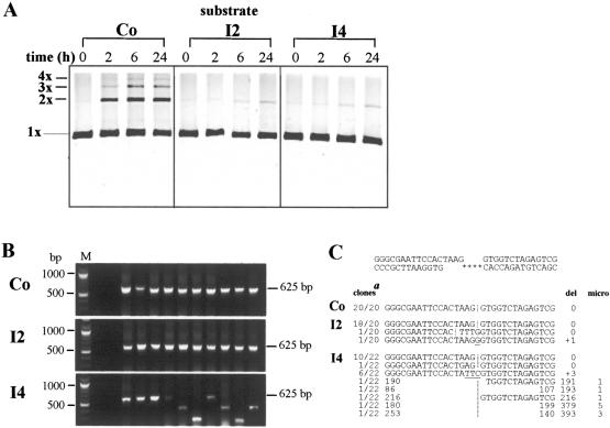
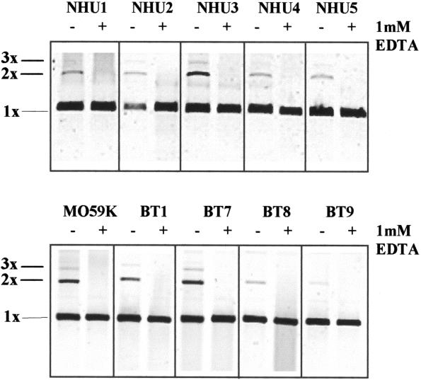
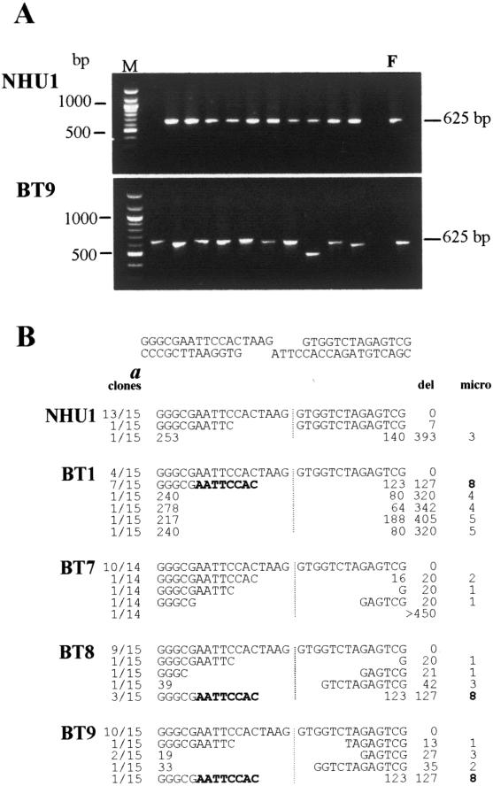
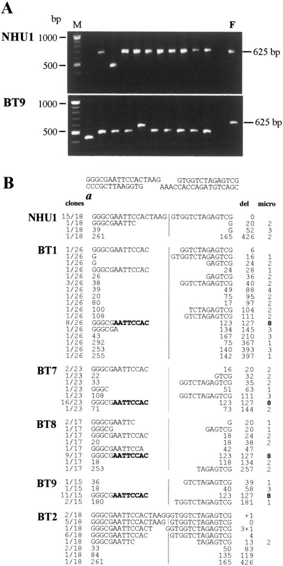
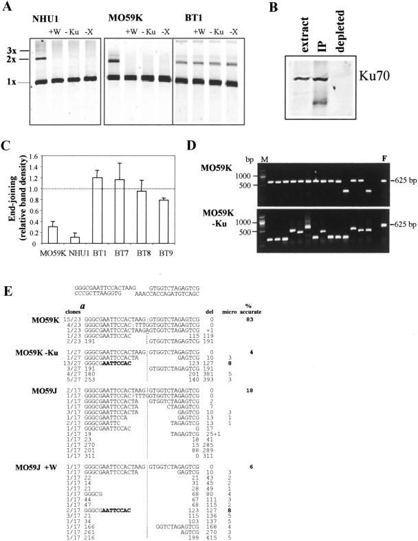
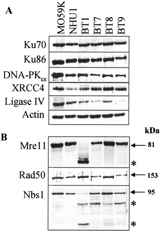
In human cells DNA double strand breaks (DSBs) can be repaired by the non-homologous end-joining (NHEJ) pathway. In a background of NHEJ deficiency, DSBs with mismatched ends can be joined by an error-prone mechanism involving joining between regions of nucleotide microhomology. The majority of joins formed from a DSB with partially incompatible 3' overhangs by cell-free extracts from human glioblastoma (MO59K) and urothelial (NHU) cell lines were accurate and produced by the overlap/fill-in of mismatched termini by NHEJ. However, repair of DSBs by extracts using tissue from four high-grade bladder carcinomas resulted in no accurate join formation. Junctions were formed by the non-random deletion of terminal nucleotides and showed a preference for annealing at a microhomology of 8 nt buried within the DNA substrate; this process was not dependent on functional Ku70, DNA-PK or XRCC4. Junctions were repaired in the same manner in MO59K extracts in which accurate NHEJ was inactivated by inhibition of Ku70 or DNA-PK(cs). These data indicate that bladder tumour extracts are unable to perform accurate NHEJ such that error-prone joining predominates. Therefore, in high-grade tumours mismatched DSBs are repaired by a highly mutagenic, microhomology-mediated, alternative end-joining pathway, a process that may contribute to genomic instability observed in bladder cancer.


Figures

Figure 1
Figure 1
End-joining with BstX1 substrates. (A) The 3′ single stranded overhang sequences of BstX1-generated plasmid DNA substrates with compatible (Co) and two (I2) or four (I4) base mismatched ends (incompatible bases shown in bold). (B) SYBR Green I detection of joining in the presence of T4 ligase or MO59K extract with the linear 3.2 kb (1×) substrates indicated. The λ2 and λ4 are 1.2 kb fragments containing ends which are compatible with I2 and I4 substrates, respectively. Incubations in the absence of 0.5 mM Mg(OAc)2 (−Mg) or 1 mM ATP (−ATP) or the presence of the DNA-PK inhibitor 10 μM wortmannin (+W), 1:50 anti-Ku70 antibody (−Ku) and 1:50 anti-XRCC4 antibody (−X) are indicated. Gels shown are representative of three independent experiments.
Figure 2
Figure 2
Accuracy of end-joining in MO59K extract. (A) End-joining reactions were incubated for 0, 2, 6 or 24 h with the plasmid DNA substrate (1×) indicated. The formation of joined linear dimers, trimers and tetramers are shown (2×, 3× and 4×). Gels shown are representative of two independent experiments. (B) PCR analysis of randomly chosen recombinant clones resulting from the cloning of DNA joins formed in the presence of MO59K extract and Co, I2 or I4 substrates. Joins formed accurately would produce a PCR product (with primers T2 and T3) of 625 bp, junctions formed by the deletion of terminal nucleotides resulted in smaller products. M, molecular mass markers. (C) Sequence analysis of joins formed with DNA substrates Co, I2 and I4, where **** indicates the sequence ATTC, AAAC and TAAG for each substrate, respectively. aTop strand of sequence is shown with the join denoted by a vertical bar, inserted nucleotides are underlined and the number of nucleotides deleted from each end of the substrate is indicated. The number of recombinants analysed (clones), the total number of nucleotides deleted or inserted to form the join (del) and microhomology (micro) at junctions are indicated. Joins are termed accurate if the sequence of DNA ends has been preserved. For incompatible substrates accurate joins are formed when 3 nt of one overhang are removed and overlap/fill-in occurs, such that the sequence information of one strand is retained. Deletions are therefore expressed as the number of nucleotides removed in addition to this processing.
Figure 3
Figure 3
End-joining in NHU and bladder tumour extracts. Joining of compatible DNA substrate by NHU (1–5), MO59K and bladder tumour (BT1, 7, 8, 9) extracts in the presence or absence of 1 mM EDTA. Plasmid DNA substrate (1×) and joined dimers and trimers (2× and 3×) are shown. Gels shown are representative of three independent experiments.
Figure 4
Figure 4
Compatible DNA substrate end-joining in bladder tumours. (A) PCR analysis of randomly chosen recombinant clones resulting from the cloning of DNA joins formed in the presence of compatible substrate with NHU1 and BT9 extracts. Joins formed accurately produced a full length PCR product of 625 bp with primers T2 and T3 (as indicated by F); junctions formed by the deletion of terminal nucleotides resulted in smaller products. M, molecular mass markers. (B) Sequence analysis of joins formed in NHU1 and bladder tumour (BT1, 7, 8, 9) extracts. Junctions formed using an 8 bp microhomology are shown in bold. aAs in Figure 2.
Figure 5
Figure 5
Error-prone end-joining in bladder tumours with incompatible DNA substrate I2. (A) PCR analysis of randomly chosen recombinant clones resulting from the cloning of DNA joins formed in the presence of incompatible substrate with NHU1 and BT9 extracts. Joins formed accurately produced a full length PCR product of 625 bp with primers T2 and T3 (as indicated by F); junctions formed by the deletion of terminal nucleotides resulted in smaller products. M, molecular mass markers. (B) Sequence analysis of joins formed in NHU1 and bladder tumour (BT1, 7, 8, 9, 2) extracts. Junctions formed using an 8 bp microhomology are shown in bold. aAs in Figure 2.
Figure 6
Figure 6
NHEJ-independent end-joining in bladder tumours and Ku70-depleted MO59K. (A) End-joining with the Co substrate in the presence of 10 μM wortmannin (+W), 1:50 anti-Ku70 antibody (−Ku) and 1:50 anti-XRCC4 antibody (−X) as indicated. Plasmid DNA substrate (1×) and joined dimers and trimers (2× and 3×) are shown. Gels shown are representative of two independent experiments. (B) Ku70 levels in 20 μg untreated MO59K sample (extract) and, following immunodepletion of Ku70 using 1:50 anti-Ku70 antibody, protein eluted from sepharose-G beads (IP) and remaining in extract (depleted). (C) PCR analysis of products joined by MO59K, NHU1 and bladder tumour (BT1, 7, 8, 9) extracts and I2 substrate in the presence of 1:20 anti-Ku70 antibody (−Ku). The level of end-joining is expressed as PCR band density in the presence of anti-Ku70 relative to that without antibody (three independent experiments, SD). (D) PCR analysis of randomly chosen recombinant clones containing DNA joins formed with incompatible I2 substrate and MO59K extract pretreated with 1:20 dilution of anti-Ku70 monoclonal antibody (−Ku). Joins formed accurately produced a full length PCR product of 625 bp with primers T2 and T3 (as indicated by F); junctions formed by the deletion of terminal nucleotides resulted in smaller products. M, molecular mass markers. (E) Sequence analysis of joins formed by MO59K and MO59J extract pretreated with 1:20 dilution of anti-Ku70 monoclonal antibody (−Ku) or 10 μM wortmannin (+W). Junctions formed using an 8 bp microhomology are shown in bold, % accurate indicates the proportion of accurate joins formed. aAs in Figure 2.
Figure 7
Figure 7
The levels of NHEJ components in bladder tumour extracts. Western blot analysis of (A) Ku70, Ku86, DNA-PKcs, XRCC4, ligase IV, actin and (B) Mre11, Rad50 and Nbs1 in MO59K, NHU1 and bladder tumour (BT1, 7, 8, 9) extracts. Molecular mass is indicated in kilodalton with proteins of aberrant size indicated by *. Blots shown are representative of three independent experiments.

References

    1. Valerie K. and Povirk,L.F. (2003) Regulation and mechanisms of mammalian double strand break repair. Oncogene, 22, 5792–5812. - PubMed
    1. Leiber M.R., Ma,Y., Pannicke,U. and Schwarz,K. (2003) Mechanism and regulation of human non-homologous DNA end-joining. Nature Rev. Mol. Cell. Biol., 4, 712–720. - PubMed
    1. Takata M., Sasaki,M.S., Sonoda,E., Morrison,C., Hashimoto,M., Utsumi,H., Yamaguchi-Iwai,Y., Shinohara,A. and Takeda,S. (1998) Homologous recombination and non-homologous end-joining pathways of DNA double-strand break repair have overlapping roles in the maintenance of chromosomal integrity in vertebrate cells. EMBO J., 17, 5497–5508. - PMC - PubMed
    1. Walker J.R., Corpina,R.A. and Goldberg,J. (2001) Structure of the Ku heterodimer bound to DNA and its implications for double-strand break repair. Nature, 412, 607–614. - PubMed
    1. Baumann P. and West,S.C. (1998) DNA end-joining catalysed by human cell free extracts. Proc. Natl Acad. Sci. USA, 95, 14066–14070. - PMC - PubMed

Publication types